Active-fluidics versus gravity-fluidics in uncomplicated cataract surgery: using optical coherence tomography angiography to estimate early changes in macular and optic disc microcirculation
Alessandra Scampoli, Emanuele Crincoli, Lorenzo Governatori, Carlo Monaco, Cosma Danilo Mancini, Federico Giannuzzi, Matteo Mario Carlà, Giulia Grieco, Stanislao Rizzo, Tomaso Caporossi

TL;DR
This study compares two cataract surgery techniques using OCTA to assess early effects on retinal and optic disc blood flow, finding that one method better preserves microcirculation.
Contribution
The study introduces OCTA as a tool to detect early microcirculation changes in cataract surgery and identifies AFS as a potentially safer technique for preserving retinal vasculature.
Findings
The GFS group showed significantly lower macular vessel density at 24 hours post-surgery compared to the AFS group.
Seven days after surgery, the GFS group had higher retinal nerve fiber layer thickness and lower vessel density in specific quadrants compared to the AFS group.
The AFS system appears to better protect retinal and optic disc microcirculation during cataract surgery.
Abstract
To compare, using optical coherence tomography angiography (OCTA), the earliest changes and damages to macular and optic disc microcirculation after active-fluidics system (AFS) and gravity-fluidics system (GFS) procedures in uncomplicated cataract surgery. We included 42 eyes affected by uncomplicated cataracts and divided them into two groups: 21 eyes were randomly assigned to an AFS group and 21 eyes were randomly assigned to a GFS group. Expert examiners performed OCTA 30 ± 10 min before surgery (T0), 30 ± 8 min after surgery (T1), 24 ± 2 h after surgery (T2) and 7 days after surgery (T3). No significant differences at T1 were detected between the groups. At T2, eyes in the GFS group exhibited a whole macula deep capillary plexus vessel density of 37.9 ± 5.8%, which was significantly lower than that of the eyes in the AFS group (42.2 ± 5.7%; p = 0.048). At T3, eyes in the GFS…
Genes, proteins, chemicals, diseases, species, mutations and cell lines named across the full text — each resolved to its canonical identifier and authoritative record.
Click any figure to enlarge with its caption.
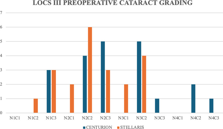 Figure 1
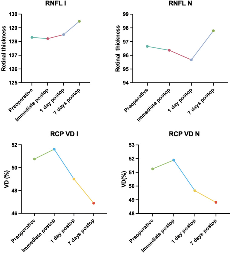 Figure 2
Peer Reviews
No public reviews on file for this paper yet. If you reviewed it on a platform where reviews are public (OpenReview, ICLR, NeurIPS, ICML), you can paste yours below so the community can read it here.
Videos
No videos yet. Explain this paper in a talk, walkthrough, or lecture? Add one.
Taxonomy
TopicsRetinal and Macular Surgery · Retinal Diseases and Treatments · Glaucoma and retinal disorders