Application of deep neural networks in automatized ventriculometry and segmentation of the aqueduct in pediatric hydrocephalus patients
Fabienne Kühne, Kilian Rüther, Christopher Güttler, Juliane C. Stöckel, Ulrich-Wilhelm Thomale, Anna Tietze, Andrea Dell’Orco

TL;DR
This paper compares two deep learning models for automatically measuring brain ventricles in children with hydrocephalus, finding that one model is more reliable and easier to use clinically.
Contribution
The study evaluates and compares VParNet and nnU-Net for pediatric ventricular segmentation, highlighting nnU-Net's advantages in handling complex cases without preprocessing.
Findings
Both VParNet and nnU-Net achieved high segmentation accuracy (DSC: 0.87–0.95) for pediatric ventricles.
nnU-Net outperformed VParNet in handling challenging cases due to no preprocessing requirements.
Aqueduct segmentation remained difficult, with nnU-Net achieving a DSC of 0.68.
Abstract
This study validated VParNet and nnU-Net for ventricular segmentation in pediatric hydrocephalus, a condition characterized by irregular and asymmetric ventricular shapes. Manual segmentation of 139 MRI scans (ages range 2.6–20.3 years) was performed for the four ventricles and the aqueduct. A five-fold cross-validation was conducted for both models. VParNet was tested with its original weights and after retraining on pediatric data. nnU-Net was extended to also segment the aqueduct. Performance was evaluated using the Dice Similarity Coefficient (DSC), Intraclass Correlation Coefficient (ICC), and Minimal Detectable Change (MDC). VParNet preprocessing failed in 20.9% of cases, requiring subject exclusion. Both models showed good to excellent segmentation accuracy and reliability (DSC: 0.87–0.95; ICC: 0.81–1.0). Retraining VParNet improved DSC scores. MDC values (0.05–3.0) indicated…
Genes, proteins, chemicals, diseases, species, mutations and cell lines named across the full text — each resolved to its canonical identifier and authoritative record.
Click any figure to enlarge with its caption.
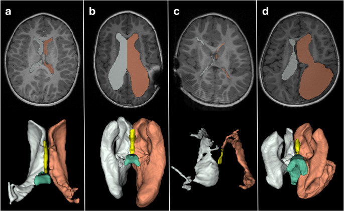 Figure 1
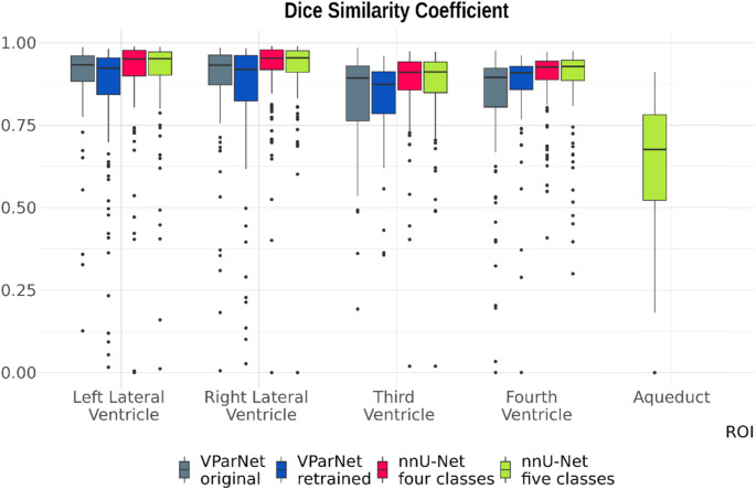 Figure 2
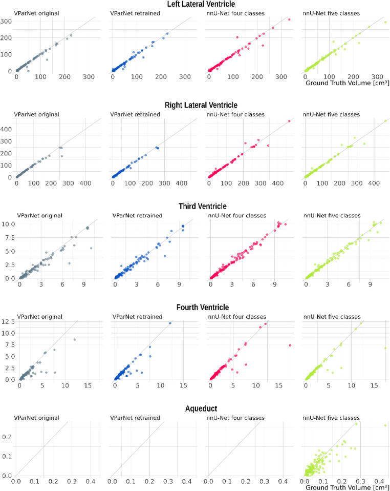 Figure 3
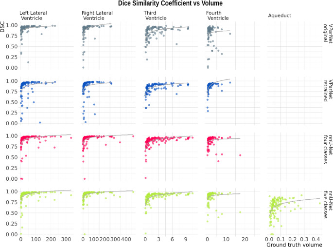 Figure 4
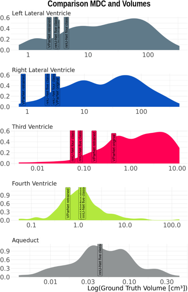 Figure 5
Peer Reviews
No public reviews on file for this paper yet. If you reviewed it on a platform where reviews are public (OpenReview, ICLR, NeurIPS, ICML), you can paste yours below so the community can read it here.
Videos
No videos yet. Explain this paper in a talk, walkthrough, or lecture? Add one.
Taxonomy
TopicsCerebrospinal fluid and hydrocephalus · Medical Image Segmentation Techniques · Fetal and Pediatric Neurological Disorders