Caudal lumbar subarachnoid diverticulum in a Cockapoo
Joe Poacher, Paul Freeman

TL;DR
A Cockapoo was diagnosed with a rare spinal condition called a subarachnoid diverticulum, which was successfully treated with surgery.
Contribution
This case report presents a rare spinal condition in a Cockapoo, highlighting its diagnostic and clinical features.
Findings
MRI identified a subarachnoid diverticulum at L6-7 containing cerebrospinal fluid.
Surgical confirmation and decompression led to full clinical resolution.
The condition initially mimicked an intervertebral disc extrusion.
Abstract
We describe a Cockapoo with a subarachnoid diverticulum (Type III), at the level of L6-7. Magnetic resonance imaging identified a circumferential dilatation of the dural sac, extending from the cranial endplate of L6 to the midbody of L7, containing T2-weighted hyperintense and T1-weighted hypointense material that suppressed on fluid-attenuated inversion recovery sequences, consistent with cerebrospinal fluid. An exploratory dorsal laminectomy confirmed a subarachnoid diverticulum (Type III), and a durotomy was performed. After surgical decompression, full clinical resolution was observed. This case had a clinical presentation that mimicked an intervertebral disc extrusion.
Genes, proteins, chemicals, diseases, species, mutations and cell lines named across the full text — each resolved to its canonical identifier and authoritative record.
Click any figure to enlarge with its caption.
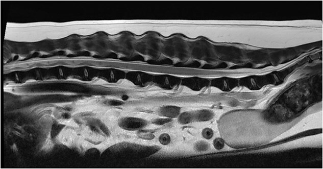 Figure 1
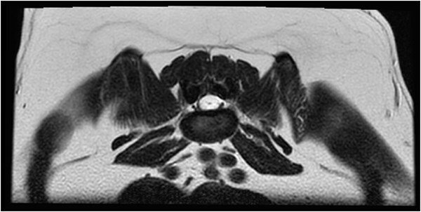 Figure 2
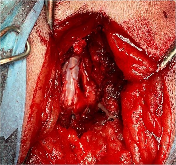 Figure 3
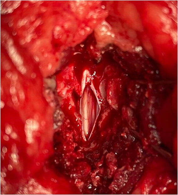 Figure 4
Peer Reviews
No public reviews on file for this paper yet. If you reviewed it on a platform where reviews are public (OpenReview, ICLR, NeurIPS, ICML), you can paste yours below so the community can read it here.
Videos
No videos yet. Explain this paper in a talk, walkthrough, or lecture? Add one.
Taxonomy
TopicsSpinal Dysraphism and Malformations · Veterinary Orthopedics and Neurology · Head and Neck Surgical Oncology