Epigenetic and O-glycosylation regulation of p66Shc mitigates mitochondrial oxidative stress in aortic dissection
Wenjun Zhang, Wanjun Liu, Xiaodan Zhong, Lei Dai, Xiaolei Liu, Shiliang Li, Hongcheng Jiang, Xingwei He, Wei Dong, Lijuan Lu, Li Zhu, Thati Madhusudhan, Hongjie Wang, Hesong Zeng

TL;DR
This study shows that aPC reduces aortic dissection by suppressing p66Shc through epigenetic and glycosylation mechanisms, protecting mitochondria from oxidative stress.
Contribution
The study reveals a novel mechanism where aPC regulates p66Shc via epigenetic and O-glycosylation pathways to mitigate mitochondrial oxidative stress in aortic dissection.
Findings
aPC downregulates p66Shc expression through epigenetic modifications.
aPC increases O-glycosylation of p66Shc at Thr29, preventing mitochondrial translocation and ROS production.
These mechanisms inhibit the progression of aortic dissection in a mouse model.
Abstract
Background: Aortic dissection (AD) is a life-threatening vascular emergency with limited effective pharmacological treatments. Recent studies have identified Src homology 2 domain-containing transforming protein C1 (p66Shc) as a crucial mediator of oxidative stress, apoptosis, and inflammation in aortic cells, thereby contributing to cellular dysfunction and vascular remodeling implicated in AD development and progression. Despite its established role in promoting vascular dysfunction and remodeling, the protective potential of targeting p66Shc in AD remains unclear. Methods: We quantified activated protein C (aPC) levels in clinical plasma samples from control subjects and AD patients using enzyme-linked immunosorbent assay (ELISA). To evaluate changes in p66Shc expression, we analyzed aortic tissues by Western blotting (WB), immunohistochemistry (IHC), and immunofluorescence (IF)…
Genes, proteins, chemicals, diseases, species, mutations and cell lines named across the full text — each resolved to its canonical identifier and authoritative record.
Click any figure to enlarge with its caption.
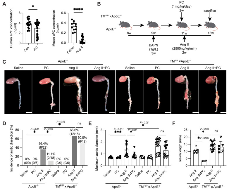 Figure 1
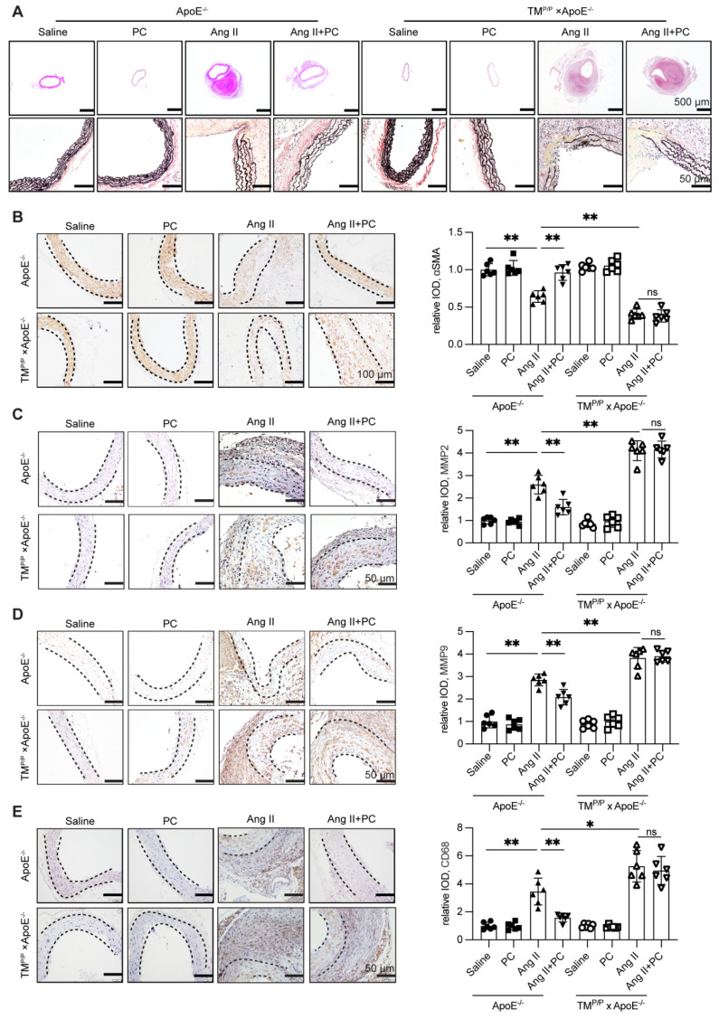 Figure 2
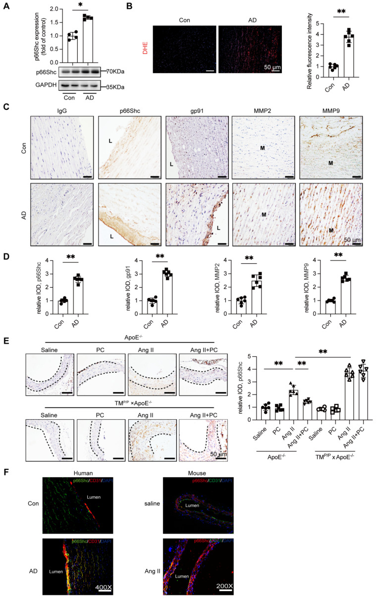 Figure 3
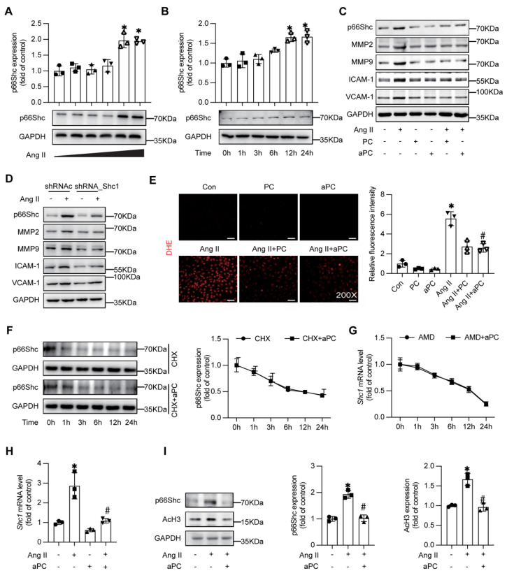 Figure 4
 Figure 5
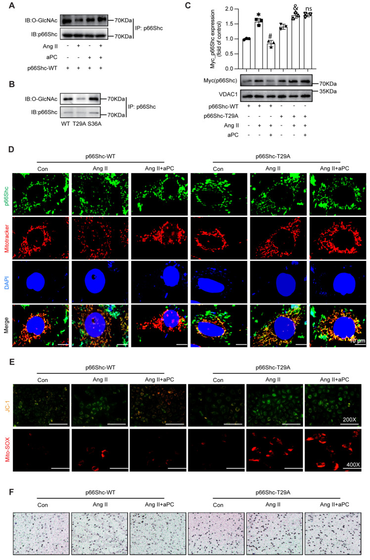 Figure 6
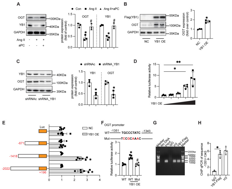 Figure 7
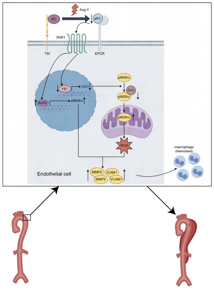 Figure 8
Peer Reviews
No public reviews on file for this paper yet. If you reviewed it on a platform where reviews are public (OpenReview, ICLR, NeurIPS, ICML), you can paste yours below so the community can read it here.
Videos
No videos yet. Explain this paper in a talk, walkthrough, or lecture? Add one.
Taxonomy
TopicsConnective tissue disorders research · Blood Coagulation and Thrombosis Mechanisms · Signaling Pathways in Disease