O-glycosylation in Cancer: Emerging Paradigms and Prospects for Precision Oncology
Junhao Wei, Shengbao Hu, Wanfang Chen, Hye Song Paek, Guohong Liu, Yunbao Pan

TL;DR
O-glycosylation influences cancer progression and treatment, offering new opportunities for precision oncology through biomarkers and therapies.
Contribution
Highlights new insights into O-glycosylation's role in cancer and its potential for personalized treatment strategies.
Findings
Aberrant O-glycosylation promotes oncogenesis through altered glycans and dysregulated enzymes.
Multi-omics and machine learning improve tumor classification using glycosylation signatures.
RNA O-glycosylation adds new regulatory layers and therapeutic possibilities.
Abstract
O-glycosylation is a key post-translational modification that profoundly shapes tumor biology by regulating cell proliferation, metastasis, and immune evasion. Aberrant O-glycosylation features truncated glycans such as Tn and sialyl-Tn antigens together with dysregulated glycosyltransferases and promotes oncogenesis in diverse malignancies. This review summarizes recent progress in elucidating the role of O-glycosylation in cancer with emphasis on its effects on cell-surface glycoproteins, intracellular signaling pathways, and emerging RNA modifications. Integration of multi-omics data and machine learning has transformed tumor classification and prognosis prediction through distinct glycosylation signatures and now supports personalized treatment strategies. Newly discovered O-glycosylation of RNA reveals additional regulatory layers and broadens the field of glycosylation research.…
Genes, proteins, chemicals, diseases, species, mutations and cell lines named across the full text — each resolved to its canonical identifier and authoritative record.
Click any figure to enlarge with its caption.
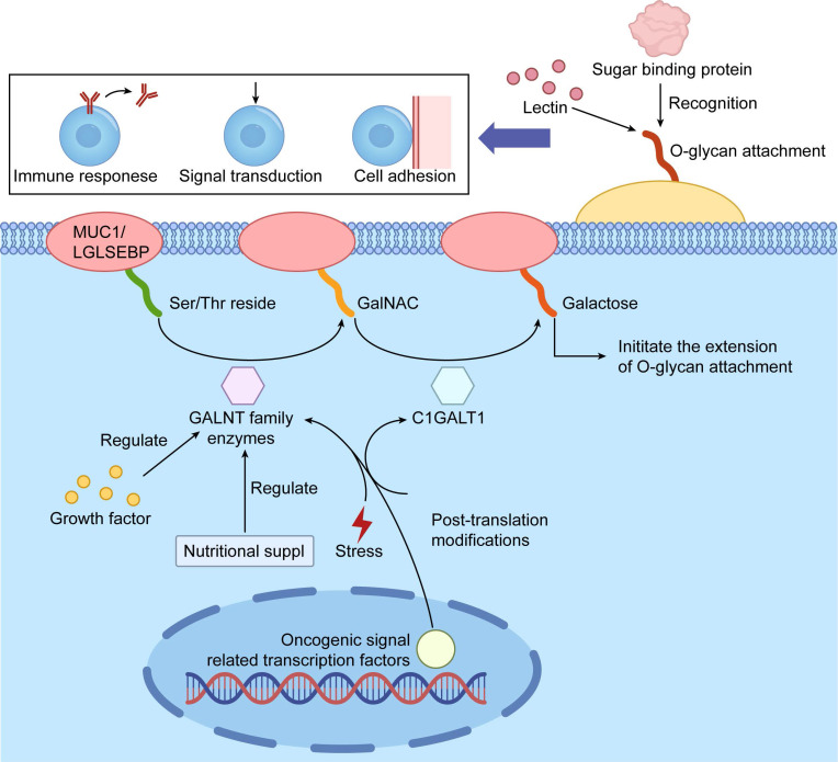 Figure 1
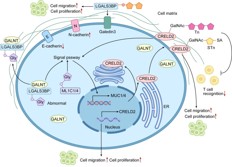 Figure 2
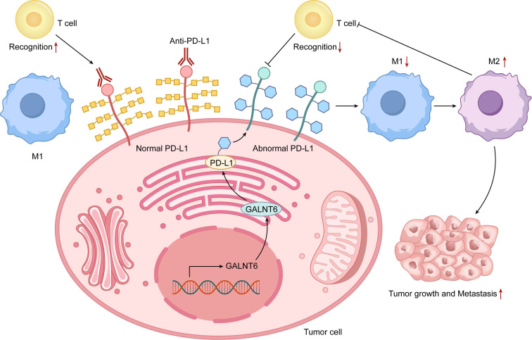 Figure 3
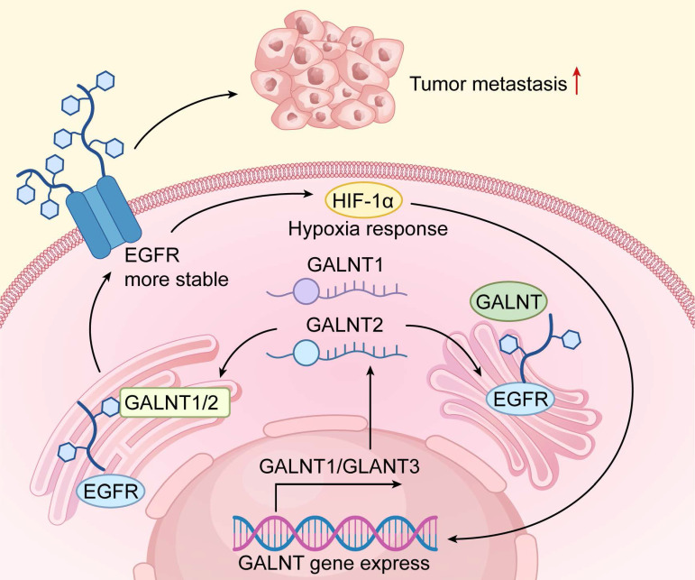 Figure 4
 Figure 5
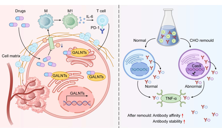 Figure 6
Peer Reviews
No public reviews on file for this paper yet. If you reviewed it on a platform where reviews are public (OpenReview, ICLR, NeurIPS, ICML), you can paste yours below so the community can read it here.
Videos
No videos yet. Explain this paper in a talk, walkthrough, or lecture? Add one.
Taxonomy
TopicsGlycosylation and Glycoproteins Research · Infant Nutrition and Health · Carbohydrate Chemistry and Synthesis
1. Introduction
O-glycosylation represents a major post-translational modification of proteins and regulates diverse biological processes including cell signaling, adhesion, and immune responses. In cancer, aberrant O-glycosylation drives malignant transformation and profoundly affects tumor progression, metastasis, and immune evasion1. Dysregulated O-glycans alter protein stability, subcellular localization, and intermolecular interactions, thereby reshaping the functional proteome of cancer cells1. The inherent complexity of O-glycosylation, encompassing site-specific alterations and the orchestration by various glycosyltransferases2, demands deeper mechanistic insight into its oncogenic functions.
In neoplastic cells, disrupted O-glycosylation triggers profound phenotypic reprogramming, promoting uncontrolled proliferation, epithelial-mesenchymal transition (EMT), invasiveness, and suppression of antitumor immunity 1, 3. These O-glycans activate key oncogenic pathways such as Wnt/β-catenin and phosphatidylinositol 3-kinase/protein kinase B (PI3K/AKT), thereby enhancing metastatic potential and therapeutic resistance4-6.
Recent advances in glycoproteomics, driven by high-resolution mass spectrometry, chemoenzymatic enrichment strategies, and multi-omics integration, have transformed our ability to interrogate site-specific O-glycosylation landscapes with unprecedented depth. These technologies have unveiled cancer-specific glyco-signatures and identified key enzymes, such as polypeptide N-acetylgalactosaminyltransferase 1 (GALNT1), as critical drivers of malignant glyco-phenotypes, highlighting the therapeutic promise of targeting glycosylation machinery to suppress tumor growth and dissemination7.
O-glycosylation interacts dynamically with other post-translational modifications, especially phosphorylation. These modifications often compete for the same or adjacent serine (Ser)/threonine (Thr) residues and generate complex regulatory networks that control protein function and cell fate in cancer8. This review integrates the latest findings on cancer-associated protein O-glycosylation, focusing on molecular mechanisms, functional impact, immune modulation, and clinical translation. By combining protein-centric and genomic views, we aim to establish a solid framework to accelerate glycosylation-based precision oncology strategies.
2. Biosynthesis and Structural Types of O-glycosylation
2.1 Basic Concepts and Classification of O-glycosylation
O-glycosylation represents a pivotal post-translational modification (PTM) characterized by the enzymatic attachment of carbohydrate moieties to the hydroxyl groups of Ser or Thr residues in proteins, facilitated by specialized glycosyltransferases9. The inherent complexity of O-glycosylation stems from its lack of consensus sequence motifs and the labile nature of its glycan cores, which hinders site identification. This modification is integral to numerous biological processes, including cell signaling, protein stability, and intercellular interactions10, 11. O-glycosylation is broadly divided into mucin-type (O-GalNAc) and O-GlcNAc subtypes. Mucin-type O-GalNAc glycosylation begins with the addition of N-acetylgalactosamine (GalNAc) to Ser/Thr residues and predominates in secreted and membrane-bound mucins that form protective barriers in epithelial tissues12. In cancer, mucin-type O-GalNAcylation holds particular clinical importance. The pathway is initiated by the polypeptide N-acetylgalactosaminyltransferase (GALNT) family, which transfer GalNAc to Ser/Thr, forming the Tn antigen. Subsequent extension to core 1 (T antigen) is catalyzed by Core 1 beta1,3-galactosyltransferase (C1GALT1) in complex with its essential chaperone Cosmc. Loss-of-function mutations or epigenetic silencing of C1GALT1 or Cosmc, or aberrant GALNT expression cause accumulation of truncated structures such as Tn and sialyl-Tn (STn) antigens. These oncofetal markers strongly associate with enhanced proliferation, immune evasion, metastasis, and poor prognosis in most epithelial cancers 3, 13, 14 (Figure 1).
2.2 Key Enzymes and Their Regulation
Mucin-type O-glycan biosynthesis depends on a coordinated enzyme network in which GALNT isoforms and C1GALT1 play central roles13. C1GALT1, or core 1 β1,3-galactosyltransferase, is indispensable for synthesizing the core 1 motif by transferring galactose to GalNAc, thereby enabling glycan chain elongation. Its functionality relies on the chaperone Cosmc for proper folding, and perturbations in C1GALT1 expression have been linked to aberrant glycosylation in cancers, driving tumor progression13. The GALNT family comprises 20 isoforms with distinct yet partially overlapping substrate specificities, peptide preferences, and subcellular localizations. This diversity enables precise spatiotemporal control of glycan initiation14.
GALNT expression is dynamically regulated by growth factors, oncogenic pathways, hypoxia, and metabolic stress, resulting in profound remodeling of the glycoproteome. Regulation occurs at transcriptional, post-transcriptional, and post-translational levels. Phosphorylation and auto-O-GlcNAcylation directly modulate enzyme activity and stability15. Dysregulated signaling often amplifies specific glycosyltransferases, yielding glycosylation profiles that support tumor proliferation, immune escape, and metastasis16, 17. For example, GALNT2 overexpression enhances O-glycosylation of growth factor receptors and sustains proliferation within hostile microenvironments 18.
Enzyme competition and cooperation among glycosyltransferases further refine substrate access and final glycan architecture. Alterations in glycosyltransferase levels can generate lectin-recognizable glycans that mediate cell adhesion, signaling, and immunity13, 19. Truncated or sialylated O-glycans on tumor surfaces mask antigens, impair immune attack, and facilitate metastasis. Consequently, these enzymes emerge as valuable diagnostic markers and therapeutic targets20.
2.3 Diversity of O-glycosylation and Tumor-Specific Glycans
Truncated O-glycans, particularly the Tn antigen and STn, represent the most clinically relevant aberrations in cancer21. These abbreviated structures are dramatically upregulated in cancers, where they correlate with aggressive phenotypes and unfavorable prognoses 22. Functionally, Tn/STn promote oncogenic signaling, inhibit apoptosis (including TRAIL-induced cell death), and drive epithelial-mesenchymal transition 23, 24. Their aberrant patterns also position them as biomarkers for early detection and immunotherapy targets25.
Glycan heterogeneity varies widely across tumor cell types and influences signaling, adhesion, and immune interactions26. Prostate cancers, for example, have unique glycosyltransferase profiles that yield distinct truncated O-glycans compared to normal prostate tissue27. Such differences modulate lectin recognition and immune cell trafficking, thereby shaping pro-tumorigenic microenvironments 28. Comprehensive mapping of O-glycan diversity is therefore essential for precision diagnostics and therapy (Table 1).
In summary, the cancer-associated shift toward truncated and aberrant O-glycans serves as both a hallmark and active driver of malignancy. Deciphering the responsible enzymes and their regulation provides critical opportunities for early detection, accurate prognostication, and development of glycosylation-directed therapeutics.
3. O-glycosylated Proteins in Tumors
3.1 Abnormal O-glycosylation of Cell Surface Glycoproteins and Tumor Progression
Dysregulated O-glycosylation of cell-surface and secreted glycoproteins is a major driver of tumor progression, primarily through altered cell adhesion, migration, and invasion. Cancer cells frequently display truncated O-glycans that profoundly change glycoprotein function. For instance, aberrant O-glycosylation of LGALS3BP enhances tumor cell binding to extracellular matrix and promotes invasion and metastasis34-36. Similarly, altered O-glycosylation of cysteine rich with epidermal growth factor (EGF)-like domains 2 (CRELD2) disrupts normal protein interactions and sustains oncogenic signaling37. These glycoproteins often bear truncated O-glycans, such as the Tn antigen and its sialylated derivatives, which are prevalent in diverse malignancies and enable immune evasion while exacerbating tumor aggressiveness43. Engagement with cognate lectins or receptors further activates proliferative and survival pathways38.
Mucins are the most extensively O-glycosylated glycoproteins and play central roles in malignancy. Overexpression and hypoglycosylation of MUC1 and MUC4 create neoepitopes that abolish apical-basolateral polarity, boost proliferation, and confer apoptosis resistance30, 44, 45. Aberrant mucin glycoforms also bind siglecs and galectins on immune and stromal cells, thereby remodeling the tumor microenvironment to favor cancer progression46-48. Together, these alterations convert cell-surface glycoproteins into active drivers of malignancy, making their modifying enzymes attractive therapeutic targets (Figure 2).
3.2 Intracellular O-GlcNAcylation and Oncogenic Signaling
O-GlcNAcylation dynamically modifies nuclear and cytoplasmic proteins and exerts major effects on oncogenic signaling. O-GlcNAcylation of enolase-1 (ENO1) alters its activity and stability, thereby reprogramming glycolytic flux in cancer cells3. In ovarian cancer, this modification enhances cell migration and invasion by shifting phosphorylation patterns49. Conversely, O-GlcNAcylation of the p53 protein influences its transcriptional efficacy and stability, potentially stabilizing the protein to favor tumorigenesis in specific contexts49, 50. This bifunctional nature underscores O-GlcNAcylation's dual role in tumorigenesis.
O-GlcNAcylation also functions as a nutrient sensor that integrates glucose, amino acid, and lipid metabolism via the hexosamine biosynthetic pathway (HBP)51. Elevated UDP-GlcNAc levels in cancer amplify O-GlcNAcylation of metabolic enzymes and transcription factors 52, 53. This adaptation not only bolsters glycolysis but also aids microenvironmental acclimation, facilitating immune circumvention. O-GlcNAcylation stabilizes PD-L1 through three mechanisms:(1) enhanced transcription via modified STAT3 and NF-κB54-56, (2) direct modification that blocks ubiquitination and proteasomal degradation56, 57, and (3) reduced endocytic recycling and lysosomal turnover, resulting in sustained surface expression58, 59. These effects make the HBP-OGT axis a central amplifier of PD-1/PD-L1 signaling.
In the immune compartment, aberrant O-GlcNAcylation in tumor-associated macrophages drives M2 polarization and immunosuppressive metabolism60, 61. Inhibition of OGT or the HBP therefore destabilizes oncogenic proteins and PD-L1 in tumor cells while simultaneously restoring antitumor immunity, providing strong rationale for combination with PD-1/PD-L1 blockade62, 63.
3.3 O-glycosylation and the Tumor Immune Microenvironment
Aberrant O-glycosylation reshapes tumor-immune crosstalk within the tumor immune microenvironment (TIME)(Figure 3). Aberrant modifications on tumor cells influence engagements with macrophages and cytotoxic T lymphocytes (CTLs)6, 64. Tn antigen expression, for instance, polarizes macrophages toward an M2 phenotype and suppresses antitumor immunity65. These glycan patterns also impair CTL recognition and killing, contributing to primary or acquired resistance to checkpoint inhibitors66, 67.
Combining glycosylation inhibitors with immune checkpoint blockade emerges as a powerful synergistic strategy68. Inhibition of GALNT6 in pancreatic cancer increases cytotoxic T-cell and macrophage infiltration and enhances immune attack66. Such approaches simultaneously restore immune surveillance and directly target tumor cells69. As understanding of glycosylation-mediated immune modulation deepens, targeting aberrant O-glycans is gaining traction as a new pillar of cancer immunotherapy (Table 2).
4. O-glycosylation-Related Genes and Their Expression and Function in Tumors
4.1 Expression Profiles and Regulation of O-glycosylation-Related Genes
Recent studies reveal widespread dysregulation of O-glycosylation genes across tumor types and highlight their central role in cancer biology75-77. In hepatocellular carcinoma (HCC), transcriptomic analyses show striking changes in GALNT family members compared with adjacent normal liver. These expression patterns faithfully mirror molecular HCC subtypes and strongly predict clinical outcome77. Similar upregulation of C1GALT1 and specific GALNTs has been documented in colorectal and breast carcinomas, where high expression correlates with aggressive behavior, immune exclusion, and poor prognosis78-80. These findings underscore the need for detailed mechanistic studies of glyco-gene regulation (Figure 4).
Transcriptional control is tightly coupled to the tumor microenvironment. Hypoxia-inducible factor-1α (HIF-1α), activated in hypoxic niches, directly transactivates several GALNTs, driving the synthesis of truncated O-glycans that facilitates metastasis81. Extensive crosstalk with Wnt/β-catenin, MYC, and other oncogenic pathways further amplifies this response82. Dysregulated O-glycosylation genes and their upstream regulators thus represent promising therapeutic targets to improve treatment efficacy (Table 3).
4.2 O-glycosylation Genes in Molecular Subtyping of Tumors
Integration of O-glycosylation gene expression with machine learning has transformed tumor classification, especially in HCC. Consensus clustering using GALNT1, GALNT2, GALNT6, and related genes defines robust molecular subclasses that differ in proliferation rate, immune infiltration, and survival83, 87, 88. GALNT1 and GALNT2 directly modulate EGFR O-glycosylation and downstream signaling strength, providing a clear mechanistic link to oncogenic driver activity83.
Single-cell analyses further resolve intratumoral heterogeneity and predict treatment response. High GALNT6 expression, for example, strongly correlates with lenvatinib resistance in HCC, while distinct glyco-subtypes show variable sensitivity to immune checkpoint blockade84. These results position O-glycosylation gene panels as clinically valuable tools for precision oncology and patient stratification.
4.3 Mutations in O-Glycosylation Genes and Tumorigenesis
Somatic mutations and epigenetic silencing of core O-glycosylation genes, most notably Core 1 beta 3-Gal-T-Specific Molecular Chaperone (COSMC /C1GALT1C1) and C1GALT1, are recurrent events in cancers and represent a direct genetic mechanism for the exposure of truncated O-glycans89. In triple-negative breast cancer (TNBC), COSMC mutations drive Tn antigen overexpression, which not only serves as a diagnostic marker but also actively reshapes the immune microenvironment41. Tn-positive tumors recruit immunosuppressive macrophages through galectin and siglec interactions while suppressing cytotoxic T-cell activity 72. These alterations confer enhanced metastatic potential and broad therapeutic resistance. Genetic disruption of O-glycosylation pathways therefore emerges as a fundamental oncogenic driver with major implications for tumor progression and clinical management.
5. O-glycosylation of RNA
5.1 Discovery and Detection of RNA O-Glycosylation
The discovery of cell-surface glycosylated RNAs (glycoRNAs) in mammalian cells has revealed a novel class of biomolecules that intersects glycobiology and RNA biology90. Although their physiological functions remain poorly defined, initial evidence points to potential roles in cell-cell communication and immune recognition.
Technological advances have driven early progress in this emerging field. The Tn-containing O-glycosylated RNAs (TnORNA) method stands out as a pioneering chemoenzymatic approach for capturing and enriching O-glycosylated RNA86, 90. Using this approach, several miRNAs including miR-103a-3p and miR-122-5p have been identified as O-glycosylated in pancreatic cancer models, with suggested effects on PI3K-Akt signaling and proliferation86. Complementary computational tools such as PONglyRNA now predict glycosylation sites on RNA with high confidence and support experimental design86, 91. Despite these advances, the stoichiometry, tissue distribution, and functional impact of RNA O-glycosylation are still inadequately characterized. Independent replication of initial findings remains essential.
5.2 O-glycosylated miRNAs in Tumors
Limited but intriguing data indicate that certain miRNAs undergo direct O-glycosylation, which may influence their stability, localization, or target engagement. In pancreatic ductal adenocarcinoma, TnORNA studies report O-glycan attachment to miR-103a-3p and miR-122-5p. These modifications correlate with increased PI3K-Akt activation and enhanced proliferation 86. Similarly, in HCC, dysregulation of the miR-424-5p/OGT axis has been implicated in oncogenic networks, though direct RNA glycosylation in this context requires verification 39.
Current evidence is primarily descriptive. The consequences of O-glycan addition to miRNAs—such as protection from degradation, altered argonaute binding, or extracellular interactions—remain unproven. The enzymes that catalyze RNA O-glycosylation or remove these marks are still unknown. Thus, while O-glycosylated miRNAs represent an exciting concept, the field is in its infancy. Assertions of clear oncogenic functions should await rigorous mechanistic validation.
5.3 RNA O-glycosylation and Tumor Signaling Pathways
RNA O-glycosylation emerges as a new frontier in cancer biology with potential links to key signaling pathways such as PI3K-Akt (Figure 5). Reported glycosylation of miR-103a-3p and miR-122-5p in pancreatic and lung adenocarcinoma correlates with sustained pathway activity and chemoresistance 40, 86. In lung cancer, GALNT14-driven protein O-glycosylation similarly boosts proliferation, prompting questions about coordinated regulation of protein and RNA glycosylation 20. By contrast, GALNT8-mediated glycosylation in breast cancer suppresses metastasis by dampening EGFR signaling and highlights context-specific effects85.
Computational tools now facilitate transcriptome-wide prediction of glycosylation sites and guide targeted investigations86. However, causal evidence connecting RNA O-glycosylation to altered signaling remains limited. The field requires prioritized efforts in independent validation, enzyme identification, and precise functional studies to distinguish correlation from true regulatory mechanisms.
6. O-glycosylation as a Target for Tumor Diagnosis and Therapy
6.1 O-glycosylation-Related Biomarkers
Tumor-associated carbohydrate antigens (TACAs) generated by aberrant O-glycosylation, including Tn, STn, and CA19-9, serve as established diagnostic and prognostic markers. Tn antigen is overexpressed in many adenocarcinomas and detectable in tissue biopsies and serum, allowing non-invasive monitoring of tumor burden and therapy response. STn similarly predicts aggressive disease and poor outcome, while CA19-9 remains the standard serum marker for pancreatic cancer 42, 92. Salivary glycoproteomics has identified distinct sialylation and fucosylation patterns in early lung cancer, supporting non-invasive screening approaches93, 94. High-resolution mass spectrometry platforms now enable site-specific and quantitative glycan analysis, driving discovery of more sensitive and specific biomarkers95, 96 (Table 4).
Clinical translation of O-glycosylation biomarkers faces substantial challenges. Glycosylation patterns exhibit profound heterogeneity at multiple levels. Inter-patient variation arises from genetics, microbiome, and metabolism100. Intra-tumor heterogeneity, driven by subclonal diversity and microenvironmental gradients such as hypoxia, creates spatially variable glyco-profiles28, 101. This mosaicism causes sampling bias in biopsies and dilutes tumor signals in liquid biopsies. Truncated glycans like Tn also appear in benign inflammation, reducing specificity102. Technical hurdles include O-glycan lability, low site occupancy, scarcity of reliable site-specific antibodies, and limited clinical compatibility of glycoproteomics workflows.
6.2 Therapeutic Targeting of O-glycosylation
Pharmacological inhibition of O-glycosylation emerges as a promising adjuvant to immunotherapy. Repurposed itraconazole suppresses glycosylation in head and neck cancer and markedly improves anti-PD-1 response by shifting macrophages to an M1 phenotype, enhancing CD8+ T-cell activity, and lowering immunosuppressive cytokines6. These benefits arise from remodeled tumor-surface glycans that disrupt inhibitory immune checkpoints.
Gene editing and enzyme modulation offer additional avenues. CRISPR/Cas9 knockout of C1GALT1 induces Tn/STn exposure, disrupts oncogenic signaling, and sensitizes colorectal and pancreatic tumors to immune clearance98. Selective glycosyltransferase inhibitors alter tumor antigen glycans to boost immune recognition103 (Figure 6). These strategies simultaneously impair tumor cell fitness and overcome immune evasion. Continued development of O-glycosylation-targeted therapies holds potential for personalized treatment across cancer types (Table 5).
6.3 O-glycosylation Engineering Cell Platforms and Biotherapeutic Optimization
Glycoengineering of O-glycosylation in producer cells has become essential for optimizing biopharmaceuticals. Specialized Chinese hamster ovary (CHO) platforms now allow precise control of O-glycan structures on recombinant proteins106. These systems produce variants with defined glycans that influence activity and pharmacokinetics. For example, etanercept analogs bearing truncated (Tn/STn) or extended (sialyl-core 1/3) O-glycans show improved TNF-α binding and therapeutic efficacy99. Such findings establish O-glycosylation as a critical quality attribute in biotherapeutic development.
Selective O-glycan modulation preserves N-glycosylation and overall protein integrity13. This approach enhances performance while enabling systematic study of glycan effects on product quality. Broader application will improve diverse biologics and deliver superior patient outcomes. Robust glycoengineered platforms are therefore vital for advancing biopharmaceutical manufacturing to modern standards.
7. Technological Advances and Future Research Directions
7.1 Development of High-Throughput Analysis Technologies
High-throughput technologies have revolutionized glycosylation research in oncology. Ultrahigh-resolution mass spectrometry, including MALDI-FT-ICR MS, enables simultaneous profiling of N- and O-glycans. These methods provide detailed structural analysis of complex glycans from cancer cell lines and tissues. A semi-automated workflow has profiled O-glycans in colorectal and pancreatic cancer cells, identifying compositions from monosaccharides to branched oligosaccharides97. Automated tools like MassyTools improve quantification accuracy using signal-to-noise and mass error criteria across large datasets97.
Glycopeptide enrichment strategies have further advanced O-glycosylation analysis. The MOTAI method combines enzymatic digestion with solid-phase extraction to boost sensitivity and coverage in tumor samples37. This approach has identified upregulated Tn/STn-glycoproteins in colorectal cancer compared with normal tissue, highlighting diagnostic potential. Sequential O-glycoprotease digestion in MOTAI isolates O-GalNAc sites and elucidates aberrant glycoprotein functions 37.
Integration with high-throughput sequencing and bioinformatics correlates glycan profiles with clinical outcomes 107. Ongoing refinements will deepen insights into glycosylation-cancer interactions and refine diagnostic paradigms107, 108.
7.2 Multi-omics and Machine Learning Applications in O-glycosylation
Multi-omics integration elucidates O-glycosylation networks in tumors. Weighted gene co-expression network analysis (WGCNA) in HCC identifies key glyco-genes and defines molecular subtypes linked to behavior and prognosis77. Subtype CS1 shows genomic heterogeneity and moderate immune infiltration. Subtype CS2 exhibits genomic stability and favorable immune profiles 77. Combining glycomics with genomics and transcriptomics reveals O-glycosylation effects on the tumor microenvironment, immune phenotypes, and outcomes, aiding biomarker and target discovery 77, 109.
Machine learning enhances O-glycosylation-based classification and prognostication110. Evaluation of 59 algorithms has yielded robust HCC prognostic models from glyco-gene expression, defining clinically relevant subtypes77. CS2 patients display superior survival and strong immune infiltration, predicting immunotherapy response. CS3 patients face poor prognosis with genomic instability, requiring alternative approaches77. These machine-learning signatures support clinical decision-making and patient stratification. Multi-omics and machine learning thus drive personalized oncology for HCC and beyond77.
7.3 New Opportunities for O-glycosylation Modification in Tumor Immunotherapy
O-glycosylation regulates tumor immunobiology by modulating immune checkpoints and cell function. Aberrant O-glycans on tumor glycoproteins stabilize PD-L1, impair T-cell activation, and promote immune evasion 56. Targeting these modifications may boost checkpoint inhibitor efficacy. Glycopeptide epitopes from CD44v6 have enabled development of selective monoclonal antibodies with enhanced tumor targeting and safety74, 111. Personalized glycopeptide vaccines mimicking TACAs could elicit strong antitumor immunity and overcome resistance104.
Sialylated O-glycans form a protective glycocalyx that shields tumors from immune attack112. Inhibitors or glycan-engineered immune cells that disrupt sialylation may synergize with immunotherapies113. Glycosylation profiles also predict immunotherapy response and guide treatment selection 114, 115. The immunomodulatory roles of O-glycosylation merit intensive study to advance cancer immunotherapy and patient outcomes.
7.4 Future Exploration in RNA Glycosylation
RNA glycosylation represents a new regulatory layer with major implications for cancer. GlycoRNAs on small RNAs modulate immune interactions and tumor progression90, 116, 117. Aberrant glycoRNA patterns associate with malignancy and resistance117, 118. Coexistence of N- and O-linked modifications on RNAs suggests interplay with protein glycosylation, influencing gene regulation and signaling86. Future studies should define glycoRNA functions and cross-talk with protein pathways to uncover therapeutic avenues.
Advanced detection technologies are essential for progress. Current methods lack sensitivity, necessitating tools like solid-phase chemoenzymatic gRNA (SPCgRNA) for selective capture and profiling 105. Bioinformatics predictors such as PONglyRNA identify RNA glycosylation sites for hypothesis testing86. Extension to long non-coding RNAs, circular RNAs, and other classes will clarify roles in disease.
Therapeutic targeting of RNA glycosylation offers novel oncology strategies. Altering glycoRNA pathways may influence RNA stability, cellular localization, and downstream signaling, thereby affecting tumor proliferation, apoptosis, and immune evasion117, 119. Multi-omics integration will decode the cancer glyco-code and enable personalized approaches. Sustained RNA glycosylation research promises key insights and innovative treatments.
8. Conclusion
Glycosylation profoundly influences tumor biology by regulating proliferation, metastasis, and immune interactions. Aberrant forms, characterized by truncated glycans such as Tn and STn antigens alongside dysregulated glycosyltransferases, actively drive oncogenesis and disease progression. Integration of multi-omics data with machine learning has transformed tumor subtyping, prognostic modeling, and treatment selection. These tools uncover cancer-specific glycosylation signatures and enable personalized interventions to improve outcomes.
The discovery of RNA O-glycosylation introduces an exciting new regulatory layer. GlycoRNAs hold promise as diagnostic markers, prognostic indicators, and therapeutic targets, warranting intensive study to integrate them into precision oncology. Therapeutically, targeted modulation of aberrant O-glycosylation, particularly in combination with immunotherapy, yields promising preclinical and early clinical results that may expand treatment efficacy.
Significant barriers persist in clinical translation. The inherent complexity of O-glycans—including low stoichiometry, heterogeneity, and technical detection challenges—necessitates continued advances in mass spectrometry, enrichment strategies, and analytical workflows. Large-scale validation studies and interdisciplinary collaboration among glycobiologists, oncologists, immunologists, bioinformaticians, and clinicians remain crucial. Overcoming these obstacles will unlock the full potential of O-glycosylation research to deliver innovative diagnostic tools and transformative therapies in precision oncology.
The reference list from the paper itself. Each links out to its DOI / PubMed record.
- 1Pinho SS Reis CA Glycosylation in cancer: mechanisms and clinical implications Nature reviews Cancer 201515540552628931410.1038/nrc 3982 · doi ↗ · pubmed ↗
- 2Hashii N Suzuki J Site-Specific O-Glycosylation Analysis by Liquid Chromatography-Mass Spectrometry with Electron-Transfer/Higher-Energy Collisional Dissociation Methods in molecular biology (Clifton, NJ)202122711697810.1007/978-1-0716-1241-5_1233908007 · doi ↗ · pubmed ↗
- 3Zhu Q Li J Sun H Fan Z Hu J Chai SO-Glc N Acylation of enolase 1 serves as a dual regulator of aerobic glycolysis and immune evasion in colorectal cancer Proceedings of the National Academy of Sciences of the United States of America 2024121 e 24083541213944638410.1073/pnas.2408354121 PMC 11536113 · doi ↗ · pubmed ↗
- 4Liu Z Liu J Dong X Hu X Jiang Y Li L Tn antigen promotes human colorectal cancer metastasis via H-Ras mediated epithelial-mesenchymal transition activation Journal of cellular and molecular medicine 2019232083923063791410.1111/jcmm.14117 PMC 6378212 · doi ↗ · pubmed ↗
- 5Rosa-Fernandes L Oba-Shinjo SM Macedo-da-Silva J Marie SKN Palmisano G Aberrant Protein Glycosylation in Brain Cancers, with Emphasis on Glioblastoma Advances in experimental medicine and biology 2022138239703602940310.1007/978-3-031-05460-0_4 · doi ↗ · pubmed ↗
- 6Lin MC Chuang YT Wu HY Hsu CL Lin NY Huang MC Targeting tumor O-glycosylation modulates cancer-immune-cell crosstalk and enhances anti-PD-1 immunotherapy in head and neck cancer Molecular oncology 202418350683745265310.1002/1878-0261.13489 PMC 10850803 · doi ↗ · pubmed ↗
- 7Zhang J Wang H Wu J Yuan C Chen S Liu SGALNT 1 Enhances Malignant Phenotype of Gastric Cancer via Modulating CD 44 Glycosylation to Activate the Wnt/β-catenin Signaling Pathway International journal of biological sciences 2022186068833643987610.7150/ijbs.73431 PMC 9682532 · doi ↗ · pubmed ↗
- 8Şener Uslupehlivan E Deveci RŞahar Uİzzetoğlu S Glycan analysis of Lamin A/C protein at G 2/M and S phases of the cell cycle Cell biochemistry and biophysics 202280689983618065810.1007/s 12013-022-01102-3 · doi ↗ · pubmed ↗