Lanthanide–carbamazepine complexes: synthesis, spectroscopic characterization, DFT Insights, molecular docking, and biological evaluation
Nora S. Mohamed, Mahmoud M. A. Mohamed, Mohamed R. Shehata, Ehab M. Abdalla

TL;DR
This paper reports the synthesis and characterization of lanthanide-carbamazepine complexes and evaluates their antimicrobial and anticancer properties.
Contribution
The study introduces novel lanthanide-carbamazepine complexes and evaluates their biological activity and binding affinity using molecular docking.
Findings
Lanthanide-carbamazepine complexes show high cytotoxicity against Hep-G2 and MCF-7 cancer cell lines.
La(III) complex demonstrated the highest antimicrobial and anticancer activity among the synthesized complexes.
Molecular docking studies revealed strong binding affinity of La(III) complex to cancer-related receptors.
Abstract
This work focuses on the synthesis of four novel lanthanide complexes: La (III), Ce (III), Nd (III), and Dy (III) with carbamazepine (CBZ). The geometric structural and bonding mode of synthesized complexes were accomplished by various analytical and spectroscopic techniques and the data reveal that the novel complexes have a 1:2 (Ln3+:CBZ) electrolytes with general formula [Ln(CBZ)2(H2O)Cl]Cl2.H2O and molecular weight: 753.83, 791.07, 777.19 and 777.43 for Ln3+ complexes respectively. The spectral studies investigated that CBZ interacts as a bidentate ligand via the amide group’s nitrogen and oxygen atoms in its moiety, and the geometric octahedral structure was confirmed by density function theory. On the other hand, PXRD data provided that the ligand and its complexes have nature that is crystalline. Antimicrobial activity was tested for their effectiveness against various…
Genes, proteins, chemicals, diseases, species, mutations and cell lines named across the full text — each resolved to its canonical identifier and authoritative record.
Click any figure to enlarge with its caption.
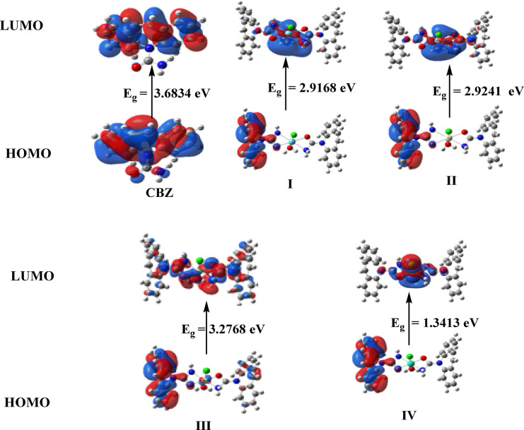 Figure 10
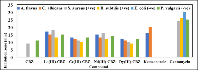 Figure 11
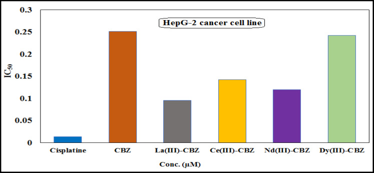 Figure 12
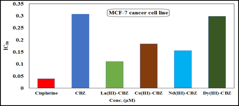 Figure 13
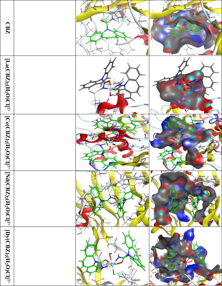 Figure 14
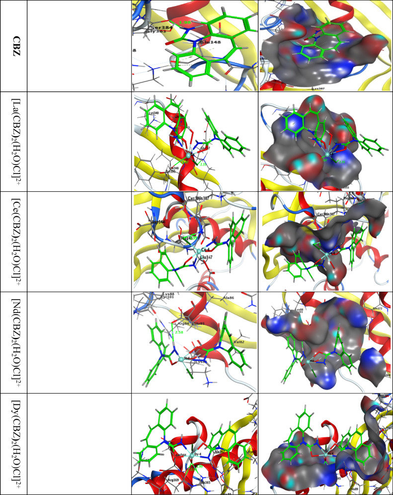 Figure 15
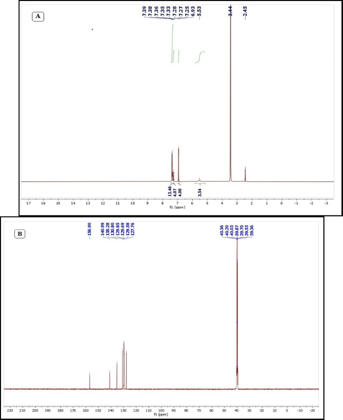 Figure 1
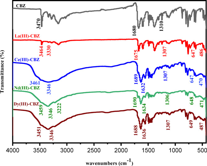 Figure 2
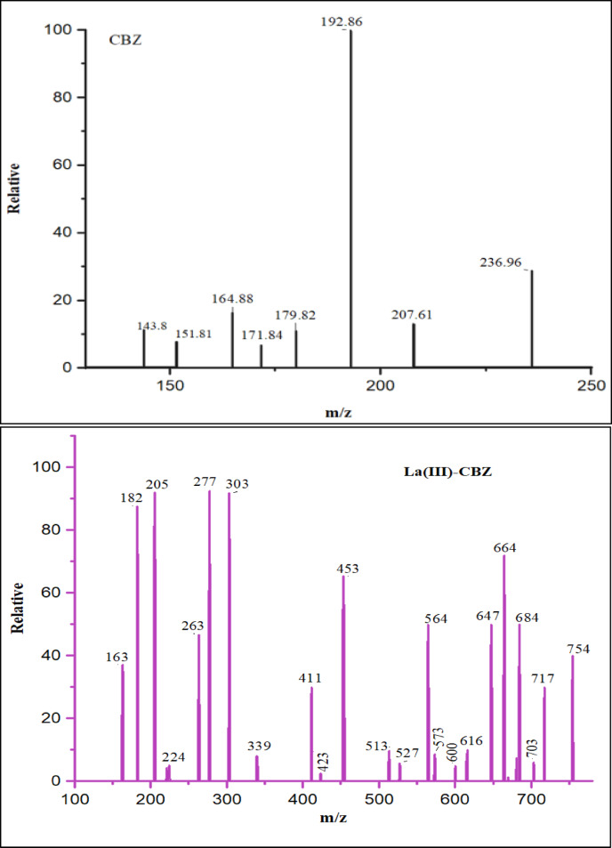 Figure 3
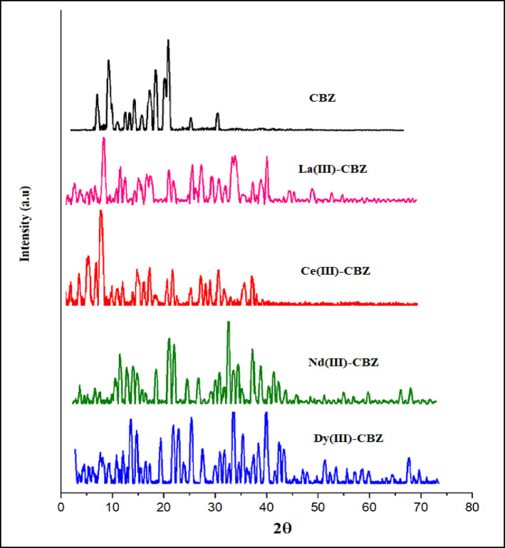 Figure 4
 Figure 5
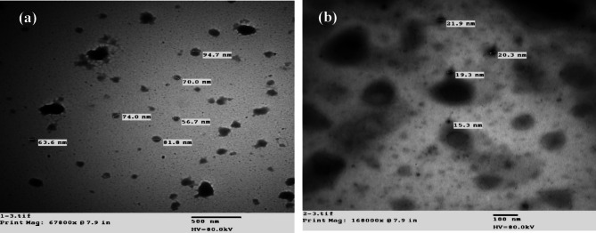 Figure 6
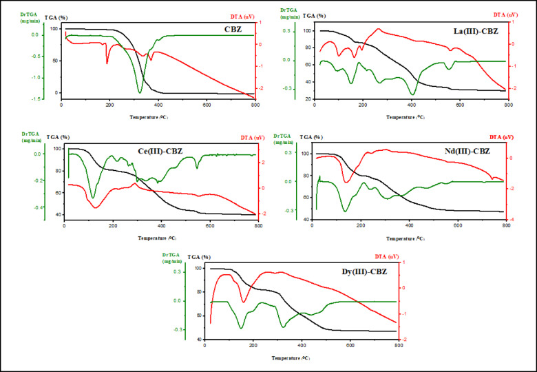 Figure 7
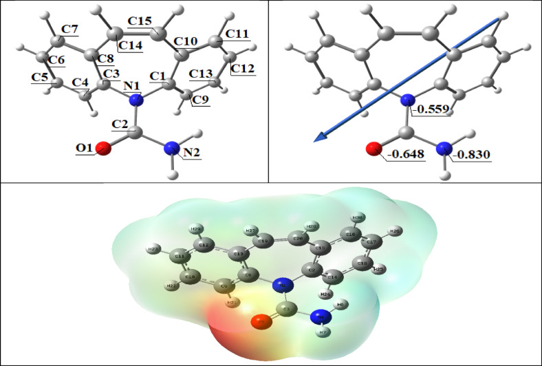 Figure 8
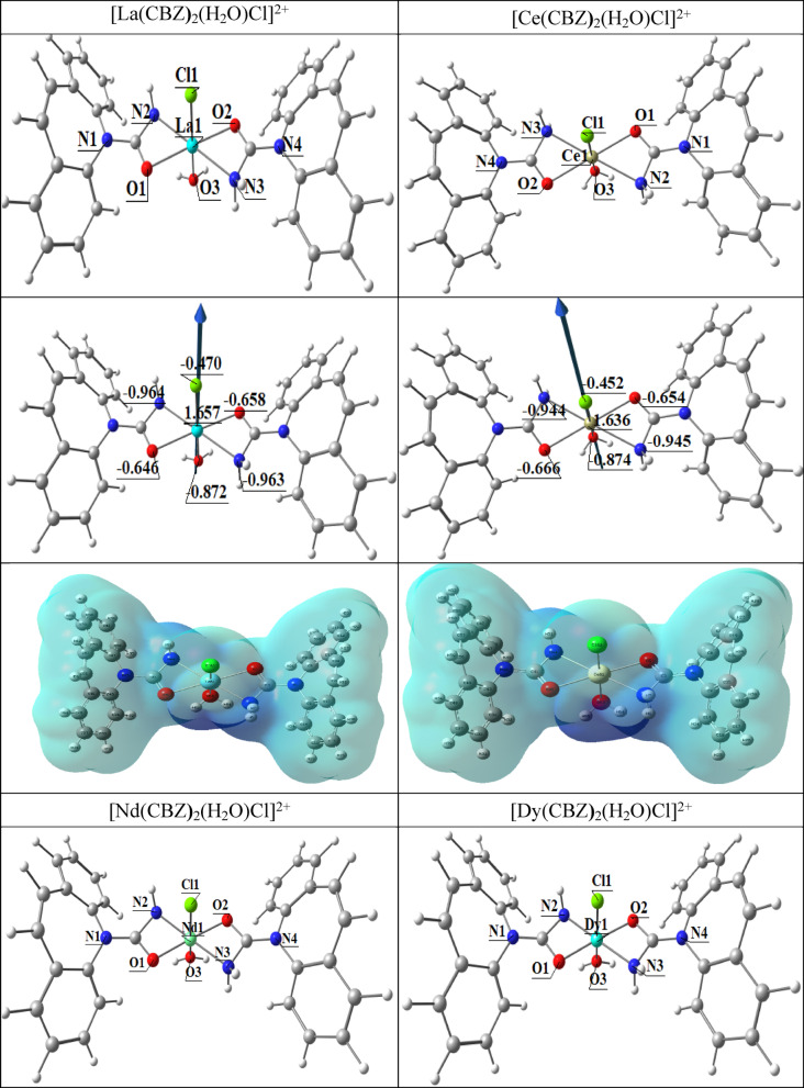 Figure 9
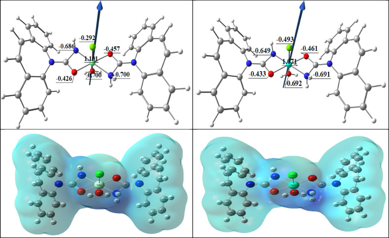 Figure 9
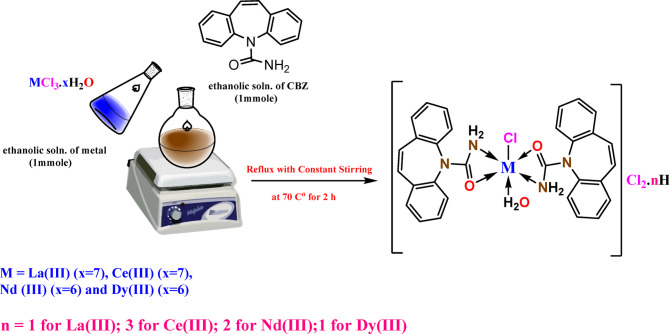 Figure 17
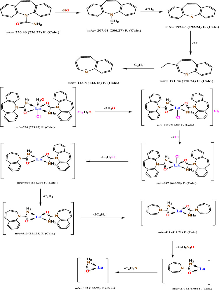 Figure 18
- —New Valley University
Peer Reviews
No public reviews on file for this paper yet. If you reviewed it on a platform where reviews are public (OpenReview, ICLR, NeurIPS, ICML), you can paste yours below so the community can read it here.
Videos
No videos yet. Explain this paper in a talk, walkthrough, or lecture? Add one.
Taxonomy
TopicsLanthanide and Transition Metal Complexes · Metal complexes synthesis and properties · Metal-Organic Frameworks: Synthesis and Applications
Introduction
Carbamazepine (CBZ) is a pharmacological compound employed for the prophylaxis and treatment of seizures, commonly indicated for several epileptic illnesses, including bipolar mood disorders, trigeminal neuralgia, and both simple and complicated seizures^1^. The medicine absorbs only at a restricted rate and is essentially insoluble in water^2^. Being a pharmaceutically active substance and a non-steroidal anti-inflammatory medication, Carbamazepine is among the most prevalent medications in the environment^3,4^^5^. It is often used as a marker for human-caused contamination of surface water sources due to wastewater^5–7^^8^. CBZ, a tricyclic iminostilbene derivative, is a crucial and extensively used medicine for the treatment of epilepsy and some psychological disorders^4^. The interaction between drugs and different metals receives significant attention in the pharmaceutical sciences and medicinal chemistry. These reactions can significantly influence the efficacy, safety, and metabolism of drugs^9^. The ability of CBZ to form compounds with metal ions has been studied, which frequently have an octahedral shape. The interaction is often accomplished through bidentate coordination between the oxygen atom of the amide group and the nitrogen of the N-H bond^5^. The consequent complexes are applicable in various uses, such as electro catalysis, spectroscopy, biochemistry, and catalytic processes^10^. One of the most important aspects of the job that scientists do all around the world is using complicated chemicals in the chemical industry; the main goal has been to enhance the efficiency and selectivity of these processes in the last years^11^. Some lanthanide elements were selected for the following reasons: Complexes of lanthanides are one of the rapidly expanding subjects of inorganic chemistry research^12^. According to a review of published research, a large number of other recently synthesized substances or their derivatives form stable complexes^13,14^^15^, also, lanthanides occupy distinctive locations in the periodic table and display an intriguing variety in their characteristics. In recent years, extensive studies have shown that lanthanides are important in many biological processes, and they are useful tools for understanding the shapes and properties of biomolecules^16,17^. The research on lanthanides demonstrated their extensive range of applications, including therapies and medical diagnostics, agriculture, industry, and materials science^18^. Due to high coordination numbers, their structural motifs, fascinating magnetic, electronic, and spectroscopic properties, compounds containing lanthanide(III) ions have garnered a lot of attention in coordination chemistry recently^19^. The lanthanide (III) ion complexes have shown pharmacological properties such anti-inflammatory, anticoagulant, antiallergic, antibacterial^20^, and anticancer properties during the 1960s. In addition, Lanthanide (III) ion complexes were used to treat burns^21^..
The synthesis and application of a carbamazepine medication to characterize various lanthanide La(III), Ce(III), Nd(III), and Dy(III) complexes are the main topics of this investigation. Their anticancer and antibacterial properties were thoroughly assessed. For the rational design and evaluation of bioinorganic compounds, computational techniques like density functional theory (DFT) and molecular docking are becoming crucial in addition to experimental methods.
Experimental
Chemicals and instruments
At room temperature and in air, all of the synthetic work was carried out. Carbamazepine (CBZ) was supplied by UP Pharma, Assiut, Egypt. All chemicals used in this study, such as LaCl_3_**·7H_2_O, CeCl_3_·7H_2_O, NdCl_3_·6H_2_O, and DyCl_3_·**6H_2_O, as well as absolute ethanol, were analytical grade and purchased from Fluka and Aldrich Chemical, which were utilized without additional purification as received. The methods and procedures utilized for the application are contained in the supporting structure (Section S1).
The metal complexes’ synthesis
The complexes were made by following a standard method (Scheme 2): equal amounts of ethanolic solutions of different metal salts (1 mmol = 0.371 g La(III); 0.372 g Ce(III); 0.358 g Nd(III); and 0.376 g Dy(III)) were mixed with an equal amount of an ethanolic solution of CBZ ligand (1 mmol, 0.236 g) in a 1:2 (Ln^3+^:CBZ) ratio, After that, the reaction mixture was refluxed at 70 °C for two hours while being constantly stirred. The formed solid complexes were filtered out and rinsed multiple times with a lot of pure EtOH to get rid of any leftover initial materials, and then they were dried in a vacuum at room temperature for 24 h^22,23^.
Scheme 1. The suggested complexation reaction of metals La (III), Nd (III), Ce (III), and Dy (III) with CBZ.
Computational study
The B3LYP functional was used in Density Functional Theory (DFT) simulations to identify the lowest energy equilibrium geometries of the CBZ ligand and its metal complexes. In the DFT, the lanthanides metal atoms in these calculations were based on the SDD basis set, whereas the C, H, N, O, and Cl atoms were based on the 6–311 + + G(d, p) basis set^24^. The software package Gaussian 09 was utilized to perform these computations.
Antimicrobial bioassay
Using the method of modified well diffusion, we tested the antimicrobial effectiveness of CBZ and its synthesized complexes against fungi (Aspergillus flavus and Candida albicans), a type of yeast; Gram-positive bacteria (Bacillus subtilis and Staphylococcus aureus); and Gram-negative bacteria (Proteus vulgaris and Escherichia coli). The standard antibacterial agent is gentamicin, and the typical antifungal agent is ketoconazole. (Section S2)^25,26^^27^,
Anticancer activity
- - Mammalian cell lines: HepG2 cells (human liver cancer cell line) and MCF-7 cells (human breast cancer cell line) were obtained from the American Type Culture Collection (ATCC, Rockville, MD).
- - Chemicals Used: MTT and trypan blue dye were purchased from Sigma (St. Louis, Mo., USA).
Fetal Bovine serum, RPMI-1640, HEPES buffer solution, L-glutamine, gentamycin, and 0.25% Trypsin-EDTA were purchased from Lonza (Belgium).
- - Cell line Propagation: The cells were grown on RPMI-1640 medium supplemented with 10% inactivated fetal calf serum and 50 µg/ml gentamycin. The cells were maintained at 37 °C in a humidified atmosphere with 5% CO_2_ and were subcultured two to three times a week.
- - Cytotoxicity evaluation using viability assay: To assess the cytotoxicity activity of CBZ and its complexes against breast and liver cancer cell lines, tumor cell lines were planted in 96-well plates at a density of 1 × 10^5^ cells/well and treated for 24 h. In triplicate, test chemicals in a range of concentrations (including 12 concentrations for cisplatin) were added after being dissolved in PBS. The MTT assay was employed to assess cell vitality following an additional 24-hour incubation. DMSO was used to dissolve the formazan crystals following the addition of the MTT solution and a four-hour incubation period. Absorbance at 590 nm was used to ascertain the viability of the cells. Plotting survival curves with GraphPad Prism software allowed for the determination of the IC_50_ values (Section S3)^28,29^.
Docking molecules
Using the MOA2022 software^30^ molecular docking studies were conducted to investigate possible binding mechanisms of the methionine adenosyl transferase in human breast cancer MCF-7 (PDB ID: 4ZVM) and the Bacillus subtilis receptor site (PDB ID: 6A4M) in liver malignancies (PDB ID: 5A19)^31,32^. The Protein Data Bank was used to get experimental crystallographic structures of the protein. Before docking, the protein was prepared by removing water molecules bound to DNA base pairs and replacing them with hydrogen. Many docking simulations were run using default parameters, and conformations were selected based on a combination of S score data, E conformation, and proper fitting with the relevant amino acids in the binding pocket; the target protein was kept rigid (the MOE docking program automatically prepare the protein and the site finder produce the best cavity appropriate for docking). Ligands (our compounds in this study) were obtained from the output of optimized DFT data.
Results and discussion
Characteristics
A variety of analytical techniques, including FTIR, UV-Vis, mass, PXRD, elemental analysis, and thermogravimetric studies, were used to describe the produced complexes. The chemical analytical data showed that the formation of complexes with the stoichiometry of 1:2 (Ln^3+^: CBZ); theses complexes are electrolytic in nature where the molar conductivity value of complexes between 201 and 223 Ω^−1^cm^2^mol^−1^ in 10^− 3^ M DMF solution^33^ Table 1, colored, stable in air, non-hygroscopic and were soluble in organic solvents as (DMF, DMSO) but insoluble in water, partially soluble in ethanol.
Table 1. Micro analytical data of CBZ and its lanthanide complexes.CompoundMol. formulaMol. Wt.Color, yield (%)Conductivity ^Ʌ^mm.p.^o^CCalculated (Found)CH N MCBZC_15_H_12_N_2_O236.27White-19276.25(76.78)5.12(5.18)11.86(12.00)-[La(CBZ)2(H_2_O)Cl]Cl_2_.H_2_OC_30_H_28_LaCl_3_N_4_O_4_753.83Off white88223> 30047.80(47.59)3.74(3.51)7.43(6.7)18.43(18.87)[Ce(CBZ)2(H_2_O)Cl]Cl_2_.3H_2_OC_30_H_32_CeCl_3_N_4_O_6_791.07Beige77.6213> 30045.55(45.34)4.08(4.02)7.08(6.88)17.71(17.12)[Nd(CBZ)2(H_2_O)Cl]Cl_2_.2H_2_OC_30_H_30_NdCl_3_N_4_O_5_777.19Pale pink73211> 30046.36(46.13)3.89(3.75)7.21(6.85)18.56(18.23)[Dy(CBZ)2(H_2_O)Cl]Cl_2_.H_2_OC_30_H_28_DyCl_3_N_4_O_4_777.43Yellow91.7201> 30046.35(47.09)3.63(3.86)7.21(7.62)20.90(20.35)Where: ^Ʌ^m represents the molar conductivity, measured in ohm^−¹^ cm^²^ mol^− 1^.
1H and13C-NMR spectra
The^1 ^H-NMR spectrum of the La(III) CBZ complex was acquired in DMSO-d_6_. A signal corresponding to the methine (CH) proton was observed at δ 5.53 ppm. A singlet at δ 6.93 ppm was assigned to the two protons of the NH_2_ group. Multiple signals in the aromatic region (δ 7.25–7.39 ppm) are attributed to the aromatic ring protons (Fig. 1 A). The ^13^C-NMR spectrum of the La(III) CBZ complex in DMSO-d_6_ (Fig. 1B) displayed signals at δ 127.76, 129.58, 129.69, 129.85, 130.80, 135.28, and 140.99 ppm, corresponding to the aromatic carbons. An additional signal at δ 156.90 ppm was assigned to the carbonyl (C = O) carbon.
Fig. 1^1^H-NMR (A) and^13^C-NMR (B) spectrum of La(III) CBZ complex in DMSO d_6_.
FTIR spectra
To assess how CBZ binds to the metal ion in the complexes, characteristic FT-IR bands were investigated within the range from 4000 to 400 cm^− 1^. The significant infrared absorption frequencies of lanthanide (III)-CBZ complexes have been displayed in Fig. 2 and given in Table 2. The spectrum of pure CBZ showed a sharp peak at 3470 cm^− 1^ corresponding to –NH_2_^2,34^, as well as an absorption band at 1680 cm^− 1^ and 1310 cm^− 1^ due to C = O of the amide group and C-N, respectively^5,35^. On complexation, the sharp peaks of the CBZ ligand at 3470 cm^− 1^ shift to lower frequencies in Complexes at 3464, 3461, 3459 and 3451 cm^− 1^ for Ln^3+^ complexes, respectively this confirm the nitrogen in the amino group coordinated with metal ions, as well as the peak related to the C = O of amide group shifted to higher and lower frequencies in all complexes (1675, 1689, 1690 and 1688 cm^− 1^) for La(III), Ce(III), Nd(III) and Dy(III) complex, respectively, which confirm the participation of the amide moiety’s C = O in the complexation reaction^36,37^. All complex spectra showed a broad band at (3330–3347) and (954–980) cm^− 1^, attributed to the ν(OH)/H_2_O confirms the presence of hydrated and coordinated water molecules^38^. New bands appeared in all complexes in the range of (647–649) and (470–487) cm^− 1^, which are attributed to ν(M–O) and ν(M–N), respectively^39–41^^42^,. Consequently, it is presumed that the CBZ ligand acts as a bidentate ligand with NO metal ion coordination sites based on the results of FT-IR spectra.
Table 2FT-IR spectra of the ligand CBZ and its compounds with lanthanides.Compoundv(OH)/H_2_Ov(NH_2_) 1^o^amine_v(C = O)amide_v(C-N)alph.v(M-O)v(M-N)CBZ—347016801310——[La(CBZ)2(H_2_O)Cl]Cl_2.H_2_O3330346416751307647486[Ce(CBZ)2(H_2_O)Cl]Cl_2.3H_2_O3346346116891307647470[Nd(CBZ)2(H_2_O)Cl]Cl_2_.2H_2_O3347345916901306648472[Dy(CBZ)2(H_2_O)Cl]Cl_2_.H_2_O3346345116881310649487
Fig. 2FT-IR spectra (cm^− 1^) of the CBZ ligand and its lanthanide complexes in the region of 4000–400 cm^− 1^.
Mass spectra
Mass spectrometry is an effective method that may be utilized for the purpose of determining unknowns, investigating the structure of molecules, and gaining insight into the fundamental principles of chemistry. We conducted mass spectrum analyses of representative complexes to validate the formula weights suggested based on elemental analyses. Fig.s 3. and fig.s S1 displays the mass spectra diagram of the ligand (CBZ) and its synthesized complexes. The molecular formula of the ligand (CBZ) (C_15_H_12_N_2_O) (M.wt. 236.27) is indicated from its mass spectrum (Fig. 3). The observed peak at m/z = 236.96 (molecular ion peak), related to C_15_H_12_N_2_O, atomic mass m/z = 236.27, the spectrum shows important fragment ions in the range m/z = 207.61 for C_15_H_12_N^+^, m/z = 192.86 for C_14_H_10_N^−^,m/z = 171.84 for C_12_H_12_N^−^ and m/z = 143.8 for C_10_H_8_N^−^ (Scheme 2). The spectrum of complex La(III)-CBZ (C_30_H_28_LaN_4_O_4_Cl_3_, atomic mass = 753.83), shows molecular ion peak [M^1+^] at m/z = 754 and a base peak at m/z = 277 for [C_7_H_8_LaN_2_O]. Additionally, the mass spectra displayed a multi-peak pattern that included a variety of fragment ions within the range of m/z = 717 for [C_30_H_24_LaN_4_O_2_Cl_3_], 647 for [C_30_H_24_LaN_4_O_2_Cl], 564 for [C_26_H_22_LaN_4_O_2_], 513 for [C_22_H_20_LaN_4_O_2_], 411 for [C_14_H_16_LaN_4_O_2_], 277 for [C_7_H_8_LaN_2_O] and 182 for [CH_3_LaNO], (Scheme 2). These peaks were exactly in line with the metal complex’ proposed chemical formula. The spectrum of complex Ce(III)-CBZ (C_30_H_32_CeN_4_O_6_Cl_3_, atomic mass = 791.07), show molecular ion peak [M^1+^] at m/z = 792, the molecular ion undergoes fragmentations at m/z = 770.53 for [C_30_H_30_CeN_4_O_5_Cl_3_], m/z = 735.6 for [C_30_H_26_CeN_4_O_3_Cl_3_], m/z = 720.22 for [C_30_H_24_CeN_4_O_2_Cl_3_], m/z = 616 for [C_30_H_24_CeN_4_O_2_], m/z = 560.57 for [C_26_H_22_CeN_4_O_2_], m/z = 247.23 for [C_2_H_10_CeN_4_O] and m/z = 230.43 for [C_2_H_9_CeN_3_O], these fragmentation patterns were perfectly match with the predicted molecular structure of the Ce(III) complex. The mass spectrum of complex Nd(III)-CBZ (C_30_H_30_NdN_4_O_5_Cl_3_, atomic mass = 777.19), exhibited a peak assigned to the molecular ion peak at m/z = 777.26 for [C_30_H_30_NdN_4_O_5_Cl_3_] and a base peak at m/z = 588.43 for [C_22_H_20_NdN_4_O_2_Cl_2_]. The pattern of several peaks in the mass spectrum reveals peaks for fragment ions in the range of m/z = 687.91 for [C_30_H_24_NdN_4_O_2_Cl_2_], 588.43 for [C_22_H_20_NdN_4_O_2_Cl_2_], 503.97 for [C_18_H_18_NdN_4_O_2_Cl], 449.04 for [C_14_H_16_NdN_4_O_2_Cl], 298.8 for [C_2_H_8_NdN_4_O_2_Cl], 190.42 for [CH_6_NdN_2_] and 159.46 for [NdNH_3_]. These data match with its suggested molecular formula. On the other hand, the molecular ion peak of complex Dy(III)-CBZ (C_30_H_28_DyN_4_O_4_Cl_3_, atomic mass = 777.43) was observed at m/z = 777.67 and a base peak at m/z = 192.78 for [CH_2_DyO] which is acceptable with its proposed molecular structure, this molecular ion undergoes fragmentation in the range of m/z = 759.83 for [C_30_H_26_DyN_4_O_3_Cl_3_], 741.2 for [C_30_H_24_DyN_4_O_2_Cl_3_], 691.4 for [C_26_H_22_DyN_4_O_2_Cl_3_], 504.3 for [C_14_H_16_DyN_4_O_2_Cl_2_], 429.16 for [C_8_H_12_DyN_4_O_2_Cl_2_], 318.57 for [C_2_H_8_DyN_4_O_2_Cl], 270.87 for [C_2_H_10_DyN_4_O] and 192.78 for [CH_2_DyO]. The proposed fragmentation pattern of Ce(III), Nd(III), and Dy(III) complexes is discussed in (Scheme S1). All the mass data are in agreement with the fragmentation pattern of the suggested complexes.
Fig. 3. Mass spectra diagram of CBZ ligand and complex La(III)-CBZ.
Scheme 2. Fragmentation pattern of CBZ ligand and complex La(III)-CBZ.
Properties of magnetic and electronic transitions
The electronic absorption spectra in DMF solution at room temperature were demonstrated from 200 to 800 nm in order to provide more relevant information on the UV-visible spectral properties of ligand (CBZ) and its metal complexes (Fig. S2). As shown in Table 3, the absorption of CBZ can be noticed at wavelengths of 295, 320, and 370 nm. It is possible to credit the first band at 295 nm to transitions of π–π*, whereas the second two bands at 320 and 370 nm can be attributed to n–π* transitions in the case of unsaturated hydrocarbons^5^. In contrast, the metal chelate’s electronic spectra demonstrate π →π* and n →π* transitions of the ligand, which are displaced to higher values upon complexation, indicating successful chelation. No spectra of the complexes exhibited bands in the visible spectrum. This is likely attributable to the weakness of the f–f bands, which are hidden by the powerful charge transfer bands^43^. Only the La(III) complex is diamagnetic, as indicated by the magnetic moments of the lanthanide (III) complexes. The other lanthanide ion complexes are paramagnetic^44^.
Table 3. The data of electronic spectra in DMF solution of CBZ ligand and lanthanide complexes.No.CompoundWavelengthλ_max_ (nm)µ_eff_BMMolar absorptivity (ε)M⁻¹∙cm⁻¹CBZC_15_H_12_N_2_O295, 320, 370—(1.65, 1.12, 0.76) ×10^3^[La(CBZ)2(H_2_O)Cl]Cl_2_·H_2_OC_30_H_28_DyCl_3_N_4_O_4_293, 338, 376Dia(1.99, 1.38, 0.74) ×10^3^[Ce(CBZ)2(H_2_O)Cl]Cl_2_·3H_2_OC_30_H_28_LaCl_3_N_4_O_4_296, 335, 3762.54(1.86, 1.23, 0.68) ×10^3^[Nd(CBZ)2(H_2_O)Cl]Cl_2_·2H_2_OC_30_H_30_NdCl_3_N_4_O_5_295, 348, 3783.62(1.74, 1.03, 0.68) ×10^3^[Dy(CBZ)2(H_2_O)Cl]Cl_2_·H_2_OC_30_H_32_CeCl_3_N_4_O_6_295, 340, 37810.65(1.78, 1.00, 0.66) ×10^3^
PXRD analysis
To gain more information related to the metal complexes’ structure, X-ray diffraction was employed. The diffractograms acquired for CBZ ligand and lanthanide complexes displayed in Fig. 4. By comparing the ligand and complexes’ diffractograms, it is evident that the ligand’s powder X-ray diffraction pattern differs entirely from that of its metal complexes; the crystalline nature of the complexes was detected. According to literature, this behavior may be a result of the presence of water molecules have been incorporated into the coordination sphere^45^. By using Debye Scherrer Eqs. 4^647^ (D = Kλ)/βCosϴ); where D is crystal size (nm), K is a Scherrer’s constant (0.95), λ is a wavelength of x-ray source (0.15406 nm), β is the width at half maximum of the diffraction peak by radians unit (FWHM), ϴ is the angle of diffraction or the peak position measured in radians^48^, we calculated the complexes’ crystallite size (D), which the estimated average crystallite size is 12.38, 10.13, 10.8, 14.01 and 1.97 nm for the ligand CBZ, the complexes of La(III)-CBZ, Ce(III)-CBZ, Nd(III)-CBZ and Dy(III)-CBZ, respectively. PXRD data of CBZ and lanthanide complexes are shown in Table 4.
Indexing of PXRD peaks using Miller indices (hkl) confirms the crystalline nature of CBZ and reveals clear structural changes upon coordination with La(III), Ce(III), Nd(III), and Dy(III) ions. The observed variations in (hkl) values indicate lattice rearrangement and partial distortion, providing strong evidence for successful complex formation.
Table 4PXRD results for CBZ ligand and lanthanide complexes.CompoundAngle2θd-spacingnmFWHMnmhklCrystallite size (D)nmCBZ12.91707^o^0.684807680.5001310015.9859022715.2679 ^o^0.579854370.7501911010.6843984624.84688 ^o^0.358054460.509420015.9693167727.20162 ^o^0.327569991.181932106.915378019La(III)-CBZ13.06668 ^o^0.677000250.6230310012.8344027233.09849 ^o^0.270433163.093882112.67870783740.81304 ^o^0.220919721.286453006.58885931847.83521 ^o^0.189999330.4716632018.42545832Ce(III)-CBZ10.33483 ^o^0.855937800.6387710012.4876060213.0746 ^o^0.676869870.7073511011.3045610320.77844 ^o^0.4273911444.042172000.18338567837.72753 ^o^0.238105270.4365532019.23077481Nd(III)-CBZ14.735 ^o^0.600703090.4688910017.0837991525.29237 ^o^0.351847881.634381114.98158584137.01814 ^o^0.242648740.4812721117.4073503442.00859 ^o^0.214904710.5136422016.56749921Dy(III)-CBZ16.76595 ^o^0.528366092.671781003.00551874626.51073 ^o^0.335947865.654181111.4434870637.50108 ^o^0.239634114.343852101.93136194444.3026 ^o^0.204294525.617132211.52700273
Fig. 4X-ray powder diffraction pattern of CBZ-ligand and lanthanide compounds.
Scanning (SEM) and transmission (TEM) electron microscope
The surface morphology and particle size of CBZ and La(III)-CBZ complex was analyzed by Scanning electron microscopy (SEM) at an accelerating voltage of 15 kV, a working distance of 10 μm and Transmission electron microscopy (TEM). The SEM images reveal significant differences between (a) carbamazepine (CBZ) and (b) its La(III) complex, as illustrated in Fig. 5; CBZ exhibits irregular, aggregated particles characterized by rough surfaces and poorly defined grain boundaries, signifying low crystallinity and weak intermolecular packing. Conversely, the La–CBZ complex displays compact, densely arranged particles with distinct forms and smoother surfaces, indicating improved structural order and heightened stability. This morphological shift offers compelling evidence for complex formation, as metal coordination markedly modifies the surface roughness and packing behavior. The TEM study of the CBZ ligand and La–CBZ complex present in the Fig. 6(a, b) where the La(III) CBZ shows that nanoscale particles with a shape that is almost spherical and little aggregation form, which the particles varied in size from 15.3 to 21.9 nm. Areas with deeper contrast are attributed to La-CBZ domains while lighter areas represent the surrounding matrix. The findings confirm the creation of homogeneous La–CBZ nanoparticles consistent with SEM and XRD data, affirming their nanocrystalline nature and successful synthesis.
Fig. 5SEM of (a): CBZ; and (b): La(III)-CBZ complex.
Fig. 6TEM images of (a): CBZ; and (b): La(III)-CBZ complex.
Thermal analysis
Thermogravimetry (TGA) is an analytical method that tracks how a substance’s weight changes over time or with temperature. Thermogravimetry requires a precise balance and a furnace with a linear temperature rise. The data can be displayed as a thermogravimetric (TGA) curve, or as a derivative thermogravimetric (DrTGA) curve, where the TGA curve’s first derivative is plotted versus time or temperature^49^. Using thermogravimetric (TGA, DrTGA and DTA) techniques (Fig. 7), the ligand’s and its complexes’ thermal properties were described throughout a temperature range of ambient temperature to 800 °C using a heating rate of 20 °C/min in an N_2_ environment. These techniques provide insight into the thermal stability of the newly synthesized complexes and determine whether any water molecules are situated within or beyond the complex’s inner coordination sphere. Table 5 lists the temperature ranges and the mass loss % for the ligand and its novel complexes. TGA curve of CBZ showed that it was completely decomposed in one step, this decomposition step occurred at the temperature range of 30–427 °C with weight loss 100% (Calc. 100%), which is related to the removal of C_15_H_12_N_2_O moiety. The DTA curve of the ligand (CBZ) shows three endothermic peaks at 186, 334, and 370 °C. TGA curve of the complex La(III)-CBZ showed two degradation steps in the temperature range 30–800 °C, the first step involves the loss of Cl_2_ gas and hydrated water molecule with mass loss 12.4% (Calc. 11.8%) at 30–162 °C, whereas the final second step occurred in the temperature range 162–570 °C with weight loss 55.7% (Calc. 55%) that corresponding to the removal of C_24_H_14_N_3_O, HCl and coordinated water molecule, leaving La + C_6_H_9_NO (33.2%) as a residue, these losses appear in the DTA data as four endothermic peaks at 98, 163, 195 and 559 and two exothermic peaks at 64 and 262 °C. TGA curve of the complex Ce(III)-CBZ showed two decomposition stages within the temperature range 30–800 °C, the first step was occurred at 30–210 °C with weight loss 19.6% (Calc. 20.4%) which corresponding to the removal of HCl, Cl_2_ gas and three water molecules of hydration, the second step was occurred at 210–566 °C with mass loss of 38.8% (Calc. 38.3%) which corresponding to removal of C_19_H_13_N_2_O and one coordinated water molecule, leaving Ce + C_11_H_10_N_2_O (41.2%) as a residue, theses mass losses were appeared in the DTA curve as two endothermic peaks at 121, 552 °C and one exothermic peak at 290 °C; on the other hand, the thermogram of the complex Nd(III)-CBZ showed that it was decomposed in two stages within the temperature range 20–800 °C. The first stage occurred at 20–210 °C with a weight loss of 20% (Calc. 20.8%) that related to the removal of HCl, Cl_2_ gas, two water molecules of hydration, and one coordinated water molecule. The second stage occurred from 210 to 555 with mass loss of 31.3% (Calc. 30.2%) which attributed to the elimination of C_15_H_11_N_2_O moiety, the Nd + C_15_H_12_N_2_O was left as a residue with a percentage (49%), the DTA curve showed three endothermic peaks at 136, 240 and 738 °C; finally, TGA curve of the complex Dy(III)-CBZ showed two decomposition steps within the temperature range 40–800 °C. The first step occurred at 40–240 °C with weight loss 17.9% (Calc. 18.5%), corresponding to the removal of HCl, Cl_2_ gas, hydrated and coordinated water molecules, the second step of decomposition occurred at 240–546 °C with mass loss 34.2% (Calc. 33.3%) related to the removal of C_17_H_11_N_2_O, leaving Dy + C_13_H_12_N_2_O (48.2%) as a residue. Based on the data provided by the DTA, it is evident that these mass losses coincide with an endothermic peak at 155 °C and 288 °C.
Table 5. Data from thermal analysis of lanthanide complexes with CBZ ligand.ComplexDegradation stepsTem. Rang (^o^C)Weight loss (%)AssignmentCalc.FoundC_15_H_12_N_2_OFirst stepTotal lossResidue150–8001001000.001001000.00Completely decomposed (C_15_H_12_N_2_O)C_30_H_28_LaCl_3_N_4_O_4_First stepSecond stepresidue30–162162–570> 57011.85533.212.455.732H_2_O_hyd_. + Cl_2_H_2_O_coord_. +HCl + C_24_H_14_N_3_OLa + C_6_H_9_NOC_30_H_32_CeCl_3_N_4_O_6_First stepSecond stepResidue30–210210–566> 56620.438.341.219.638.841.63H_2_O_hyd_. + Cl_2_ + HClH_2_O_coord_. + C_19_H_13_N_2_OCe + C_11_H_10_N_2_OC_30_H_30_NdCl_3_N_4_O_5_First stepSecond stepresidue20–210210–555> 55520.830.2492031.348.72H_2_O_hyd_. + Cl_2_ + HCl + H_2_O_coord_.C_15_H_11_N_2_ONd + C_15_H_12_N_2_OC_30_H_28_DyCl_3_N_4_O_4_First stepSecond stepResidue40–240240–546> 54618.533.348.217.934.248H_2_O_hyd_. + Cl_2_ + HCl + H_2_O_coord_.C_17_H_11_N_2_ODy + C_13_H_12_N_2_O
Fig. 7. Thermal graphs for lanthanide complexes with CBZ ligands.
Theoretical studies
Molecular DFT calculation of the (CBZ)
Figure 8 shows the configurations with the lowest energy as the ligand’s ideal structures. According to Natural Bond Orbital Analysis’s (NBO) natural charges, the more negatively charged active sites are O1 (−0.648), N1 (−0.559), and N2 (−0.830). Metal ions may coordinate with atoms O1 and N2.
Fig. 8. The optimized ligand structure, the dipole moment vector, the natural charges on atoms, and the molecular electrostatic potential (MEP) surface by density function B3LYP/6–311 + + g(d, p).
Molecular DFT complex calculation
Figure 9 displays the complexes [La(CBZ)2(H_2_O)Cl]^2+^ (I), [Ce(CBZ)2(H_2_O)Cl]^2+^ (II), [Nd(CBZ)2(H_2_O)Cl]^2+^ (III), and [Dy(CBZ)2(H_2_O)Cl]^2+^ (IV) optimal lowest energy forms; respectively. For every complex, the metal atoms create six-coordinated deformed octahedral structures. The atoms O1, N2, O2, and N3 are deviated by −3.906°, −5.634, −4.782, and − 4.225° in the complexes [La(CBZ)2(H_2_O)Cl], [Ce(CBZ)2(H_2_O)Cl], [Nd(CBZ)2(H_2_O)Cl], and [Dy(CBZ)2(H_2_O)Cl]; respectively, Table 6. Also Fig. 9. displays the complexes’ [La(CBZ)2(H_2_O)Cl]^2+^ (I), [Ce(CBZ)2(H_2_O)Cl]^2+^ (II), [Nd(CBZ)2(H_2_O)Cl]^2+^ (III), and [Dy(CBZ)2(H_2_O)Cl]^2+^ (IV) optimal structures, the natural charges on their coordinated atoms and the dipole moment vector. The atoms’ natural charges from the NBO analysis were coordinated for [La(CBZ)2(H_2_O)Cl]^2+^ are {La(+ 1.657), N2(−0.964), N3(−0.963), O1(−0.646), O2(−0.658), O3(−0.872) and Cl(−0.470)}, for [Ce(CBZ)2(H_2_O)Cl]^2+^ are {Ce(+ 1.636), N2(−0.945), N3(−0.944), O1(−0.654), O2(−0.666), O3(−0.847) and Cl(−0.452), for [Nd(CBZ)2(H_2_O)Cl]^2+^ are {Nd(+ 1.101), N2(−0.686), N3(−0.700), O1(−0.426), O2(−0.457), O3(−0.700) and Cl(−0.292) and for [Dy(CBZ)2(H_2_O)Cl]^2+^ are {Dy(+ 1.071), N2(−0.649), N3(−0.691), O1(−0.433), O2(−0.461), O3(−0.692) and Cl(−0.493)}.
Table 7 displays the calculated total energy, the dipole moment, and the energies of the ligands and complexes’ highest occupied molecular orbital (HOMO) and lowest unoccupied molecular orbital (LUMO). The energy gap (Eg) equals E_LUMO_ – E_HOMO_ is less in the case of complexes than that of the ligand because of chelation of the ligand to metal ions, and the bigger negative values of the complexes’ total energy relative to the free ligand indicate that the complexes are more stable than the free ligand, Table 7. The lower quantity of Eg in complexes when compared to the ligand clarifies the charge transfer interactions during compound formation. (Fig. 10).
Reactivity studies
Numerous reactivity descriptors derived from HOMO and LUMO energies have been proposed to help understand various aspects of reactivity in chemical processes. Electron affinity (A), hardness (η), ionization potential (I), chemical potential (µ), softness (S), electronegativity (χ), and electrophilicity index (ω) are some of these. Table 7.
Table 6. Significant optimal bond angles (°) and bond lengths (Å) of the complexes [La(CBZ**)2(H_2_O)Cl]^2+^ (I), [Ce(CBZ)2(H_2_O)Cl]^2+^ (II), [Nd(CBZ)2(H_2_O)Cl]^2+^ (III), and [Dy(CBZ)**2(H_2_O)Cl]^2+^ (IV).Bond lengthsIIIIIIIVM-O12.3492.3531.9141.936M-O22.3482.3521.9741.964M-N22.4212.4942.0261.937M-N32.4162.4852.0942.035M-Cl2.7232.6742.6052.470M-O32.5172.5521.8871.847 Angles
I
II
III
IV O1-M-N257.9656.8363.0863.13O2-M-N358.0356.9461.6961.59O1-M-N3121.3122.5116.1116.1O2-M-N2122.6123.8119.1119.2O3-M-O188.8591.3492.1991.80O3-M-O288.7288.3187.8087.58O3-M-N291.0491.3489.0687.93O3-M-N390.3490.2295.6495.63Cl-M-O191.3092.0288.0988.45Cl-M-O291.1391.8791.9192.17Cl-M-N289.2389.1191.2192.14Cl-M-N389.3889.3484.0984.31Cl-M-O3179.7179.5179.7179.7O1-M-O2177.5176.1177.8177.6N2-M-N3178.4178.3175.3176.4O1-N2-O2-N3−3.906*−5.634*−4.782*−4.225**dihedral angle.
Fig. 9. The dipole moment vector, the optimal structures, and the natural charges on coordinated atoms of [La(CBZ**)2(H_2_O)Cl]^2+^ (I), [Ce(CBZ)2(H_2_O)Cl]^2+^ (II), [Nd(CBZ)2(H_2_O)Cl]^2+^ (III), and [Dy(CBZ)**2(H_2_O)Cl]^2+^ (IV) complexes.
Table 7. Calculated ligand and complex energies and characteristics [La(CBZ**)2(H_2_O)Cl]^2+^ (I), [Ce(CBZ)2(H_2_O)Cl]^2+^ (II), [Nd(CBZ)2(H_2_O)Cl]^2+^ (III), and [Dy(CBZ)**2(H_2_O)Cl]^2+^ (IV).PropertyCBZ IIIIIIIVE(a.u.)−763.769−2499.059−2538.569−2624.790−2765.206HOMO (eV)−6.2004−10.9067−10.9048 −11.0846 −11.0204LUMO (eV)−1.7761 −7.8195 −7.6372 −9.1480−9.2862E_g_(eV)4.42433.08723.26761.93661.7342Dipole moment (Debye)3.81929.36929.12778.52077.0687I=-E_HOMO_6.200410.906710.904811.084611.0204A=-E_LUMO_1.77617.81957.63729.1489.2862χ=(I +A)/23.98829.36319.271010.116310.1533η=(I -A)/22.21211.54361.63380.96830.8671S=1/2η0.22600.32390.30600.51640.5766μ=-χ−3.9882−9.3631−9.2710−10.1163−10.1533ω=μ^2^/2η3.595128.397126.304152.845059.4450
Fig. 10. Ligand’s HOMO and LUMO charge density maps, [La(CBZ**)2(H_2_O)Cl]^2+^ (I), [Ce(CBZ)2(H_2_O)Cl]^2+^ (II), [Nd(CBZ)2(H_2_O)Cl]^2+^ (III), and [Dy(CBZ)**2(H_2_O)Cl]^2+^ (IV).
Biological processes
Antimicrobial efficacy
The antibacterial and antifungal properties of the ligand and its lanthanide complexes against a range of pathogenic microbes, including two Gram-positive bacteria (Bacillus subtilis and Staphylococcus aureus), two Gram-negative bacteria (Proteus vulgaris and Escherichia coli), and anti-fungal efficacy (Candida albicans and Aspergillus flavus), were evaluated using the diffusion agar technique^50^ Fig. 11. The antibacterial efficiency and mean zone of inhibition in millimeters produced by ligand and its complexes on a range of harmful pathogens are shown in Table S1. Based on the acquired results; we notice that, the ligand (CBZ) and its synthesized complexes have no antibacterial properties with Escherichia coli (Gram-negative bacteria), on the other hand, all complexes have antibacterial activity against Bacillus subtilis (Grame positive bacteria) with inhibition zone (13, 10, 12, and 9 mm for the complexes of La(III), Ce(III), Nd(III), and Dy(III), respectively) but ligand (CBZ) showed no antibacterial activity against Bacillus subtilis. There was no antifungal effect of the ligand towards Aspergillus flavus and Candida albicans. The complexes have been arranged according to their antibacterial and antifungal activity as follows: La-CBZ > Nd-CBZ > Ce-CBZ > Dy-CBZ > CBZ. Among all complexes, the La (III) complex demonstrated high antibacterial and antifungal activity, with its inhibitory zone significantly larger than that of ketoconazole against Aspergillus flavus. In contrast, the Nd (III) complex exhibited an inhibition zone nearly equal to that of ketoconazole, measuring 15 mm compared to 16 mm for ketoconazole.
Fig. 11CBZ ligand and lanthanide complexes’ antimicrobial properties.
Analyzing the anticancer activity in vitro
Using the MTT assay, the anticancer efficacy of CBZ ligand and its lanthanide complexes [La(III), Ce(III), Nd(III), and Dy(III)] was found in vitro on two distinct human cancer cell lines (Hep-G2 and MCF-7)^51^. The data’s cytotoxicity of CBZ and the newly synthesized complexes versus liver and breast cancer cell lines at dosages of 3.9, 7.8, 15.6, 31.25, 62.5, 125, 250, and 500 µg/ml is mentioned in Table S2. The IC_50_ values of CBZ and its complexes against Hep-G2 and MCF-7, compared with cisplatin as a reference, are shown in Figs. 12 and 13. From the obtained results; it is showed that the La(III) complex (IC_50_ = 0.095 and 0.110 µM) demonstrated a high degree of activity against Hep-G2 and MCF-7 cancer cell lines, the IC_50_ values showed that all synthesized complexes have high ability rather than free ligand to inhibit the growth of all breast and human liver cancer cell lines compared with cisplatin as a standard drug (IC_50_ = 0.013 and 0.039 µM). The cytotoxic activity of CBZ and the new complexes can be ordered depending on their IC_50_ values as follows: La-CBZ > Nd-CBZ > Ce-CBZ > Dy-CBZ > CBZ. The complexes that have been prepared are more effective towards the human cancer cell lines, which are considered a good inhibitor, dependent on their concentration, which may have a significant impact on cancer treatment.
Fig. 12CBZ ligand and its lanthanide complexes’ IC_50_ values towards the HepG-2 cell line.
Fig. 13CBZ ligand and its lanthanide complexes’ IC_50_ values towards the MCF-7 cell line.
Molecular Docking of complexes
Using molecular docking, ligand (CBZ), La(III), Ce(III), Nd(III), and Dy(III) complexes with Bacillus subtilis receptor proteins (PDB ID: 6A4M) and the receptor of methionine adenosyltransferases in hepatic carcinoma (PDB ID: 5A19) and human breast cancer MCF-7 (PDB ID: 4zvm) provide important insights into their possible antibacterial and anticancer mechanisms. These simulations demonstrate how the metal complexes affect the activity of important bacterial enzymes or membrane proteins by interacting with their active sites. Through hydrogen bonding, π-π stacking, and coordination interactions with amino acid residues, the ligand (CBZ) enhances the binding affinity. Higher binding energies are frequently seen in metal complexes involving Dy(III) and Ce(III), indicating more persistent interactions with the bacterial and cancer receptors. These results suggest that the mechanism of action probably involves disruption of essential biochemical pathways in B. subtilis (PDB ID: 6A4M) and the methionine adenosyltransferase receptor in hepatic carcinoma (PDB ID: 5A19) and human breast cancer MCF-7 (PDB ID: 4zvm), thereby impairing its growth and survival. The concept that these complexes can be useful for antibacterial applications in addition to their capacity to combat cancer is supported by the docking data.
The ligand’s binding free energy and complexes with the Bacillus subtilis receptor (PDB ID: 6A4M) are found to be − 7.2, −18.1, −15.0, −17.4 and − 13.6 kcal/mol for CBZ, [La(CBZ**)2(H_2_O)Cl]^2+^, [Ce(CBZ)2(H_2_O)Cl]^2+^, [Nd(CBZ)2(H_2_O)Cl]^2+^, and [Dy(CBZ)**2(H_2_O)Cl]^2+^; respectively, Table 8. The interaction is stronger when the binding energy is more negative.
CBZ˂[Dy(CBZ**)2(H_2_O)Cl]^2+^˂[Ce(CBZ)2(H_2_O)Cl]^2+^˂[Nd(CBZ)2(H_2_O)Cl]^2+^˂[La(CBZ)**2(H_2_O)Cl]^2+^.
Table 8. Docking interaction data computations of CBZ, [La(CBZ**)2(H_2_O)Cl]^2+^, [Ce(CBZ)2(H_2_O)Cl]^2+^, [Nd(CBZ)2(H_2_O)Cl]^2+^, and [Dy(CBZ)2(H_2_O)Cl]^2+^ with the active sites of Bacillus subtilis (PDB ID: 6A4M).ReceptorInteractionDistance(Å)E (kcal/mol)S scoreRMSDCBZN 4OD1 ASP 115H-donor2.89 (2.01)−3.6N 4O ILE 116H-donor3.35 (2.46)−1.0O 5N PHE 118H-acceptor3.33 (2.46)−1.1−5.4771.0157-ringCD ARG 36pi-H4.09−1.06-ringCD ARG 36pi-H4.15−0.5[La(CBZ*)2(H_2_O)Cl]^2+^N 4OE2 GLU 306Ionic2.96−11.7−9.1311.122N 43OXT GLU 309Ionic3.17−6.4[Ce(CBZ**)2(H_2_O)Cl]^2+^N 1OE1 GLU 114Ionic3.61−1.5N 1OE2 GLU 114Ionic3.63−1.4N 4OE1 GLU 114Ionic2.91−5.15.8611.539N 4OE2 GLU 114Ionic3.57−1.6CE 60NH2 ARG 143Ionic2.87−5.4[Nd(CBZ)2(H_2_O)Cl]^2+^N 4OD1 ASP 37H-donor3.24 (2.50)−2.4O 61N ASP 37H-acceptor2.90 (1.90)−5.2N 4OD1 ASP 37Ionic3.24−4.0−6.9071.217N 4OD2 ASP 37Ionic3.08−5.06-ringN ALA 34pi-H3.90−0.8[Dy(CBZ)**2(H_2_O)Cl]^2+^CL 60NH1 ARG 229H-acceptor3.05 (2.30)−4.5N 4OE2 GLU 119Ionic3.19−2.3N 43OE2 GLU 226Ionic3.46−1.1−5.4381.345DY 59NH1 ARG 229Ionic3.85−0.8DY 59NH2 ARG 229Ionic2.89−4.36-ringN ALA 120pi-H4.11−0.6
- H-bond lengths are enclosed in brackets.
Fig. 14displays the 2D and 3D plots showing how CBZ, [La(CBZ)2(H_2_O)Cl]^2+^, [Ce(CBZ)2(H_2_O)Cl]^2+^, [Nd(CBZ)2(H_2_O)Cl]^2+^, and [Dy(CBZ)2(H_2_O)Cl]^2+^ interact with the active site of the Bacillus subtilis receptor (PDB ID: 6A4M).
Figure 14. Plots in two and three dimensions showing how CBZ, [La(CBZ)2(H_2_O)Cl]^2+^, [Ce(CBZ)2(H_2_O)Cl]^2+^, [Nd(CBZ)2(H_2_O)Cl]^2+^, and [Dy(CBZ)2(H_2_O)Cl]^2+^ interact with the Bacillus subtilis active site (PDB ID: 6A4M). Dotted curves illustrate hydrophobic interactions with amino acid residues.
- The free energy of the CBZ ligand’s binding and complexing with the methionine adenosyltransferase receptor in human breast cancer MCF-7 (PDB ID: 4zvm) and hepatic carcinoma (PDB ID: 5A19) is shown to be (−2.8, −17.9, −13.9, −14.6 and − 13.7) and (−2.4, −15.8, −12.7, −13.7 and − 11.3) kcal/mol for the 1for CBZ, [La(CBZ**)2(H_2_O)Cl]^2+^, [Ce(CBZ)2(H_2_O)Cl]^2+^, [Nd(CBZ)2(H_2_O)Cl]^2+^, and [Dy(CBZ)**2(H_2_O)Cl]^2+^; respectively, (Table 9 and S3). Greater negative binding energies result in stronger interactions.
CBZ˂[Dy(CBZ**)2(H_2_O)Cl]^2+^˂[Ce(CBZ)2(H_2_O)Cl]^2+^˂[Nd(CBZ)2(H_2_O)Cl]^2+^˂[La(CBZ)**2(H_2_O)Cl]^2+^.
Table 9CBZ, [La(CBZ**)2(H_2_O)Cl]^2+^, [Ce(CBZ)2(H_2_O)Cl]^2+^, [Nd(CBZ)2(H_2_O)Cl]^2+^, and [Dy(CBZ)2(H_2_O)Cl]^2+^ Docking interaction data calculations with the active sites of the liver cancer protein receptor (PDB ID: 5A19).ReceptorInteractionDistance(Å)E (kcal/mol)S scoreRMSDCBZN 4O GLY 381H-donor2.99(1.98)−1.27-ringNZ LYS 303pi- cation4.91−0.8−4.3261.2346-ringNZ LYS 303pi-cation3.66−0.8[La(CBZ*)2(H_2_O)Cl]^2+^LA 59OXT TYR 395H-donor2.47−8.6CL 60NZ LYS 351H-acceptor3.36 (2.39)−2.5N 4OXT TYR 395Ionic3.20−2.3−7.2331.5416-ringNZ LYS 340pi-cation4.05−0.86-ringNZ LYS 340pi-cation3.23−2.66-ringNZ LYS 351pi-cation4.44−1.1[Ce(CBZ**)2(H_2_O)Cl]^2+^CL 61NZ LYS 307H-acceptor2.91 (1.99)−7.9CL 61SG CYS 149H-donor4.31−0.9−6.2701.157CE 60NZ LYS 307Ionic2.92−5.1[Nd(CBZ)2(H_2_O)Cl]^2+^N 43OE1 GLU 85H-donor3.60 (2.58)−2.1−6.3761.371N 43OE1 GLU 85Ionic3.60−12.5[Dy(CBZ)**2(H_2_O)Cl]^2+^O 61NH1 ARG 169H-acceptor2.97 (1.99)−8.3O 61CD ARG 169H-acceptor3.47 (2.39)−0.6N 4OE2 GLU 27Ionic3.98−0.6−5.1791.098DY 59NE ARG 169Ionic3.79−1.0DY 59NH1 ARG 169Ionic3.38−2.46-ringCE2 TYR 377pi-H4.32−0.8
- H-bond lengths are enclosed in brackets.
Fig.s 15 and S3 display The two-dimensional and three-dimensional representations of CBZ interaction, [La(CBZ**)2(H_2_O)Cl]^2+^, [Ce(CBZ)2(H_2_O)Cl]^2+^, [Nd(CBZ)2(H_2_O)Cl]^2+^, and [Dy(CBZ)**2(H_2_O)Cl]^2+^ with the active site of the receptor of breast cancer MCF-7 (PDB ID: 4zvm) and hepatocellular carcinoma protein (PDB ID: 5A19).
Fig. 15. Plots in two and three dimensions that illustrate the interactions between CBZ, [La(CBZ)2(H_2_O)Cl]^2+^, [Ce(CBZ)2(H_2_O)Cl]^2+^, [Nd(CBZ)2(H_2_O)Cl]^2+^, and [Dy(CBZ)2(H_2_O)Cl]^2+^ with the liver cancer protein receptor’s active site (PDB ID: 5A19). Hydrophobic interactions with amino acid residues are depicted by dotted curves.
Conclusions
Different lanthanide complexes, specifically [La(CBZ)2(H_2_O)Cl]Cl_2_∙H_2_O, [Ce(CBZ)2(H_2_O)Cl]Cl_2_∙3H_2_O, [Nd(CBZ)2(H_2_O)Cl]Cl_2_∙2H_2_O, and [Dy(CBZ)2(H_2_O)Cl]Cl_2_∙H_2_O, have been synthesized in a 1:2 molar ratio (Ln^3+^: CBZ) and characterized using various techniques. These octahedral geometry complexes have the carbamazepine ligand coordinating as a bidentate ligand through the nitrogen and oxygen of the amide group. Molar conductivity indicated their electrolytic behavior. Thermogravimetric analysis (TGA) validated the molecular formulas and thermal stability. DFT data confirmed that the novel stable complexes have good electronic characteristics for use in coordination chemistry’s application. These complexes have demonstrated significant activity regarding their antibacterial and antifungal properties, which La (III) complex in particular exhibiting the highest antibacterial and antifungal activity. The complexes studied have demonstrated a high potential for cytotoxic effect against a variety of cancer cell lines, including MCF-7 and HepG-2. It has been demonstrated that the La (III) complex revealed the highest activity towards the MCF-7 and HepG-2 cancer cell lines in contrast with cisplatin as a standard drug and the activity arrange as follow: La-CBZ > Nd-CBZ > Ce-CBZ > Dy-CBZ > CBZ. This suggests potential use as antimicrobial agents and alternative metal-based anticancer drugs, also indicating possible applications in bioimaging and drug delivery due to their unique electronic and coordination properties.
Supplementary Information
Below is the link to the electronic supplementary material.
Supplementary Material 1
The reference list from the paper itself. Each links out to its DOI / PubMed record.
- 1Prajapati, S., Gohel, M. & Patel, L. Studies to enhance dissolution properties of carbamazepine. Indian J. Pharm. Sci., 69 (3), 427-430 (2007).
- 2Frisch et al. M. J., Gaussian 09, Revision D. 01; Gaussian, Gaussian 09, Revision D Vol. 01 (Gaussian; Inc., 2013).
- 3Farhan, N. et al. Engineered for impact: multifunctional Co (II), Ni (II), Cu (II), and cd (II) complexes of 2-Aminobenzothiazole with potent Antitumor, Antibacterial, and antioxidant actions supported by theoretical approaches. J. Mol. Struct.1351 (1), 144295 (2026).
- 4Alamri, H. M. et al. Liver, breast and colon Anticancer, DNA binding and antimigration investigation of Mono-and binuclear metal complexes with theoretical studies.Sci. Afr.30, e 02991 (2025).
- 5Azam, M. et al. Chiral anionic binuclear zinc complexes based on diaminocyclohexane ligand and their in vitro antiproliferative studies, Inorg. Chem. Commun. 46, 73–80 73–80 (2014).