Spontaneous isolated celiac artery dissection: a rare mimicker of pancreatic mass masquerading as neoplasm
Muhammad Umair Khalid, Hassan Siddiki

Abstract
Genes, proteins, chemicals, diseases, species, mutations and cell lines named across the full text — each resolved to its canonical identifier and authoritative record.
Click any figure to enlarge with its caption.
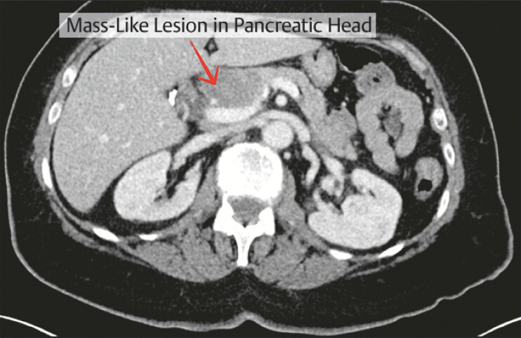 Fig. 1
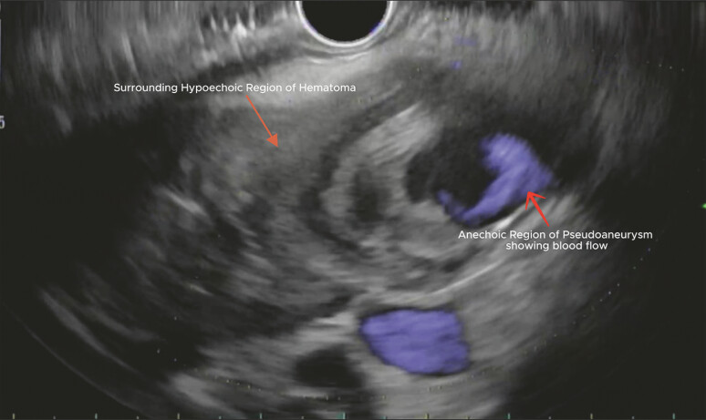 Fig. 2
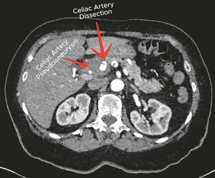 Fig. 3
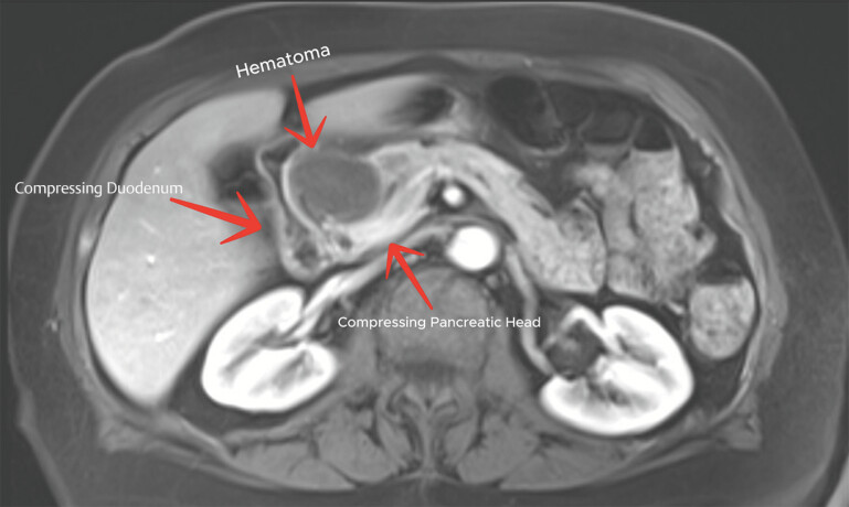 Fig. 4
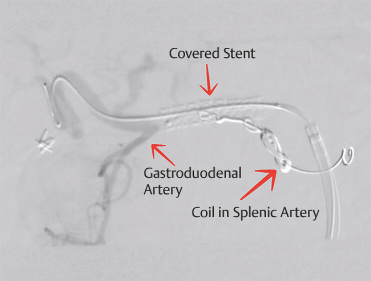 Fig. 5
Peer Reviews
No public reviews on file for this paper yet. If you reviewed it on a platform where reviews are public (OpenReview, ICLR, NeurIPS, ICML), you can paste yours below so the community can read it here.
Videos
No videos yet. Explain this paper in a talk, walkthrough, or lecture? Add one.
Taxonomy
TopicsAbdominal vascular conditions and treatments · Pancreatitis Pathology and Treatment · Infectious Aortic and Vascular Conditions
A 77-year-old woman presented with a 3-month history of intermittent, nonradiating epigastric pain that became persistent and more severe over the 2 days prior to admission.
Initial computed tomographic (CT) imaging demonstrated a mass-like lesion in the pancreatic head which raised the concern for a pancreatic neoplasm ( Fig. 1 ). Advanced endoscopy was subsequently performed for further evaluation with endoscopic ultrasound (EUS) ( Video 1 ). EUS revealed an intimal tear in the celiac artery. A heterogeneous hypoechoic lesion adjacent to the pancreatic head with an internal anechoic component ( Fig. 2 ) was also visualized. This characteristic “donut” appearance 1 was more consistent with a pseudoaneurysm surrounded by hematoma rather than a solid pancreatic mass.
Initial abdominal CT demonstrating a mass-like lesion in the pancreatic head. CT, computed tomography.
Endoscopic ultrasound (EUS) with Doppler showing hypoechoic hematoma with a central anechoic pseudoaneurysm with blood flow.
Diagnosis and management of spontaneous isolated celiac artery dissection (SICAD) with endoscopic ultrasound features mimicking a pancreatic mass.Video 1
Fine-needle aspiration was deferred due to the high risk of hemorrhage, and further vascular imaging was recommended. CT angiography confirmed a celiac artery dissection with an associated pseudoaneurysm at the celiac artery bifurcation ( Fig. 3 ). Magnetic resonance imaging further excluded a pancreatic mass and confirmed the aneurysm and dissection ( Fig. 4 ).
CT angiography depicting celiac artery dissection with an associated hematoma. CT, computed tomography.
MRI demonstrating hematoma compressing duodenum and pancreatic head. MRI, magnetic resonance imaging.
Vascular surgery subsequently managed the patient endovascularly. The splenic artery was embolized by a coil and a covered stent, extending from the abdominal aorta across the celiac trunk into the common hepatic artery, was placed ( Fig. 5 ). The patient had an uncomplicated post-operative course and was discharged the following day.
A final fluoroscopic image after endovascular management.
Spontaneous isolated celiac artery dissection (SICAD) is a rare condition which involves tear of the intima of the celiac artery without the involvement of aorta 2 . This case demonstrates key findings that the endoscopist should recognize to avoid missing a life-threatening diagnosis which needs urgent treatment and without treatment the patient can have catastrophic outcomes. CT angiography remains the most sensitive test for diagnoses 3 and standard CT with IV contrast can mischaracterize vascular lesions as masses, as in our case 4 .
Endoscopy_UCTN_Code_CCL_1AF_2AZ
The reference list from the paper itself. Each links out to its DOI / PubMed record.
- 1Varadarajulu S Eloubeidi MA Diagnosis of an aneurysm masquerading as a pancreatic-cyst lesion at EUS Gastrointest Endosc 20076572172510.1016/j.gie.2006.08.03717327129 · doi ↗ · pubmed ↗
- 2Vaidya S Dighe M Spontaneous celiac artery dissection and its management J Radiol Case Rep 20104303310.3941/jrcr.v 4i 4.40822470724 PMC 3303389 · doi ↗ · pubmed ↗
- 3Chaer RA Abularrage CJ Coleman DM The Society for Vascular Surgery clinical practice guidelines on the management of visceral aneurysms J Vasc Surg 2020723 S 39S 10.1016/j.jvs.2020.01.03932201007 · doi ↗ · pubmed ↗
- 4Itoh K Kamiya Y Ohno NA case of pancreaticoduodenal artery aneurysm causing pancreatic pseudotumour and duodenal obstruction Eur J Gastroenterol Hepatol 20021445746110.1097/00042737-200204000-0002311943965 · doi ↗ · pubmed ↗