Monocyte‐Differentiation‐Activated Fluorescent “Scout” Probe for Precise in Vivo Detection of Vulnerable Plaque
Zechuan Li, Jiankai Dong, Zhengkun Liu, Chaoke Zhang, Jisen Li, Ying Tao, Ding Yang, Yansong Liu, Haoting Chen, Lu Liu, Jingsen Ji, Feng Cao, Dan Ding, Qian Liu, Chenxing Fu, Weisheng Guo

TL;DR
A new fluorescent probe uses monocytes to detect dangerous heart plaque by hitchhiking and activating only at vulnerable sites, improving diagnostic accuracy.
Contribution
A novel monocyte-based fluorescent probe that activates specifically during monocyte differentiation in vulnerable plaque environments.
Findings
The MDAF probe hitchhikes monocytes to vulnerable plaque and activates via legumain during differentiation.
The probe generates a high signal-to-noise ratio fluorescence switch in vulnerable lesions in Apoe−/− mice.
FLECT imaging with MDAF overcomes depth limitations of conventional fluorescence methods.
Abstract
Precise identification of vulnerable plaque (VAP) is essential for the prevention of acute cardiovascular diseases, yet current molecular probes are hampered by poor VAP lesion penetration and high background. Here, the innate tropism of circulating inflammatory monocytes for VAP, and their differentiation‐driven expression of legumain (Lgmn) in response to the VAP microenvironment is exploited. A monocyte differentiation‐activated fluorescent (MDAF) probe is conceived that hitchhikes monocytes to precisely migrate to VAP and is activated by Lgmn during monocyte differentiation. This activation triggers in situ self‐assembly, resulting in spatiotemporally controlled aggregation‐induced emission (AIE) fluorescence signals, and turning the monocyte itself into an on‐site “scout” that reports plaque instability. In Apoe−/− mice bearing both vulnerable and stable plaques, the MDAF produces…
Genes, proteins, chemicals, diseases, species, mutations and cell lines named across the full text — each resolved to its canonical identifier and authoritative record.
Click any figure to enlarge with its caption.
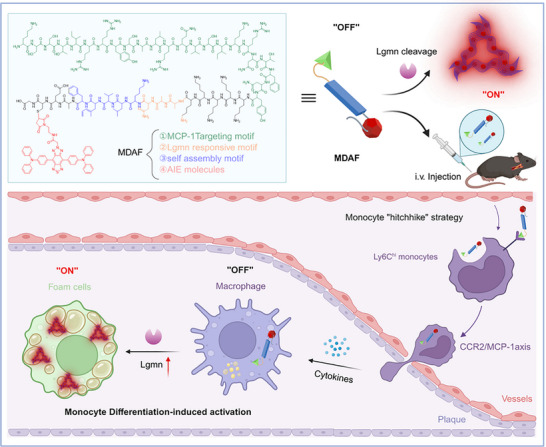 Figure 1
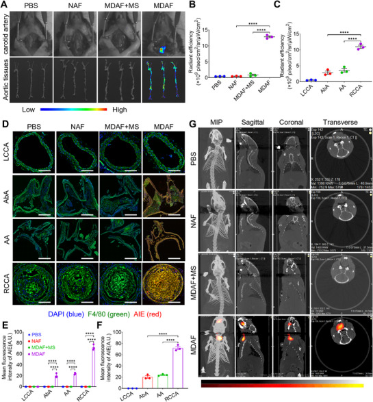 Figure 2
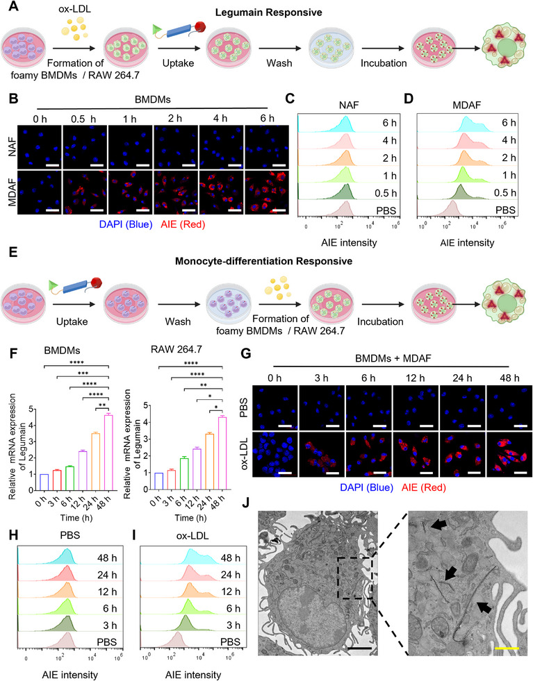 Figure 3
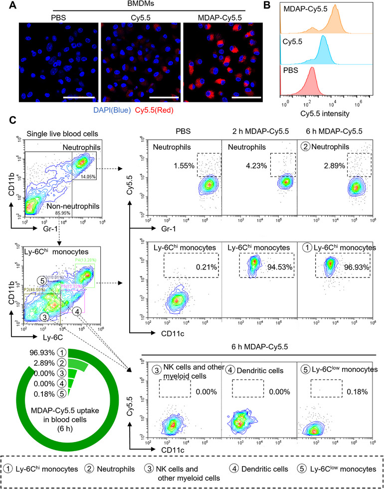 Figure 4
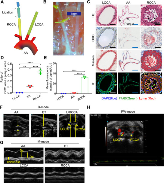 Figure 5
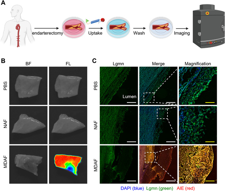 Figure 6
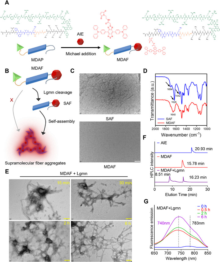 Figure 7
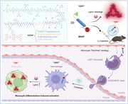 Figure 8
Peer Reviews
No public reviews on file for this paper yet. If you reviewed it on a platform where reviews are public (OpenReview, ICLR, NeurIPS, ICML), you can paste yours below so the community can read it here.
Videos
No videos yet. Explain this paper in a talk, walkthrough, or lecture? Add one.
Taxonomy
TopicsNanoplatforms for cancer theranostics · Immune cells in cancer · Cell Adhesion Molecules Research