Intraosseous epidermoid cyst in the digit of an atopic dog with chronic pododermatitis: case report
Cristiane Deon Figueiredo, Daniela Flores Fernandes, Juliana Maciel Cassali Vieira, Renata Bianco Demartini

TL;DR
This case report describes a rare intraosseous epidermoid cyst in a dog with atopic dermatitis and chronic pododermatitis.
Contribution
The novelty lies in documenting a rare intraosseous epidermoid cyst in a dog with atopic dermatitis.
Findings
Intraosseous epidermoid cysts are rare in dogs and can mimic malignant tumors.
The cyst was found in a dog with chronic pododermatitis and atopic dermatitis.
Accurate diagnosis is crucial to avoid unnecessary aggressive treatment.
Abstract
Epidermoid cysts are non-neoplastic lesions characterized by a cavity lined by stratified squamous keratinized epithelium and filled with lamellar keratin. While dermal epidermoid cysts are commonly observed in dogs, the intraosseous form is rarely reported. These cysts must be differentiated from other digital lesions involving bone, particularly malignant tumors, as the latter often require more extensive surgical intervention for definitive management. This report describes a case of an intraosseous epidermoid cyst in a dog diagnosed with atopic dermatitis and chronic pododermatitis.
Genes, proteins, chemicals, diseases, species, mutations and cell lines named across the full text — each resolved to its canonical identifier and authoritative record.
Click any figure to enlarge with its caption.
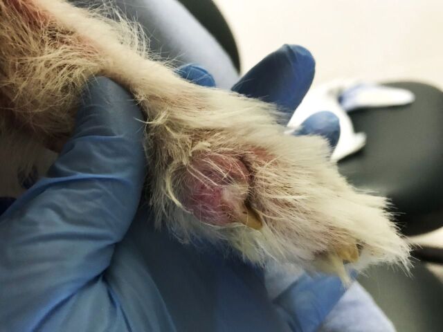 Figure 1
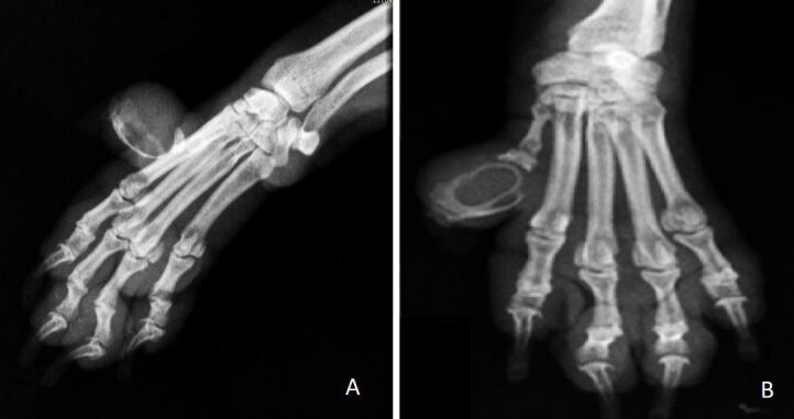 Figure 2
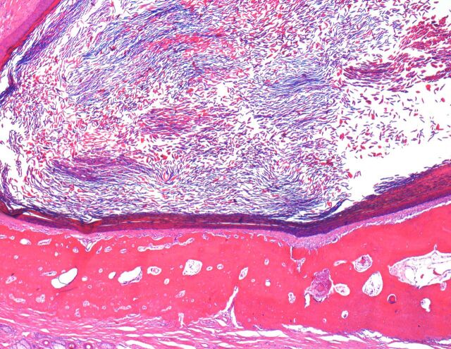 Figure 3
Peer Reviews
No public reviews on file for this paper yet. If you reviewed it on a platform where reviews are public (OpenReview, ICLR, NeurIPS, ICML), you can paste yours below so the community can read it here.
Videos
No videos yet. Explain this paper in a talk, walkthrough, or lecture? Add one.
Taxonomy
TopicsTeratomas and Epidermoid Cysts · Veterinary Oncology Research · Veterinary Medicine and Surgery