Multimodal approach to characterize surgically removed epileptogenic zone from patients with focal drug‐resistant epilepsy: From operating room to wet lab
Jenni Kyyriäinen, Adriana Della Pietra, Mastaneh Torkamani‐Azar, Mireia Gómez‐Budia, Polina Abushik, Nataliia Novosolova, Henri Eronen, Omar Narvaez, Ekaterina Paasonen, Vera Lezhneva, Anssi Pelkonen, Liudmila Saveleva, Antti Huotarinen, Ilya Belevich, Eija Jokitalo

TL;DR
A new protocol for studying brain tissue from epilepsy surgery helps understand how epilepsy develops and could improve diagnosis and treatment.
Contribution
A systematic protocol for multimodal analysis of resected epileptogenic tissue to identify therapeutic targets and improve patient care.
Findings
A protocol was developed to preserve and analyze resected tissue using imaging, electrophysiology, and molecular biology.
The protocol enables co-registration of multimodal data with in vivo MRI to map epileptogenic zones.
The approach allows for identifying structural, functional, and molecular characteristics of epileptogenic tissue.
Abstract
We have established a comprehensive sample handling protocol designed for the multiscale assessment of epileptogenic tissue. This protocol aims to identify novel therapeutic targets and enhance the diagnosis and stratification of patients with drug‐resistant epilepsy, thereby optimizing their treatment with anti‐seizure medications and surgical interventions. Patients with drug‐resistant focal epilepsy, recommended for surgical treatment, are recruited after detailed multidisciplinary preoperative evaluation at the Epilepsy Center at Kuopio University Hospital in Finland. A day before the resective surgery, patients undergo magnetic resonance imaging (MRI) including advanced methodologies. During the surgery, each piece of resected tissue is placed under oxygenation on ice‐cold artificial cerebral spinal fluid solution. The pieces are then immediately transported to the laboratory,…
Genes, proteins, chemicals, diseases, species, mutations and cell lines named across the full text — each resolved to its canonical identifier and authoritative record.
Click any figure to enlarge with its caption.
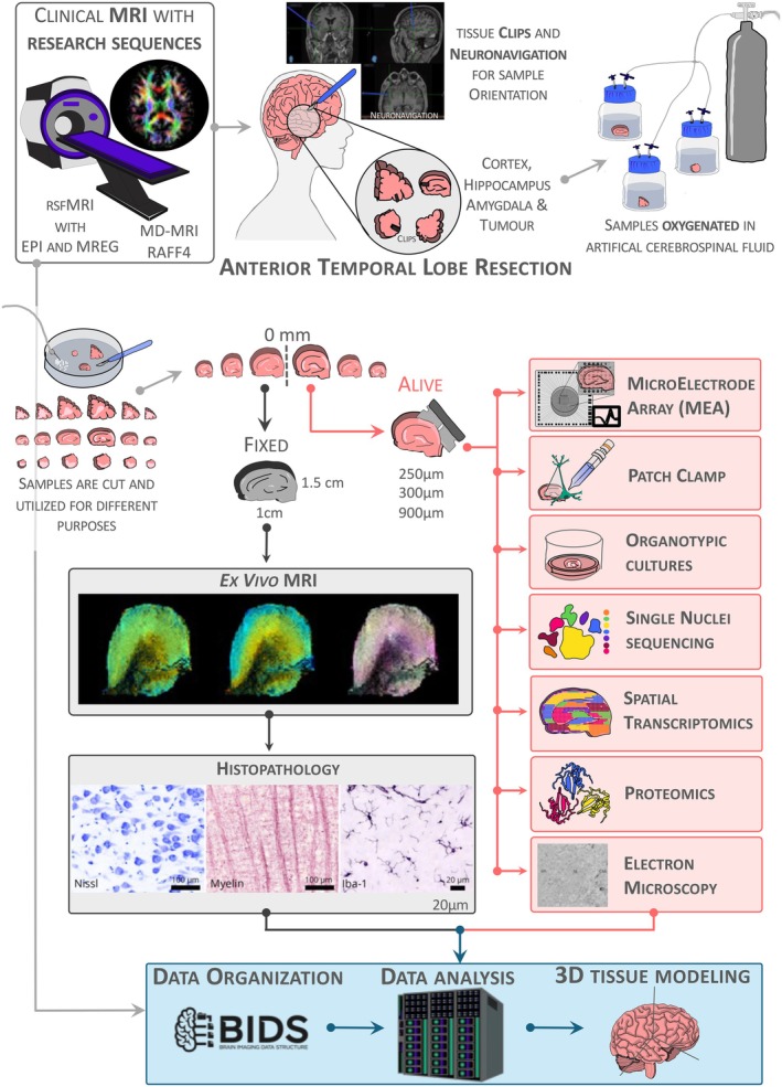 Figure 1
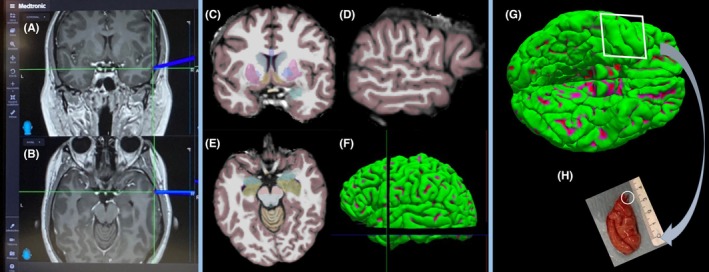 Figure 2
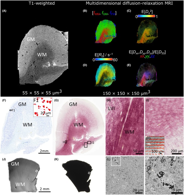 Figure 3
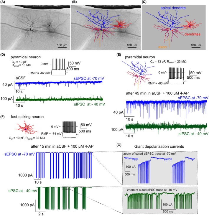 Figure 4
 Figure 5
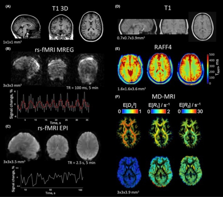 Figure 6
Peer Reviews
No public reviews on file for this paper yet. If you reviewed it on a platform where reviews are public (OpenReview, ICLR, NeurIPS, ICML), you can paste yours below so the community can read it here.
Videos
No videos yet. Explain this paper in a talk, walkthrough, or lecture? Add one.
Taxonomy
TopicsEpilepsy research and treatment · Cerebrospinal fluid and hydrocephalus · Glioma Diagnosis and Treatment