Atomic force microscopy-based topographical imaging of SARS-CoV-2 as part of a tripartite strategy for RNA virus characterization
Tanja Deckert-Gaudig, Xiaobin Yao, Erwan Darussalam, Franziska Hornung, Pablo Carravilla, Ziliang Zhao, Kourosh Rezaei, Christian Eggeling, Stefanie Deinhardt-Emmer, Volker Deckert

TL;DR
This study combines atomic force microscopy and fluorescence imaging to accurately identify SARS-CoV-2 particles based on their size, shape, and molecular features.
Contribution
A novel tripartite imaging strategy that integrates AFM and double-staining fluorescence for high-specificity RNA virus identification.
Findings
SARS-CoV-2 particles were identified with a height range of 60–100 nm using AFM and fluorescence correlation.
The method distinguishes intact SARS-CoV-2 from hollow particles, fragments, and artifacts.
The approach is broadly applicable to other RNA viruses for high-specificity detection.
Abstract
The pre-selection of virus particles based on size and morphology is a crucial step toward rapid and reliable virus identification. Pre-selecting virus particles based on size and morphology represents a critical step toward rapid and reliable identification, which is particulary important in clinical settings when novel virus variants emerge. Although conventional fluorescence imaging enables visualization of specific viral structures via labeling, it does not allow for reliable differentiation of structurally similar particles. In this study, we present a combined imaging approach that integrates atomic force microscopy (AFM) and double-staining fluorescence microscopy to identify SARS-CoV-2 as a model RNA-virus from other sample constituents. Initially, topographical imaging via AFM enables high-resolution visualization of individual virus particles, providing detailed information…
Genes, proteins, chemicals, diseases, species, mutations and cell lines named across the full text — each resolved to its canonical identifier and authoritative record.
Click any figure to enlarge with its caption.
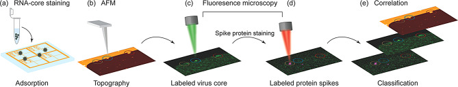 Figure 1
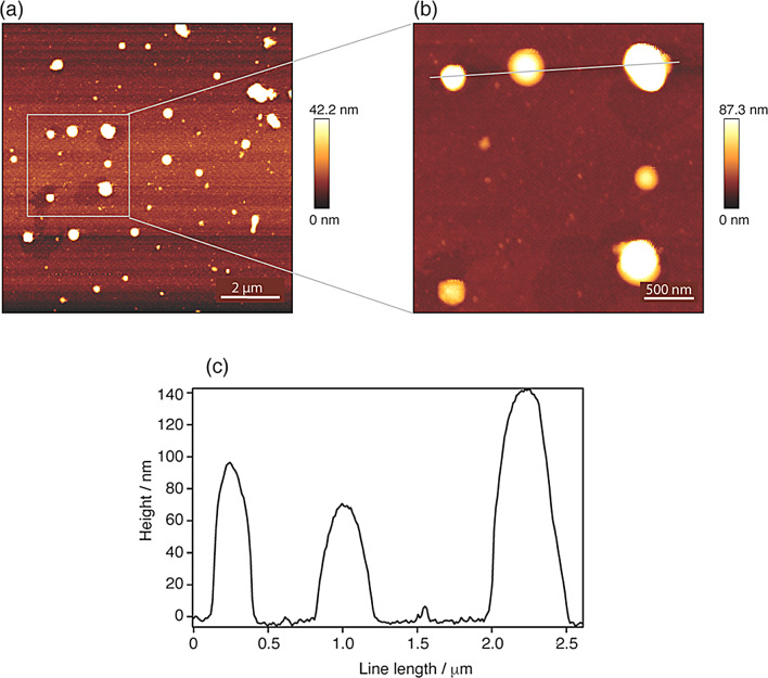 Figure 2
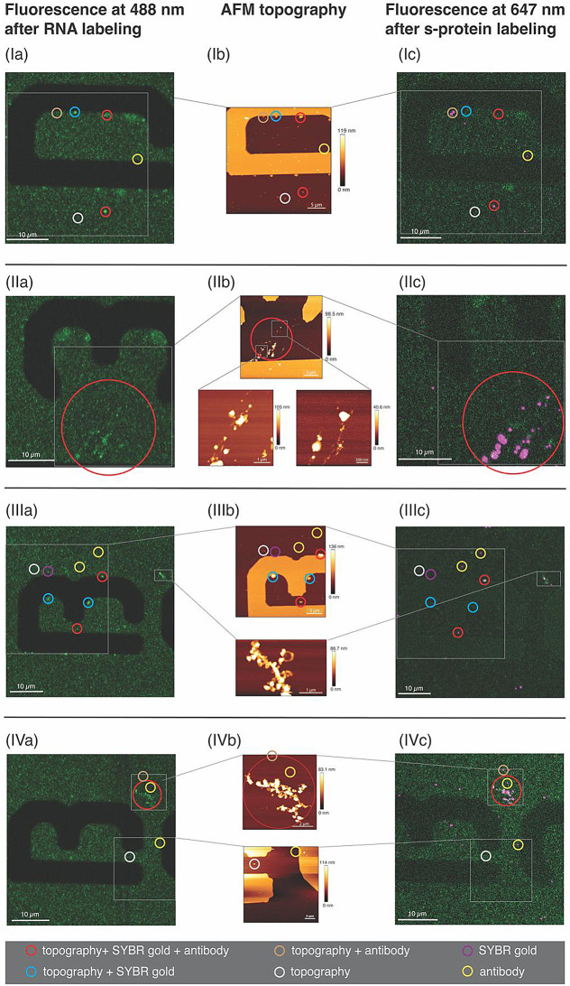 Figure 3
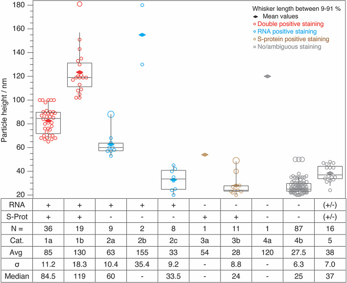 Figure 4
Peer Reviews
No public reviews on file for this paper yet. If you reviewed it on a platform where reviews are public (OpenReview, ICLR, NeurIPS, ICML), you can paste yours below so the community can read it here.
Videos
No videos yet. Explain this paper in a talk, walkthrough, or lecture? Add one.
Taxonomy
TopicsForce Microscopy Techniques and Applications · SARS-CoV-2 and COVID-19 Research · SARS-CoV-2 detection and testing