GLUT1 expression, lymphocyte distribution and CD3+ T-cell metabolic subsets as predictive markers of response to immunotherapy in advanced melanoma
Elizabeth C. Paver, Tuba N. Gide, Zarwa Yaseen, Paola Cornejo-Paramo, Peter Ferguson, Nigel G. Maher, Alexander M. Menzies, Matteo S. Carlino, Ines Pires da Silva, Jeff Holst, Georgina V. Long, Richard A. Scolyer, James S. Wilmott

TL;DR
This study shows that the presence of glycolysis in melanoma tumors, marked by GLUT1 and GLUT3, is linked to T-cell infiltration and better response to immunotherapy.
Contribution
The study introduces new predictive markers (GLUT1/GLUT3 and T-cell spatial distribution) for immunotherapy response in advanced melanoma.
Findings
GLUT1+ melanoma regions have fewer CD3+ T-cells compared to GLUT1- regions.
Responders to immunotherapy have higher proportions of GLUT1+ and GLUT3+ T-cells.
CD3+ T-cells near GLUT1+ melanoma cells are associated with longer progression-free survival.
Abstract
Glycolysis, commonly used by malignant tumors for energy production, results in acidification of the tumor microenvironment (TME) through the secretion and accumulation of lactic acid. Acidosis is a potent inhibitor of immune cell function and may therefore affect T-cell infiltration and the efficacy of immunotherapy. This study aimed to characterize the metabolic tumor microenvironment and its association with lymphocyte distribution in patients with advanced melanoma treated with immune checkpoint blockade (ICB) therapies. Pre-treatment formalin-fixed, paraffin-embedded metastatic melanoma specimens from 45 patients treated with anti-PD-1 ± anti-CTLA-4 ICB were included in this study. Patients with progression-free survival (PFS) ≥ 6mo were categorized as responders (n = 23), while non-responders had a PFS < 6mo (n = 22). Two custom multiplex immunofluorescence panels were developed…
Genes, proteins, chemicals, diseases, species, mutations and cell lines named across the full text — each resolved to its canonical identifier and authoritative record.
Click any figure to enlarge with its caption.
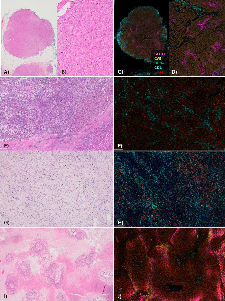 Figure 1
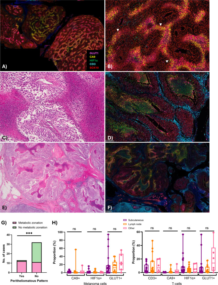 Figure 2
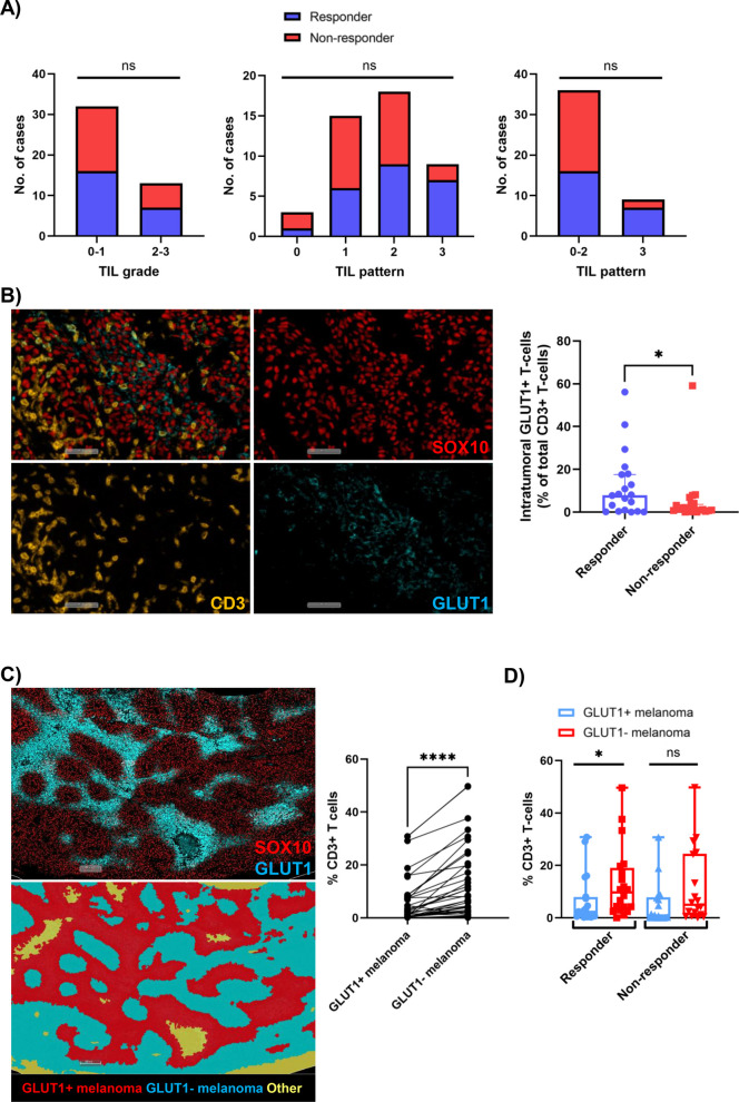 Figure 3
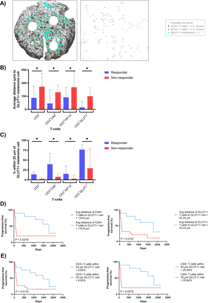 Figure 4
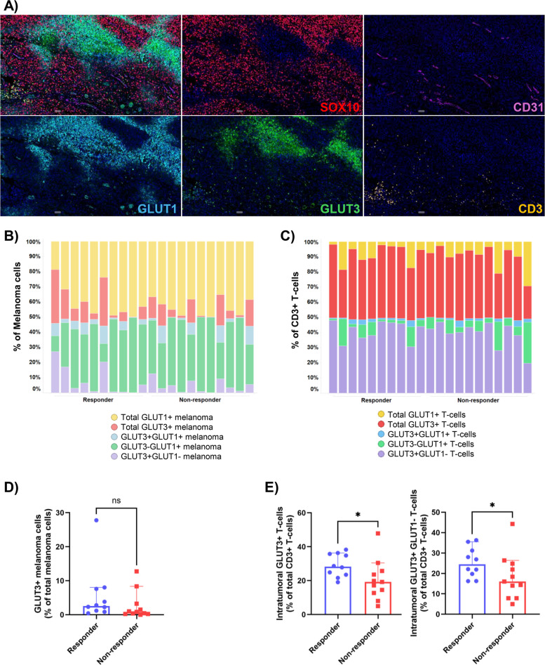 Figure 5
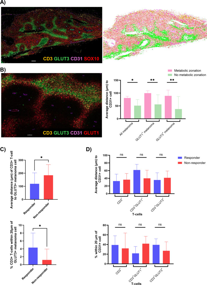 Figure 6
Peer Reviews
No public reviews on file for this paper yet. If you reviewed it on a platform where reviews are public (OpenReview, ICLR, NeurIPS, ICML), you can paste yours below so the community can read it here.
Videos
No videos yet. Explain this paper in a talk, walkthrough, or lecture? Add one.
Taxonomy
TopicsCancer Immunotherapy and Biomarkers · Cancer, Hypoxia, and Metabolism · Immune cells in cancer