Label-free fluorescence lifetime imaging can distinguish cancer from healthy tissue in spontaneously occurring canine oral tumors
Stephanie Goldschmidt, Laura Marcu, Katjana Ehrlich, Mohamed Abul Hassan, Iris Rivas, Andrew Birkeland, Xiangnan Zhou, Julien Bec, Alba Alfonso Garcia, Shuai Chen, Yichu Chen, Yash Tipirneni, Max Kampe, Abigail Weir, Abraham Morales, Christine Ly, Robert Rebhun, Brian G. Murphy

TL;DR
Label-free fluorescence lifetime imaging can accurately tell cancer from healthy tissue in dogs' mouths without needing special markers.
Contribution
The study shows that label-free FLIm is sufficient for cancer detection in dogs, and adding exogenous markers does not significantly improve accuracy.
Findings
Fluorescence emission parameters significantly differ between cancer and healthy tissues in both autofluorescence and PpIX channels.
Autofluorescence features, especially lifetimes and intensity ratios, provided the strongest in vivo discrimination.
Exogenous markers like 5-ALA–induced PpIX do not markedly improve diagnostic accuracy in FLIm.
Abstract
Post-surgical local recurrence of oral cancer remains unacceptably high across species due to the lack of non-invasive tools capable of accurately delineating tumor from healthy tissue. Label-free fluorescence lifetime imaging (FLIm) has shown moderate-to-high success in human head and neck squamous cell carcinoma, but it is unclear whether diagnostic accuracy can be enhanced by incorporating exogenous fluorophores that selectively accumulate in cancer. This study evaluated the performance of 5-ALA induced Protoporphyrin IX (PpIX) fluorescence and autofluorescence FLIm features to discriminate epithelial cancer and healthy tissues in a spontaneous large animal model of disease (15 pet dogs). Fluorescence emission parameters (e.g. lifetimes, intensity ratios, phasors and Laguerre coefficients) differed significantly (p < 0.001) between cancer and healthy tissues in both autofluorescence…
Genes, proteins, chemicals, diseases, species, mutations and cell lines named across the full text — each resolved to its canonical identifier and authoritative record.
Click any figure to enlarge with its caption.
 Figure 1
 Figure 2
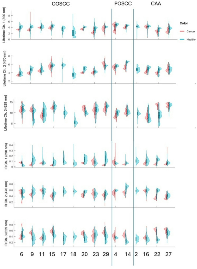 Figure 3
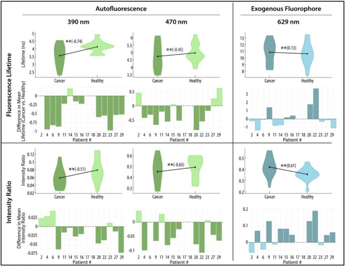 Figure 4
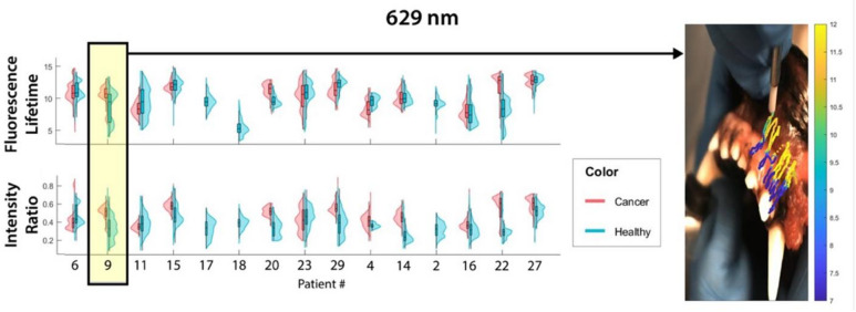 Figure 5
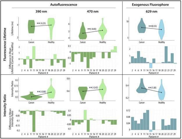 Figure 6
 Figure 7
 Figure 8
Peer Reviews
No public reviews on file for this paper yet. If you reviewed it on a platform where reviews are public (OpenReview, ICLR, NeurIPS, ICML), you can paste yours below so the community can read it here.
Videos
No videos yet. Explain this paper in a talk, walkthrough, or lecture? Add one.
Taxonomy
TopicsPhotodynamic Therapy Research Studies · Advanced Fluorescence Microscopy Techniques · Nanoplatforms for cancer theranostics