Decoding the mechanisms of acupuncture by neuroimaging: an integrated review from networks to molecules
Shiping Liu, Yan Bai, Jie Liu, Xia Chen, Peizhu Lv, Yulin Wang, Dandan Wang, Shun Wang

TL;DR
This review explores how acupuncture affects the brain and nervous system using neuroimaging, aiming to better understand its mechanisms and limitations.
Contribution
The paper proposes a multi-level framework linking acupuncture's peripheral stimulation to clinical outcomes via brain networks and neurochemistry.
Findings
Acupuncture modulates large-scale brain networks like the Default Mode Network and Salience Network.
Neuroimaging reveals neurochemical changes and neural oscillation effects associated with acupuncture.
Non-specific effects, such as placebo responses, are significant and require careful distinction from specific acupuncture effects.
Abstract
Acupuncture, a cornerstone of Traditional Chinese Medicine (TCM), is widely used for conditions like chronic pain and functional disorders, yet its neurobiological mechanisms are not fully understood. This review synthesizes findings from multimodal neuroimaging–including functional magnetic resonance imaging (fMRI), electroencephalography (EEG), and positron emission tomography (PET)–to examine the central nervous system and neurochemical correlates of acupuncture. We summarize reports of its modulatory effects on large-scale brain networks (e.g., Default Mode Network, Salience Network) and neural oscillations, alongside evidence of neurochemical changes. Importantly, we also address the methodological limitations, inconsistent results, and significant role of non-specific (e.g., placebo) effects prevalent in this literature (Chen B. et al., 2023; Yu et al., 2024). Building on this…
Genes, proteins, chemicals, diseases, species, mutations and cell lines named across the full text — each resolved to its canonical identifier and authoritative record.
Click any figure to enlarge with its caption.
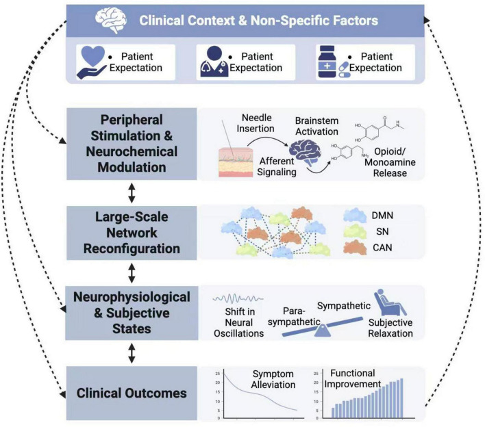 FIGURE 1
Peer Reviews
No public reviews on file for this paper yet. If you reviewed it on a platform where reviews are public (OpenReview, ICLR, NeurIPS, ICML), you can paste yours below so the community can read it here.
Videos
No videos yet. Explain this paper in a talk, walkthrough, or lecture? Add one.
Taxonomy
TopicsAcupuncture Treatment Research Studies · Traditional Chinese Medicine Studies · Functional Brain Connectivity Studies
Introduction
1
Clinical efficacy and the mechanistic challenge
1.1
With a history spanning millennia, acupuncture is globally utilized for conditions such as chronic low back pain (Wen et al., 2021), various pain syndromes (Pock and Niemtzow, 2021), Alzheimer’s disease (Cai and Yang, 2020), and functional dyspepsia (Dong et al., 2022). Despite its clinical application, the biological mechanisms underlying its effects remain incompletely defined. While traditional concepts like “Qi” and “meridians” offer one explanatory framework, a gap persists with modern biomedical understanding (Hui et al., 2000). Closing this gap through robust mechanistic evidence is essential for acupuncture’s integration into contemporary evidence-based medicine.
Neuroimaging: a window into central correlates
1.2
Non-invasive neuroimaging has opened a valuable window into the brain’s response to acupuncture (Napadow et al., 2009). Early research focused on identifying activated or deactivated brain regions (Fang et al., 2009). The field has since progressed toward a systems-level approach, investigating how acupuncture might influence the dynamics of large-scale brain networks and neural circuits (Dhond et al., 2007; Napadow et al., 2013). Yet, interpreting these neuroimaging findings demands careful acknowledgment of their inherent correlational nature and the various confounds present in acupuncture studies.
Review scope and critical perspective
1.3
This review provides a comprehensive and critical overview of current neuroimaging evidence concerning acupuncture mechanisms. We will: (1) Detail the insights and constraints of established techniques (fMRI, EEG) in revealing brain network and neural dynamics; (2) Discuss the emerging, though often preliminary, role of molecular imaging (PET) and novel technologies in probing neurochemical substrates; (3) Integrate these findings into a coherent, multi-level framework, while explicitly addressing the predominance of correlational data and the challenges in establishing causality; and (4) Identify key methodological issues, contradictory findings, and outline future directions that prioritize scientific rigor.
Established neuroimaging techniques: mapping macroscopic brain dynamics
2
Functional magnetic resonance imaging (fMRI)
2.1
Functional magnetic resonance imaging, particularly using the blood oxygenation level-dependent (BOLD) signal, has been extensively employed to map brain responses to acupuncture with high spatial resolution.
Studies suggest that needling at specific acupoints, such as ST36 (Zusanli) and LI4 (Hegu), can modulate activity within a distributed network involving the limbic system (e.g., amygdala, hippocampus), paralimbic regions (e.g., anterior cingulate cortex, insula), and subcortical structures (e.g., hypothalamus) (Chae et al., 2013; Hui et al., 2000). This network is integral to pain perception, emotion, and autonomic control, potentially underpinning acupuncture’s diverse effects.
Resting-state fMRI (rs-fMRI) research indicates that acupuncture may alter functional connectivity within and between large-scale intrinsic brain networks. A commonly observed finding is a decrease in Default Mode Network (DMN) connectivity following acupuncture (Fan et al., 2020; Wang et al., 2017). Since the DMN is active during self-referential thought, its deactivation aligns with subjective reports of relaxation post-treatment (Dhond et al., 2008). Acupuncture has also been shown to modulate the functional connectivity of the Salience Network (SN). For instance, in patients with low back pain, the analgesic effect of real acupuncture (with needle sensation) was specifically associated with decreased connectivity between the posterior insula (a key node of the SN) and the default mode network, highlighting the role of SN in processing salient somatosensory stimuli like Deqi (Lee et al., 2019). And may influence the Central Autonomic Network (CAN), relevant to visceral regulation (Li et al., 2024; Sun et al., 2021).
These fMRI findings must be considered alongside significant methodological constraints and inconsistent results. The BOLD signal is an indirect measure of neural activity. More critically, the specificity of acupuncture’s effects on brain networks is debated. Reviews indicate that while some studies report distinct patterns for real versus sham acupuncture, others demonstrate considerable overlap, with sham procedures often eliciting similar, if somewhat attenuated, brain modulation (Chen B. et al., 2023; Kelly et al., 2024). This highlights the substantial impact of non-specific factors like expectation and general somatosensory stimulation. Additional challenges include motion artifacts during needling, frequently small sample sizes, a historical lack of study pre-registration, and the inherent difficulty in designing truly inert control interventions.
Electroencephalography (EEG)
2.2
Electroencephalography measures neuronal electrical activity directly with millisecond resolution, capturing rapid dynamics.
Acupuncture has been associated with changes in oscillatory power across frequency bands. An increase in alpha power (8–13 Hz), particularly over parietal and occipital areas, is a frequently reported finding linked to states of relaxed wakefulness (Yu et al., 2024). Changes in beta (13–30 Hz) and gamma (>30 Hz) activity have also been noted, possibly related to somatosensory processing and attention (Nierhaus et al., 2015). The temporal precision of EEG allows analysis of event-related potentials (ERPs) synchronized to needle manipulation.
Variability and Interpretation: Considerable variability exists across EEG studies in the topography, direction, and consistency of these oscillatory changes. Outcomes are influenced by needling parameters, subject state, and control conditions. This heterogeneity means EEG responses to acupuncture are not uniform. As with fMRI, a central challenge remains distinguishing effects specific to acupuncture from generalized responses to sensory stimulation and attentional shifts.
Complementary use and shared constraints of fMRI and EEG
2.3
Combining fMRI and EEG can powerfully correlate spatial networks with temporal dynamics. Concurrent EEG-fMRI studies may link fast oscillations with slower hemodynamic changes (Liang et al., 2024; Zhang et al., 2025). However, this multimodal approach also inherits and combines the limitations of each technique, adding complexity to experimental design and data interpretation.
Emerging molecular and functional imaging approaches
3
While fMRI and EEG map macroscopic correlates, they lack biochemical specificity. Newer molecular and functional imaging technologies promise greater specificity but remain at early stages of application in acupuncture research.
Positron emission tomography (PET)
3.1
Positron emission tomography enables quantitative measurement of specific neurochemical processes via radiotracers. Though limited in number, PET studies offer more direct biochemical insights.
A key application is investigating neurotransmitter systems. For example, using the radioligand [^11^C]carfentanil, PET has shown that verum acupuncture can alter μ-opioid receptor availability in certain brain regions compared to sham, supporting a role for the opioid system in some contexts of acupuncture analgesia (Harris et al., 2009). PET with ^1^8F-fluorodeoxyglucose (^1^8F-FDG), a measure of cerebral glucose metabolism, has suggested that electroacupuncture can modulate metabolism in brain regions relevant to conditions like Alzheimer’s disease in animal and human studies (Xu et al., 2020a,b).
Considerations: PET studies in acupuncture are few and resource-intensive. Their findings, while valuable, are not yet consistent across all populations or paradigms. They demonstrate the potential for neurochemical modulation but do not establish a definitive neurochemical signature for acupuncture.
Novel technologies and integrative approaches
3.2
Several emerging technologies offer new avenues for investigation, though their contributions are currently preliminary.
Functional near-infrared spectroscopy (fNIRS): This portable tool monitors cortical hemodynamics in clinical settings. Studies have used fNIRS to observe altered prefrontal activation during scalp acupuncture in stroke patients (Lin et al., 2025; Zhong et al., 2025). Hyperscanning studies also report increased inter-brain synchrony between practitioner and patient during needling (Chen L. et al., 2023), hinting at a neural basis for the therapeutic interaction. However, it is important to note that fNIRS is limited by its shallow penetration depth and remains susceptible to systemic physiological confounds unrelated to neural activity.
Bio-inspired and nanobiosensors: Integrating micro/nanotechnology with acupuncture needles has produced innovative biosensors. For instance, a transistor-based neuroprobe modeled on an acupuncture needle enabled real-time neurotransmitter monitoring in vivo (Zhou et al., 2022). Similarly, microneedle biosensors have monitored molecular release (e.g., hydrogen sulfide) in animal models after electroacupuncture (Pei et al., 2025). While these engineered devices represent a transformative toolset for basic science with high potential, a critical perspective is necessary: their use in acupuncture research is currently almost entirely confined to preclinical animal models. Translation to human studies faces major challenges regarding safety, biocompatibility, and practicality. Therefore, data from these platforms should be viewed as hypothesis-generating for human mechanisms rather than as direct evidence. They primarily illustrate technological possibilities rather than confirm established human pathophysiology.
Multimodal integration and artificial intelligence: Combining data from multiple imaging modalities and applying AI (e.g., machine learning) is a growing trend to overcome single-method limitations. AI may help identify complex patterns, predict treatment response from baseline scans, or assist with data processing (Ma et al., 2024; Zhang et al., 2025). Methodological frameworks like “mFusion” have been proposed to bridge imaging and genetic data via neurotransmitter systems (Cao et al., 2024). However, a note of caution is warranted: AI applications in acupuncture neuroimaging are still nascent. Risks include overfitting models to small, heterogeneous datasets and the limited interpretability of derived features, the so-called “black box” problem. At present, AI’s primary value lies in exploratory analysis and generating new, testable hypotheses rather than in providing definitive biomarkers.
Toward an integrative multi-level framework
4
A central aim of this field is to integrate multimodal findings into a coherent explanatory framework while progressively testing causal hypotheses.
Integrating multimodal evidence critically
4.1
Evidence convergence across modalities can strengthen inferences but does not equal proof of cause. Truly multimodal studies–like combined PET-fMRI to link neurochemical release with network changes, or concurrent EEG-fMRI to trace temporal sequences–are rare but crucial for advancing beyond correlation.
A proposed multi-level framework
4.2
Synthesizing the correlative evidence, we propose a multi-level framework that outlines a hypothesized pathway from peripheral stimulation to clinical outcome (Figure 1). This framework organizes existing findings into a plausible sequence while noting that causal connections require further validation.
A proposed multi-level framework of acupuncture mechanisms (arrows indicate hypothesized relationships based on current evidence; establishing definitive causal links is a primary objective for future research).
The proposed sequence starts with peripheral needle insertion, evoking local biochemical and neural responses (Wu et al., 2024; Zhang et al., 2008). Signals are relayed to brainstem regions like the periaqueductal gray (PAG) (Napadow et al., 2009). Evidence from ligand studies suggests this may trigger neurochemical modulation, including release of endogenous opioids and monoamines (Cui et al., 1999; Harris et al., 2009). We hypothesize that these neurochemical shifts could contribute to a reconfiguration of large-scale brain networks. For instance, opioid release might be involved in altering connectivity within the descending pain pathway and affective networks (Wang et al., 2017; Yu et al., 2020). Such network-level changes, which include modulation of the DMN and SN, are correlated with subjective reports of altered self-awareness, attention, and autonomic state (Dhond et al., 2008; Lee et al., 2019; Sun et al., 2021; Yu et al., 2024). These network dynamics are reflected in shifts in neural oscillations, such as increased alpha power (Nierhaus et al., 2015; Yu et al., 2024). The ultimate clinical benefit likely arises from the combined influence of these processes.
It is crucial to maintain a key distinction here: this framework is built largely on correlations observed across different studies. Demonstrating that needle-induced neurochemical release directly causes a specific network change, which then directly causes a specific clinical improvement, remains an unmet goal. The model therefore serves as a working guide for designing future research that can test these proposed links, for example through pharmacological challenges combined with neuroimaging.
Future directions and ongoing challenges
5
Technical and analytical advances
5.1
Advances in imaging hardware (e.g., ultra-high-field MRI, next-generation PET) will enhance resolution and sensitivity. AI and computational models offer promising tools for analyzing complex datasets and predicting outcomes (Ma et al., 2024; Zhang et al., 2025). Their application, however, must be grounded in methodologically sound studies to avoid generating misleading results.
Methodological priorities
5.2
The credibility of future research hinges on addressing current shortcomings. A critical priority is enhancing rigor and standardization. The field urgently needs consensus on standardized protocols for acupoint location, needling technique, and–most critically–the selection and reporting of control conditions (sham/placebo). Pre-registration of trials and analytical plans is vital to improve reproducibility and reduce bias.
Simultaneously, future studies must proactively confront non-specific effects. Employing robust experimental designs, such as validated sham needles and effective blinding strategies, is essential to disentangle the specific physiological effects of needling from the powerful contextual, placebo, and somatosensory effects inherent in the therapeutic encounter.
Furthermore, research should systematically account for individual differences to pave the way for personalized approaches. This includes integrating factors like genetic polymorphisms [e.g., in the COMT gene, known to affect pain modulation (Ho et al., 2020)], psychological traits, and baseline brain architecture. Pretreatment neuroimaging has already shown preliminary value in predicting clinical response to acupuncture (Yu et al., 2020).
The path to translation
5.3
A major translational goal is the development of validated neuroimaging biomarkers capable of predicting treatment response or objectively verifying target engagement. Achieving this will require a concerted effort through large-scale, longitudinal, multicenter trials that incorporate neuroimaging as a core component. Fostering closer collaboration between neuroscientists, methodologies, and clinical acupuncturists is equally essential to ensure that research questions remain both biologically insightful and directly relevant to clinical practice.
Conclusion
6
Neuroimaging has significantly advanced the exploration of acupuncture’s mechanisms, shifting the discourse toward identifiable neurobiological correlates. Established techniques have mapped its influence on brain network dynamics and electrical activity, while molecular imaging has begun to illuminate potential neurochemical underpinnings. Integrating these strands of evidence allows for the construction of a multi-level framework that hypothesizes how needling might link to neurobiological events and clinical outcomes. Nevertheless, the current evidence landscape is characterized by correlation, heterogeneity, and notable methodological limitations. The clinical utility of acupuncture demands a research trajectory that places scientific rigor at its core. Moving forward, the field must leverage advanced technologies within robust experimental designs, rigorously address non-specific effects, and actively test the causal relationships within proposed mechanistic frameworks. By following this rigorous and transparent path, research can work toward a solidly evidence-based understanding of acupuncture’s place in integrative healthcare.
The reference list from the paper itself. Each links out to its DOI / PubMed record.
- 1Cai M. Yang E. (2020). Effect of combined electroacupuncture and selegiline treatment in Alzheimer’s disease: An animal model. Front. Pharmacol. 11:606480. 10.3389/fphar.2020.606480 33362561 PMC 7758426 · doi ↗ · pubmed ↗
- 2Cao L. Wang Z. Yuan Z. Luo Q. (2024). m Fusion: A multiscale fusion method bridging neuroimages to genes through neurotransmissions in mental health disorders. Commun. Biol. 7:1699. 10.1038/s 42003-024-07404-x 39719509 PMC 11668864 · doi ↗ · pubmed ↗
- 3Chae Y. Chang D. Lee S. Jung W. Lee I. Jackson S. (2013). Inserting needles into the body: A meta-analysis of brain activity associated with acupuncture needle stimulation. J. Pain 14 215–222. 10.1016/j.jpain.2012.11.011 23395475 · doi ↗ · pubmed ↗
- 4Chen B. Guo Q. Zhang Q. Di Z. Zhang Q. (2023). Revealing the central mechanism of acupuncture for primary dysmenorrhea based on neuroimaging: A narrative review. Pain Res. Manag. 2023:8307249. 10.1155/2023/8307249 36852393 PMC 9966569 · doi ↗ · pubmed ↗
- 5Chen L. Qu Y. Cao J. Liu T. Gong Y. Tian Z. (2023). The increased inter-brain neural synchronization in prefrontal cortex between simulated patient and acupuncturist during acupuncture stimulation: Evidence from functional near-infrared spectroscopy hyperscanning. Hum. Brain Mapp. 44 980–988. 10.1002/hbm.26120 36255178 PMC 9875919 · doi ↗ · pubmed ↗
- 6Cui M. Feng Y. Mc Adoo D. Willis W. (1999). Periaqueductal gray stimulation-induced inhibition of nociceptive dorsal horn neurons in rats is associated with the release of norepinephrine, serotonin, and amino acids. J. Pharmacol. Exp. Ther. 289 868–876. 10.1016/S 0022-3565(99)38213-810215665 · doi ↗ · pubmed ↗
- 7Dhond R. Kettner N. Napadow V. (2007). Neuroimaging acupuncture effects in the human brain. J. Altern. Complement. Med. 13 603–616. 10.1089/acm.2007.7040 17718643 · doi ↗ · pubmed ↗
- 8Dhond R. Yeh C. Park K. Kettner N. Napadow V. (2008). Acupuncture modulates resting state connectivity in default and sensorimotor brain networks. Pain 136 407–418. 10.1016/j.pain.2008.01.011 18337009 PMC 2440647 · doi ↗ · pubmed ↗