Impact of Ethylene Oxide Sterilization on PEDOT:PSS Electrophysiology Electrodes
Ali Maziz, Clement Cointe, Benjamin Reig, Christian Bergaud

TL;DR
This study shows that ethylene oxide sterilization does not harm PEDOT:PSS electrodes used in medical electrophysiology devices.
Contribution
The first comprehensive evaluation of ethylene oxide sterilization effects on PEDOT:PSS electrodes for biomedical applications.
Findings
EtO sterilization caused minimal changes in surface topography and no detectable alteration in electrical or electrochemical performance.
Impedance spectroscopy and cyclic voltammetry showed EtO-treated electrodes retained properties similar to untreated controls.
EtO-sterilized PEDOT:PSS coatings showed robust long-term stability with negligible degradation over extended operation.
Abstract
Poly(3,4-ethylenedioxythiophene)–polystyrene sulfonate (PEDOT:PSS) is widely used to fabricate conductive organic coatings for electrodes in electrophysiology. As these devices move toward clinical translation, establishing sterilization methods that preserve their functional properties is essential. Ethylene oxide (EtO) is routinely used for sterilizing heat- and moisture-sensitive medical devices due to its high penetration efficiency and low thermal load. However, the absence of systematic studies evaluating its impact on PEDOT:PSS raises concerns about the compatibility of EtO sterilization with organic electrophysiology interfaces. Here, we report the first comprehensive evaluation of EtO sterilization on PEDOT:PSS electrodes electrochemically deposited onto cortical interfaces designed for intraoperative monitoring and stimulation. EtO exposure induced only minimal changes in…
Genes, proteins, chemicals, diseases, species, mutations and cell lines named across the full text — each resolved to its canonical identifier and authoritative record.
Click any figure to enlarge with its caption.
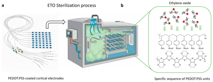 Figure 1
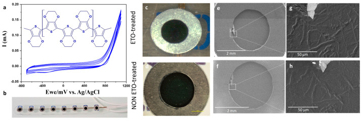 Figure 2
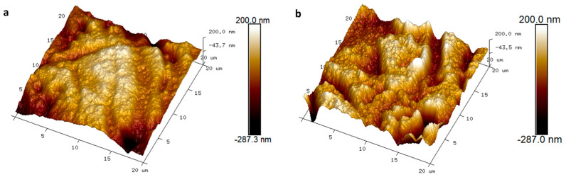 Figure 3
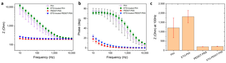 Figure 4
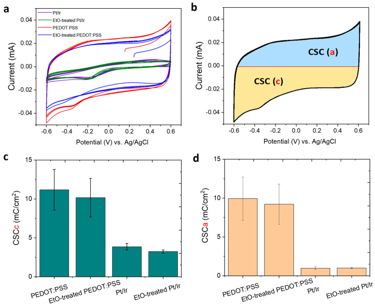 Figure 5
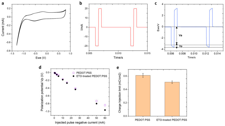 Figure 6
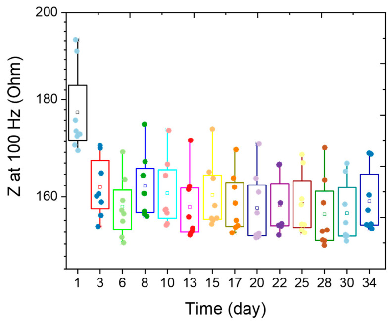 Figure 7
- —Agence Nationale de la Recherche (France)
Peer Reviews
No public reviews on file for this paper yet. If you reviewed it on a platform where reviews are public (OpenReview, ICLR, NeurIPS, ICML), you can paste yours below so the community can read it here.
Videos
No videos yet. Explain this paper in a talk, walkthrough, or lecture? Add one.
Taxonomy
TopicsConducting polymers and applications · Neuroscience and Neural Engineering · Advanced Sensor and Energy Harvesting Materials
1. Introduction
The development of reliable materials for long-term neural recording and stimulation remains a central challenge in neurotechnology. Conventional electrode arrays are typically fabricated using noble metals such as gold (Au), platinum (Pt), and iridium (Ir) [1], which have enabled decades of clinical applications, including cochlear implants, deep brain stimulation for Parkinson’s disease, and motor function restoration [2,3,4,5]. Despite their long history of use, metal-based neural interfaces still face significant limitations. Their stiffness and mechanical mismatch with soft neural tissue contribute to chronic inflammation and scar formation, while their high intrinsic electrochemical impedance restricts safe charge injection and limits recording fidelity [6]. For chronic neural interfaces, stability of material properties and electrical performance is crucial; however, metallic materials interact poorly with surrounding tissues due to persistent mechanical, electrical, and biological mismatches [7,8].
To address these challenges, significant effort has been devoted to developing soft, biointegrated coatings using organic and carbon-based nanomaterials. Conducting polymers (CPs) [9,10], carbon nanotubes (CNTs) [11,12], and graphene [13] have emerged as promising candidates for creating compliant, high-performance neural interfaces with reduced immune response and enhanced electrochemical characteristics, including low impedance, low power consumption, and high charge-storage capacity [14]. Among these, CP nanostructures, and in particular poly(3,4-ethylenedioxythiophene) doped with polystyrene sulfonate (PEDOT:PSS) [15,16,17,18], represent one of the most impactful advancements. PEDOT:PSS offers a unique combination of low impedance, high charge-injection capacity, mechanical softness, and excellent biocompatibility [19,20,21]. It can be easily micro- and nanopatterned [22,23,24], doped or functionally modified [25,26,27,28], and supports mixed ionic–electronic conduction [29,30], enabling both efficient neural stimulation and high-quality recording of neuronal activity [31]. Consequently, PEDOT:PSS-coated electrodes exhibit superior electrical properties compared to bare metals, including higher charge-transfer capabilities at the neuroprosthetic interface and improved recording fidelity in both in vitro and in vivo studies [32,33,34,35].
As PEDOT:PSS-based devices progress toward clinical translation, ensuring robust, biocompatible sterilization becomes a fundamental requirement. Implantable medical devices must undergo complete sterilization prior to use, and common methods include autoclaving, ultraviolet (UV) irradiation, gamma irradiation, hydrogen peroxide gas plasma, and ethylene oxide (EtO). The first clinical demonstration of PEDOT:PSS-based electrocorticography (ECoG) recording, using the EtO-sterilized NeuroGrid array to capture cortical action potentials in rodents and humans, has highlighted the potential of these materials for clinical neurophysiology [36]. However, the effects of sterilization on PEDOT:PSS remain poorly understood. Sterilization processes may alter the polymer’s morphology, electrical behavior, or electrochemical stability, thereby compromising device performance. To date, no systematic studies have evaluated how EtO sterilization impacts the functional properties of PEDOT:PSS electrodes, raising critical questions about the feasibility of safely translating these interfaces into clinical use.
In this work, we present the first comprehensive investigation of the influence of EtO sterilization on PEDOT:PSS-coated electrocorticography electrodes (Figure 1). We show that EtO treatment preserves the morphology of PEDOT:PSS films and induces only minimal changes in electrical and electrochemical performance. Furthermore, EtO-sterilized electrodes maintain long-term stability under accelerated lifetime testing. These findings demonstrate that EtO constitutes a compatible and reliable sterilization method for PEDOT:PSS-based neural interfaces, representing an important step toward their future adoption in clinical electrophysiology.
2. Materials and Methods
2.1. Materials
Commercial cortical electrodes were used as reference devices in this study (DIXI Medical, Chaudefontaine, France, cortex strip electrodes, product ID C10-06BIOM). Each flexible strip consisted of eight numbered Pt/Ir contacts arranged linearly, with a contact diameter of 2.5 mm and an inter-contact spacing of 10 mm. The electrodes had a total strip width of 10 mm and a thickness below 0.8 mm, allowing close conformity to the cortical surface. The contacts were dome-shaped, and micro-perforations between strips facilitated handling and adaptation to the brain surface. The electrodes were equipped with integrated touch-proof DIN 42,802 connectors, ensuring compatibility with standard clinical recording and stimulation systems.
3,4-Ethylenedioxythiophene (EDOT) and poly(sodium 4-styrenesulfonate) (NaPSS, average M_w_ ≈ 70,000) were purchased from Sigma-Aldrich (Burlington, MA, USA) and used as received. Deionized water (18 MΩ·cm) was used for the preparation of all solutions and throughout all experiments. Platinum and silver wires were purchased from World Precision Instruments (WPI, Sarasota, FL, USA).
2.2. Electrochemical Deposition of PEDOT:PSS
The surfaces of all PtIr electrodes were initially cleaned in 0.5 M H_2_SO_4_ by performing multiple electrochemical oxidation-reduction cycles. The applied potential was cycled between −0.4 V and 1.6 V versus an Ag/AgCl reference electrode, with a platinum wire serving as the counter electrode, at a scan rate of 200 mV·s^−1^ using a Biologic potentiostat (BioLogic VMP3). Following surface activation, the electrodes were rinsed with deionized water and thoroughly dried. PEDOT:PSS was subsequently deposited on the electrodes using the cyclic voltammetry (CV) technique. Each electrode underwent four polymerization CV cycles between −0.7 V and 1.1 V at a scan rate of 10 mV·s^−1^ versus Ag/AgCl. After deposition, the electrodes were rinsed with deionized water and immersed in 0.7% (w/v) NaCl aqueous solution for at least 30 min prior to further characterization.
2.3. Ethylene Oxide Sterilization of PEDOT:PSS Electrodes
PEDOT:PSS cortical electrodes were sterilized using ethylene oxide (EtO) gas by an external certified provider (Steriservices, Bernay, France). Sterilization was performed in an industrial EtO chamber with a total volume of 11 m^3^ and an effective working volume of 6.9 m^3^. The maximum mass of EtO injected per cycle was 8.0 kg, supplied as a gas mixture of 90% ethylene oxide and 10% carbon dioxide. The sterilization cycle comprised a conditioning phase of 180 min, followed by a gas exposure phase of 240 min. During exposure, the temperature was maintained between 30 and 50 °C, and relative humidity was controlled at a minimum of 50%. Following sterilization, all electrode strips were subjected to a mandatory post-sterilization aeration (desorption) step in which devices were held in a controlled chamber for a minimum of 24 h to ensure removal of residual ethylene oxide, in accordance with the provider’s validated cycle. The process was designed to achieve a minimum Sterility Assurance Level (SAL) of 10^−6^.
2.4. Electrochemical Characterizations
All electrochemical characterizations (CV, EIS and voltage transient responses) were performed using a three-electrode cell, with the cortical electrodes serving as working electrodes (WEs), a thick platinum wire (2 mm diameter, ~5 mm^2^, WPI, 99.99%) as the counter electrode (CE), and a chloritized silver wire Ag/AgCl (0.5 mm diameter) as the reference electrode (RE).
For all reported metrics, mean ± standard deviation (SD) values were calculated across independent PEDOT:PSS-coated electrodes (n = 8 per flexible strip). Repeated measurements on the same electrode were also performed to verify reproducibility and stability. For pre- and post-EtO sterilization comparisons, the same electrodes were measured before and after sterilization, making these paired measurements. This approach ensures that the reported SD reflects electrode-to-electrode variability, providing a robust representation of experimental reproducibility.
Electrochemical data acquisition and curation were performed using EC-Lab software V11.71.2 (Bio-Logic Science Instruments, Seyssinet-Pariset, France). Experimental data processing and statistical analysis were carried out using OriginPro 2021 (OriginLab Corporation, Northampton, MA, USA).
2.4.1. Electrochemical Impedance Spectroscopy (EIS)
Impedance measurements were conducted by Electrochemical Impedance Spectroscopy (EIS) using a Bio-Logic VMP3 potentiostat (BioLogic, Seyssinet-Pariset, France) in 0.7% (w/v) NaCl aqueous solution. Measurements were performed over the frequency range of 10 Hz to 10 kHz using a 10 mV AC signal at 0 V versus Ag/AgCl.
2.4.2. Cyclic Voltammetry (CV)
Capacitance measurements were performed by cyclic voltammetry (CV) using the low-current channel of the Bio-Logic VMP3 potentiostat. Scans were carried out between 0.6 V and −0.6 V versus Ag/AgCl in 0.7% (w/v) NaCl aqueous solution at a scan rate of 200 mV·s^−1^. Electrodes were cycled until a stable voltammogram was obtained, typically by the fourth cycle.
2.4.3. Voltage Transient Response
To estimate the charge injection limit, voltage transient measurements were carried out at different input currents by applying charge-balanced biphasic current pulse (from 0.5 to 60 mA) waveforms at 10 Hz, with pulse durations of 500 µs, using a Bio-Logic VSP3 potentiostat. The negative polarization potential (V_p_) was calculated by subtracting the initial access voltage (V_a_) due to solution resistance from the total voltage (V_max_). The charge injection limits were calculated by multiplying the current amplitude and pulse duration before polarization potential reaches the water reduction limit (−1.0 V), divided by the geometric surface area of the electrode [37,38].
2.5. Scanning Electron Microscopy (SEM)
The morphology of the PEDOT:PSS coatings was examined using a HITACHI S-4800 cold field-emission high-resolution scanning electron microscope (SEM) (Hitachi, Ltd., Tokyo, Japan) operated at 800 V and a beam current of 2 µA. For sSEM analysis, multiple independent electrodes (n = 3) per condition (untreated and EtO-treated) were analyzed.
2.6. Atomic Force Microscopy (AFM)
AFM imaging and force spectroscopy of PEDOT:PSS deposited on Pt/Ir electrodes were performed in contact mode using a Bruker ICON system equipped with a Nanoscope V controller (Veeco Instruments Inc., Plainview, NY, USA). SI_3_N_4_ AFM probes (MLCT, Veeco Instruments, Plainview, NY, USA) with a pyramidal tip and an opening angle of 35° were used for all measurements. Topography and surface potential analyses of the PEDOT:PSS coatings were conducted in tapping mode and via Kelvin probe force microscopy (KFM) mapping under ambient conditions. For topography analysis, multiple independent electrodes (n = 3) per condition (untreated and EtO-treated) were analyzed. For each electrode, several scan regions were measured, including 20 × 20 μm^2^ and 5 × 5 μm^2^ areas, to assess surface uniformity and reproducibility. Root mean square (RMS) roughness values were extracted from these scans and represent typical values across electrodes and scan areas.
3. Results
3.1. PEDOT:PSS Electrochemical Deposition
To evaluate the impact of EtO sterilization on the electrochemical properties of PEDOT:PSS, we employed macroscopic electrocorticography (ECoG) electrodes (DIXI Medical, cortex strip electrodes, product ID C10-06BIOM). Each flexible strip consisted of eight numbered Pt/Ir contacts arranged linearly, with a contact diameter of 2.5 mm and an inter-contact spacing of 10 mm, as in Figure 2b. Our study focuses on the clinical translation potential of PEDOT:PSS by examining the effects of EtO sterilization on electrode performance. This simple design allowed direct comparisons between PEDOT:PSS-modified electrodes and standard PtIr electrodes in terms of structure, morphology, electrochemical behavior, and stability, both before and after sterilization.
The PtIr electrodes were coated with PEDOT:PSS, a conducting polymer recognized for its excellent biocompatibility, high electrical conductivity, and superior charge-injection capabilities. We have previously demonstrated that electrochemical polymerization can deposit uniform, stable, and mechanically robust PEDOT:PSS films directly onto metal electrode sites [39]. In contrast to aqueous PEDOT:PSS dispersions typically processed into thin films, electrodeposition occurs potentiodynamically (Figure 2a), via a chain-propagation mechanism on the PtIr surface. This process produces a smooth and porous three-dimensional surface structure resulting from nucleation and growth of the polymer. The resulting PEDOT:PSS coatings had a thickness of approximately 400 nm. PEDOT:PSS electrodes subjected to EtO sterilization (Figure 1) were characterized before and after treatment to assess potential changes in electrochemical activity and surface morphology, providing a direct evaluation of sterilization compatibility for clinical applications.
3.2. Morphology
To assess the structural integrity of PEDOT:PSS electrodes and their interfaces with the underlying metal contacts, a critical safety consideration for clinical use, we investigated the morphological stability of the films before and after EtO sterilization. Optical microscopy revealed no visible cracking or noticeable changes in surface morphology following sterilization (Figure 2c,d). To further examine surface stability, SEM was performed on both untreated and EtO-treated PEDOT:PSS electrodes. As shown in Figure 2e,f, SEM images confirm that EtO sterilization does not alter the overall morphology of the polymer surface, consistent with optical observations. A minor manufacturing defect was present at the periphery of one electrode, allowing for direct comparison of the same area before (Figure 2g) and after (Figure 2h) sterilization. Importantly, this defect did not propagate to other electrodes. The surface of EtO-treated PEDOT:PSS appeared relatively smooth, while the untreated films displayed slightly less uniform structures.
AFM further quantified the surface topography. As shown in Figure 3, EtO-treated PEDOT:PSS exhibited a slightly smoother surface compared to untreated films. Nonetheless, the overall surface roughness was not significantly different, with a root mean square roughness of 71 nm for untreated PEDOT:PSS and 68 nm for EtO-treated films. These results indicate that EtO sterilization preserves the morphological integrity of PEDOT:PSS coatings, supporting their suitability for clinical applications.
3.3. Electrochemical Impedance Spectroscopy
We next evaluated the effect of EtO sterilization on the electrical and electrochemical properties of PEDOT:PSS electrodes using electrochemical impedance spectroscopy (EIS), cyclic voltammetry (CV), and charge storage capacity measurements. Assessing potential alterations in electrode performance after EtO sterilization is critical to ensure the functional reliability of PEDOT:PSS coatings. Non-coated PtIr electrodes of comparable diameter were used as controls.
Figure 4 shows Bode plots of impedance magnitude and phase angle for electrodes before and after sterilization, for both PEDOT:PSS-coated and bare PtIr electrodes. Across the physiologically relevant frequency range (10 Hz–10 kHz), PEDOT:PSS electrodes exhibited substantially lower impedance than PtIr electrodes, consistent with their enhanced electrochemical properties. After EtO sterilization, PEDOT:PSS electrodes showed only minor increases in impedance, with the average value at 100 Hz rising from 195 ± 4 Ω to 211 ± 7 Ω (Figure 4c). Bare PtIr electrodes displayed a similar trend, with average impedance increasing from 1270 ± 340 Ω to 1810 ± 188 Ω. The phase angle also increased across all frequencies following sterilization, consistent with the observed impedance changes (Figure 4b). To further quantify these effects, we calculated the relative change in impedance at 10 Hz, 100 Hz, and 1 kHz, as well as across the full 10 Hz–10 kHz spectrum, presented as Z_post_/Z_pre_. These analyses reveal that the largest impact of EtO sterilization occurs at low frequencies, where the ratio Z_post_/Z_pre_ was 1.31 ± 0.08 at 10 Hz. At 100 Hz and 1 kHz, the corresponding ratios were 1.08 ± 0.03 and 1.04 ± 0.03, respectively. Overall, these results indicate that EtO sterilization induces only minor changes in PEDOT:PSS impedance, particularly at frequencies relevant for neural recording and stimulation, supporting the compatibility of this sterilization method for clinical applications.
3.4. Charge Storage Capacity (CSC)
We next investigated the effect of EtO sterilization on the charge storage capacity (CSC) of PEDOT:PSS electrodes, a key metric of their ability to deliver charge during neural stimulation. Cyclic voltammetry (CV) was performed in 0.7% (w/v) NaCl aqueous solution within a potential window of −0.6 to +0.6 V (Figure 5a). The cathodal (CSCc) and anodal (CSCa) charge storage capacities were calculated by integrating the cathodal and anodal currents, respectively (Figure 5b). Histograms of CSC values (Figure 5c,d) show minor differences between EtO-treated and untreated PEDOT:PSS electrodes. EtO sterilization resulted in a modest decrease of approximately 9% in CSCc and 7% in CSCa. This slight reduction reflects a small decrease in the current response to voltage ramping, but the overall charge storage ability of the electrodes remained substantially higher than that of bare PtIr controls. Specifically, PtIr electrodes exhibited average CSC values of 3.2 mC·cm^−2^ (CSCc) and 0.98 mC·cm^−2^ (CSCa), whereas PEDOT:PSS-coated electrodes achieved 10.2 mC·cm^−2^ (CSCc) and 9.2 mC·cm^−2^ (CSCa).
The marked improvement in charge storage is attributed to the high surface area and porous structure of the PEDOT:PSS coating, which facilitates efficient electrolyte ion diffusion. Importantly, despite the minor reduction after EtO sterilization, PEDOT:PSS electrodes maintain a substantial electrochemical advantage over metallic electrodes, demonstrating their robustness and suitability for clinical neural interface applications.
3.5. Electrical Stimulation
While CSC provides a measure of the total charge an electrode can store under slow voltage ramps, it does not fully represent performance under neural stimulation conditions, which typically involve cathodal-first, biphasic current pulses of millisecond duration. During such fast stimulation, only a fraction of the total CSC is accessible. Therefore, we assessed the charge injection limit (CIL) of PEDOT:PSS electrodes to evaluate their functional performance before and after EtO sterilization. CIL is defined as the maximum charge that can be injected into the solution during a stimulation pulse without exceeding the water electrolysis limits. To determine safe polarization levels, the water window of PEDOT:PSS electrodes was measured in 0.7% (w/v) NaCl aqueous solution using cyclic voltammetry at 200 mV/s versus an Ag/AgCl reference electrode (Figure 6a). Water reduction and oxidation potentials were found at −1 V and 0.6 V, respectively. Based on these limits, a conservative polarization potential of −0.85 V was selected as the safe cathodic polarization threshold, ensuring operation within the water window and avoiding the onset of water electrolysis. Next, the voltage excursions in response to biphasic, cathodic first, current pulses were recorded with a 500 µs pulse width in 0.7% (w/v) NaCl (Figure 6b,c). Using a range of pulse current intensities, we defined the CIL as the amount of charge injected which caused polarization of the electrode beyond its water hydrolysis window. Figure 6d compares the electrode V_p_ measured during cathodal-first biphasic current pulses for untreated and EtO-treated PEDOT:PSS electrodes. For a given injected current, EtO-treated PEDOT:PSS electrodes exhibit a slightly more negative polarization potential, resulting in a modestly higher absolute V_p_ compared to untreated electrodes. This indicates a marginally increased voltage excursion during fast current pulsing following EtO sterilization. Importantly, despite this small increase in polarization, the V_p_ values for EtO-treated electrodes remain well within the defined water window, with the safe polarization limit set at −0.85 V just prior to water reduction. Consequently, the observed shift does not compromise electrochemical safety. Instead, it translates into only a minor reduction in the calculated charge injection, as shown in Figure 6e. The calculated CIL for PEDOT:PSS and EtO-treated PEDOT:PSS electrodes was 0.61 ± 0.2 mC/cm^2^ and 0.50 ± 0.1 mC/cm^2^, respectively. Overall, the results demonstrate that EtO sterilization induces only minimal changes in the interfacial charge-transfer dynamics of PEDOT:PSS under sub-millisecond stimulation conditions, preserving the electrode’s ability to inject charge efficiently and safely.
3.6. Long-Term Stability of EtO-Treated PEDOT:PSS Electrodes
To evaluate the long-term stability of EtO-treated PEDOT:PSS electrodes, the devices were immersed in 0.7% (w/v) NaCl aqueous solution at 37 °C for 34 days. EIS measurements (10 Hz–10 kHz) were performed approximately every two days. Figure 7 presents the impedance evolution at 100 Hz for all electrodes over the course of the study. The initial impedance of PEDOT:PSS electrodes measured at room temperature (20 °C) in 0.7% (w/v) NaCl aqueous solution was 177 ± 10 Ω. Upon incubation at 37 °C, the median impedance decreased to 161 ± 5 Ω, consistent with enhanced mobility of charge carriers at physiological temperature. Throughout the 34-day period, the electrodes exhibited remarkable stability, with median impedance values at 100 Hz remaining around 159 ± 5 Ω at 37 °C. These results demonstrate that EtO-treated PEDOT:PSS electrodes maintain stable electrochemical properties under prolonged exposure to physiological conditions, highlighting their robustness and suitability for long-term neural interface applications.
4. Discussion
The clinical translation of PEDOT:PSS-based neural interfaces requires sterilization methods that preserve the structural, electrical, and electrochemical properties of the polymer coatings. EtO sterilization is widely used for heat- and moisture-sensitive medical devices, yet its compatibility with CPs has remained largely unexplored. In this work, we provide the first evaluation of the effects of EtO sterilization on electrochemically deposited PEDOT:PSS electrodes intended for clinical electrophysiology, demonstrating that EtO treatment preserves both morphological integrity and functional performance.
Morphological analyses reveal that EtO sterilization does not induce macroscopic or microscopic damage to PEDOT:PSS coatings (Figure 2). Optical microscopy and SEM imaging show no evidence of cracking, delamination, or film discontinuities after sterilization. AFM measurements further confirm that surface roughness remains essentially unchanged, with only a slight smoothing of the PEDOT surface observed after EtO exposure (Figure 3). This minor change may reflect subtle polymer chain relaxation or limited surface reorganization induced during the sterilization process, potentially facilitated by transient exposure to reactive gases or residual moisture. Importantly, these effects do not compromise film integrity or adhesion to the underlying PtIr substrate, which is critical for ensuring safety and reliability in clinical electrodes.
EIS demonstrates that EtO sterilization has only a minimal impact on the electrical properties of PEDOT:PSS electrodes. A modest increase in impedance is observed, particularly at low frequencies, with an average increase of approximately 8% at 100 Hz (Figure 4). This frequency range is most sensitive to interfacial phenomena, suggesting that EtO sterilization may slightly alter ion transport or interfacial capacitance at the polymer-electrolyte interface. Nevertheless, PEDOT:PSS electrodes retain impedance values that are substantially lower than those of bare PtIr electrodes across the entire frequency spectrum relevant for neural recording and stimulation. From a practical perspective, the post-sterilization impedance remains well within the optimal range for high-quality electrophysiological measurements. Consistent with the impedance results, cyclic voltammetry reveals only a modest reduction in charge storage capacity following EtO sterilization. The observed decreases of approximately 9% in cathodal CSC and 7% in anodal CSC remain small compared to the large enhancement provided by PEDOT:PSS relative to metallic electrodes (Figure 5). These changes may arise from slight reductions in electroactive surface area or minor alterations in redox accessibility within the polymer matrix. However, the overall electrochemical reversibility and capacitive behavior of the PEDOT:PSS films are preserved, indicating that EtO sterilization does not induce significant chemical degradation or loss of electroactivity. Importantly, functional stimulation performance, assessed through charge injection limit measurements under physiologically relevant pulsed conditions, is largely unaffected by EtO sterilization (Figure 6). The small (~14%) decrease in CIL remains within experimental variability and does not meaningfully restrict the safe stimulation window. Because CIL directly determines the maximum charge that can be delivered without triggering irreversible electrochemical reactions such as water electrolysis, preservation of high CIL values is essential for clinical neural stimulation. These results confirm that EtO-sterilized PEDOT:PSS electrodes maintain their ability to safely deliver sub-millisecond-scale biphasic current pulses. Long-term stability under physiological conditions is another critical requirement for clinical neural interfaces. Accelerated aging experiments at 37 °C show that EtO-treated PEDOT:PSS electrodes exhibit highly stable impedance over more than one month of continuous immersion (Figure 7). This stability indicates that EtO sterilization does not introduce latent defects or chemical instabilities that could compromise long-term performance in vivo.
Taken together, these findings demonstrate that EtO sterilization is fully compatible with electrochemically deposited PEDOT:PSS electrodes. Unlike sterilization methods such as gamma irradiation or high-temperature autoclaving, which can induce polymer chain scission, oxidation, or delamination, EtO preserves the mixed ionic–electronic conduction properties that underlie the superior electrochemical performance of PEDOT:PSS. This study fills a critical gap in the literature by providing quantitative, multi-modal evidence supporting the use of EtO sterilization for organic conducting polymer-based neural interfaces.
5. Conclusions
In this study, we provide a comprehensive evaluation of the effects of EtO sterilization on PEDOT:PSS-coated electrophysiology electrodes. We demonstrate that electrochemically deposited PEDOT:PSS on PtIr electrodes can be effectively sterilized using EtO without appreciable alterations to film morphology. Electrical and electrochemical properties, including impedance, charge storage capacity, and charge injection limit, were only minimally affected, and long-term stability in physiological conditions was preserved. These findings establish that EtO sterilization, a method widely available in clinical laboratories, is fully compatible with PEDOT:PSS-based biomedical devices. By confirming the structural, electrochemical, and functional integrity of PEDOT:PSS electrodes after sterilization, this work represents a crucial step toward the safe translation of organic conducting polymer interfaces into clinical neuroengineering applications. While this work focuses on macroscopic ECoG electrodes, the conclusions are expected to be broadly applicable to PEDOT:PSS-coated microelectrode arrays and other bioelectronic devices fabricated by electrochemical deposition. Future studies may explore the effects of repeated sterilization cycles, alternative PEDOT formulations, or molecular-level chemical changes induced by EtO exposure. Nonetheless, the present results establish a robust and clinically relevant foundation for the safe translation of PEDOT:PSS-based electrophysiology interfaces.
The reference list from the paper itself. Each links out to its DOI / PubMed record.
- 1Cogan S.F. Neural stimulation and recording electrodes Annu. Rev. Biomed. Eng.20081027530910.1146/annurev.bioeng.10.061807.16051818429704 · doi ↗ · pubmed ↗
- 2Chapin J.K. Moxon K.A. Markowitz R.S. Nicolelis M.A. Real-time control of a robot arm using simultaneously recorded neurons in the motor cortex Nat. Neurosci.1999266467010.1038/1022310404201 · doi ↗ · pubmed ↗
- 3Wessberg J. Stambaugh C.R. Kralik J.D. Beck P.D. Laubach M. Chapin J.K. Kim J. Biggs S.J. Srinivasan M.A. Nicolelis M.A. Real-time prediction of hand trajectory by ensembles of cortical neurons in primates Nature 200040836136510.1038/3504258211099043 · doi ↗ · pubmed ↗
- 4Hatsopoulos N.G. Donoghue J.P. The science of neural interface systems Annu. Rev. Neurosci.20093224926610.1146/annurev.neuro.051508.13524119400719 PMC 2921719 · doi ↗ · pubmed ↗
- 5Benabid A.L. Chabardes S. Mitrofanis J. Pollak P. Deep brain stimulation of the subthalamic nucleus for the treatment of Parkinson’s disease Lancet Neurol.20098678110.1016/S 1474-4422(08)70291-619081516 · doi ↗ · pubmed ↗
- 6Sharafkhani N. Kouzani A.Z. Adams S.D. Long J.M. Lissorgues G. Rousseau L. Orwa J.O. Neural tissue-microelectrode interaction: Brain micromotion, electrical impedance, and flexible microelectrode insertion J. Neurosci. Methods 202236510938810.1016/j.jneumeth.2021.10938834678387 · doi ↗ · pubmed ↗
- 7Polikov V.S. Tresco P.A. Reichert W.M. Response of brain tissue to chronically implanted neural electrodes J. Neurosci. Methods 200514811810.1016/j.jneumeth.2005.08.01516198003 · doi ↗ · pubmed ↗
- 8Salatino J.W. Ludwig K.A. Kozai T.D. Purcell E.K. Glial responses to implanted electrodes in the brain Nat. Biomed. Eng.2017186287710.1038/s 41551-017-0154-130505625 PMC 6261524 · doi ↗ · pubmed ↗