Concentrated Colloidal Dispersion of Nickelladithiolene Coordination Nanosheet Realized by an Alkylated Modulator
Naoya Fukui, Yu Endo, Miyu Ito, Kenji Takada, Hiroaki Maeda, Hiroshi Nishihara

TL;DR
A new modulator ligand improves the stability and concentration of a conductive nickel-based nanosheet dispersion, suitable for industrial applications.
Contribution
A novel modulator ligand, CL1, enables a more stable and concentrated colloidal dispersion of Ni3BHT nanosheets.
Findings
CL1 is incorporated both at the termini and within the interior of Ni3BHT nanoflakes.
The Ni3BHT ink achieves highest conductivity and stability at a CL1/BHT ratio of 0.3.
Abstract
Nickelladithiolene nanosheet, Ni3BHT, is a two-dimensional material composed of nickel ions and benzenehexathiol (BHT). Ni3BHT has attracted considerable attention owing to its electrical conductivity. Although conventional Ni3BHT is obtained as a solid film or powder, recent studies have explored methods for handling Ni3BHT as a liquid ink, which facilitates industrial applications. One such method involves adding a modulator ligand to control the morphology of Ni3BHT. In this study, we developed a novel modulator ligand, 4,5-dihexylbenzene-1,2-dithiol (CL1), which afforded a more stable and concentrated Ni3BHT dispersion than those previously reported. Further investigations suggest that CL1 is incorporated not only at the termini but also within the interior of the Ni3BHT nanoflakes, based on the consistent interpretation of spectroscopic and morphological data, in the dispersion via…
Genes, proteins, chemicals, diseases, species, mutations and cell lines named across the full text — each resolved to its canonical identifier and authoritative record.
Click any figure to enlarge with its caption.
 Figure 1
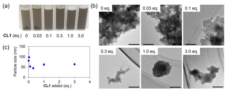 Figure 2
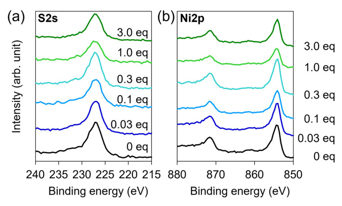 Figure 3
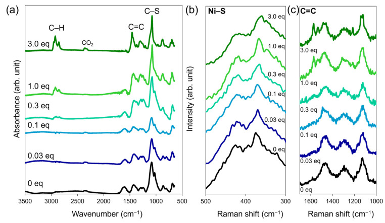 Figure 4
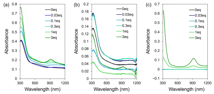 Figure 5
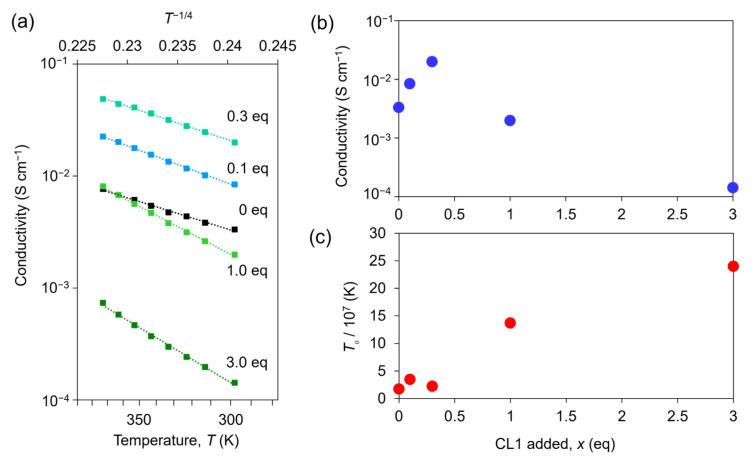 Figure 6
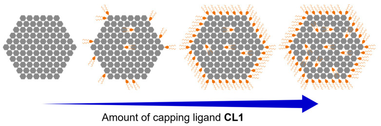 Figure 7
- —JSPS KAKENHI
- —White Rock Foundation
Peer Reviews
No public reviews on file for this paper yet. If you reviewed it on a platform where reviews are public (OpenReview, ICLR, NeurIPS, ICML), you can paste yours below so the community can read it here.
Videos
No videos yet. Explain this paper in a talk, walkthrough, or lecture? Add one.
Taxonomy
TopicsConducting polymers and applications · Organic and Molecular Conductors Research · Supercapacitor Materials and Fabrication
1. Introduction
Metal–organic frameworks (MOFs) are composed of metal ions coordinated by organic molecules, which serve as linkers [1,2,3,4,5,6,7,8,9]. They have attracted considerable attention because of their interesting properties and high structural and functional designability. Among them, two-dimensional frameworks in which planar ligand molecules are linked by planar complex moieties are called coordination nanosheets (CONASHs) [10,11,12,13,14,15,16,17,18,19,20]. The presence of delocalized π–d conjugated electronic states endows CONASHs with high electrical conductivity, placing it among the representative conductive metal–organic frameworks [12]. Significant efforts have been made towards its application in electronic devices [15], electrochemical catalysis [21,22,23,24], energy storage [25,26], and sensors [27,28,29,30].
CONASHs usually take the form of continuous films synthesized at a liquid–liquid interface or a powder obtained by precipitation. In these macroscopic forms, their properties reflect bulk, macroscopic characteristics. However, nanoparticles of inorganic materials, such as quantum dots, exhibit remarkable properties different from those of macroscopic materials [31,32]. Furthermore, nanoparticles can be dispersed in a solvent, facilitating easy handling. The fascinating properties of such inorganic nanoparticles have motivated us to synthesize nanoscale CONASHs (nanoflakes), owing to their potential chemical and physical properties. Our recent study revealed that nanoparticles of Ni_3_BHT can be obtained using benzene-1,2-dithiol as an additive that suppresses the further polymerization of Ni_3_BHT [33,34]. These Ni_3_BHT nanoparticles served as more efficient co-catalyst for water splitting than μm-sized Ni_3_BHT sheets. However, studies on CONASHs are still in their infancy, and further research is required.
In this study, we synthesized Ni_3_BHT nanoparticles modified with 4,5-dihexylbenzene-1,2-dithiol, CL1, as a capping ligand. The obtained nanoparticles were terminated with alkyl chains from the capping ligand, thereby improving the stability of the nanoparticles dispersed in the organic solvent. Various measurements indicate that CL1 plays dual roles in the formation of Ni_3_BHT nanoparticles: partial substitution of BHT ligands within the coordination network and modification of the flake termini, stabilizing the dispersion when an adequate amount of CL1 is added to the reaction system. In contrast, when the amount of CL1 was insufficient, alkyl modification of the Ni_3_BHT nanoparticles was imperfect, leading to aggregation. The Ni_3_BHT dispersion serves as a conductive ink, facilitating future industrial applications.
2. Materials and Methods
- Materials
All chemicals were purchased from Tokyo Chemical Industry Co., Ltd. (Tokyo, Japan), Kanto Chemical Co. (Tokyo, Japan), Sigma-Aldrich Co. LLC (St. Louis, MO, USA), Wako Pure Chemical Industries Ltd. (Osaka, Japan), and Taiyo Nippon Sanso Corp. (Tokyo, Japan), unless otherwise stated. All samples were used without further purification. Water was purified by using a Milli-Q purification system (Merck KGaA, Darmstadt, Germany). 1,2-dibromo-4,5-dihexylbenzene [35] and benzenehexathiol [36] were synthesized according to previously reported methods.
- Instruments
Transmission electron microscopy (TEM) images were collected at 100 kV using an H-7650 microscope (Hitachi, Tokyo, Japan). TEM samples were prepared by drop-casting a sample dispersion diluted 50 times on a copper grid with supporting carbon films (Nisshin EM, Tokyo, Japan). Dynamic light scattering (DLS) was performed at a scattering angle of 173° at 25 °C and analyzed under the assumption of a broad dispersion using SZ-100 (HORIBA, Kyoto, Japan) after diluting the sample dispersion 50 times with a mixed solvent (tetrahydrofuran (THF): MeOH = 1:1 (v/v)). The viscosity of the mixed solvent was estimated by interpolating values from literature [37]. Powder X-ray diffraction (PXRD) measurements were performed at BL44B2 of SPring-8 at a wavelength of 0.8 Å. PXRD patterns were reproduced using VESTA software (Ver. 3.5.8) [38]. X-ray photoelectron spectroscopy (XPS) measurements were performed using a PHI 5000 VersaProbe and VersaProbe III (ULVAC-PHI, Kanagawa, Japan). Al Kα (15 kV, 25 W) was used as the X-ray source, and the beam was focused on an area of 100 μm^2^. The XP spectra were analyzed using MultiPak Software (Ver. 9.9.3) and standardized using a C 1s peak at 284.6 eV. Infrared absorption (IR) spectra were collected using an FT/IR-6100 spectrophotometer (JASCO, Tokyo, Japan) and attenuated total reflection (ATR) method. The background signal was subtracted using the software. Raman spectra were collected using an NRS-5500 (JASCO, Tokyo, Japan) instrument with a 532 nm excitation laser. Ultraviolet-visible-near-infrared absorption (UV-Vis-NIR) spectra were collected using a V-700 (JASCO, Tokyo, Japan) instrument in a 10 mm quartz glass cell with an integrating sphere. Centrifugation was performed using a CS150GXII centrifuge (Eppendorf Himac Technologies Co., Ltd., Ibaraki, Japan). Electrical conductivity measurements were performed under Ar using a custom-ordered probe system within a glove box (Oyama, Hyogo, Japan) connected to a Keithley 2450 SourceMeter (Beaverton, OR, USA) in the two-probe mode. Samples for the electrical conductivity measurements were prepared by drop-casting the sample dispersion onto an interdigitated gold electrode (GEOMATEC, Kanagawa, Japan). Atomic force microscopy (AFM) images were collected using an Agilent Technologies 5500 Scanning Probe Microscope (Santa Clara, CA, USA) and processed using Gwyddion 2.69 [39].
- Synthesis of 1,2-bis(methylthio)-4,5-dihexylbenzene (1)
1,2-Dibromo-4,5-dihexylbenzene (3.342 g, 9.87 mmol) and excess sodium methanethiolate (6.2 g, 88 mmol) were added to 50 mL of dry 1,3-dimethyl-2-imidazolidinone. The mixture was stirred and refluxed overnight in a nitrogen atmosphere. This resulted in a brownish-yellow suspension. After cooling to room temperature, 6 mL iodomethane was added dropwise. Hexane and ethyl acetate (v/v = ~ 4:1) were added to the reaction mixture, and the mixture was repeatedly washed with water, followed by evaporation. The crude material was purified using silica gel column chromatography (hexane: ethyl acetate = 8:1), and the second band was collected. A colorless oil was obtained (649 mg, 23%). ^1^H-NMR (400 MHz, CDCl_3_, ppm): δ = 0.90 (t, J = 6.78 Hz, 6H), 1.27–1.42 (m, 12H), 1.50–1.59 (m, 4H), 2.45 (s, 6H), 2.56 (t, J = 7.98 Hz, 4H), 7.01 (s, 2H). ^13^C-NMR (100 MHz, CDCl_3_, ppm): δ = 14.23, 16.83, 22.76, 29.52, 31.44, 31.85, 32.51, 128.63, 134.40, 139.08.
- Synthesis of 4,5-dihexylbenzene-1,2-dithiol (CL1)
All processes except for collection were performed using the Schlenk technique. An excess amount of sodium (0.5 g, 22 mmol) was dissolved in liquid ammonia (~100 mL) and cooled in a dry ice/acetone bath. The ammonia solution turned dark blue. 1 (301.35 mg, 0.972 mmol) dissolved in degassed THF (4 mL) was added dropwise to liquid ammonia and stirred for 4 h. Degassed methanol (2 mL) was carefully added to liquid ammonia to quench the excess sodium. When the reaction mixture was allowed to warm to room temperature, the blue color disappeared. The product was diluted with 90 mL of degassed water and washed repeatedly with degassed diethyl ether. Ten milliliters of degassed HCl aq. (35 wt%) was added, and the solution was extracted with diethyl ether under an ambient atmosphere. The organic phase was dried with Na_2_SO_4_ and evaporated to obtain a greenish-yellow oil (200 mg, 72%). ^1^H-NMR (400 MHz, CDCl_3_, ppm): δ = 0.89 (t, J = 7.03 Hz, 6H), 1.25–1.39 (m, 12H), 1.47–1.55 (m, 4H), 2.49 (t, J = 8.01 Hz, 4H), 3.64 (s, 2H), 7.15 (s, 2H). ^13^C-NMR (100 MHz, CDCl_3_, ppm): δ =14.25, 22.75, 29.48, 31.27, 31.84, 32.24, 127.81, 132.08, 140.09.
- Synthesis of Ni_3_BHT-x
Ni_3_BHT-x was synthesized by adding 10 mL of a 4.5 mM Ni(OAc)2 methanol solution to 10 mL of a stirred solution containing 1.5 mM benzenehexathiol (BHT) and 1.5 × x mM CL1 THF under an inert atmosphere at room temperature. The reaction proceeded immediately, yielding a dark brown dispersion, with or without black precipitation. For all measurements except UV–Vis–NIR spectroscopy, the resulting Ni_3_BHT-x were collected by evaporating the solvent to promote aggregation, followed by filtration through a polytetrafluoroethylene (PTFE) membrane with a pore size of 450 nm. The collected black solids (Ni_3_BHT-x) were thoroughly washed with THF and methanol and dried in vacuo.
- Synthesis of Ni_3_BHT-BDT-x
Ni_3_BHT-BDT-x was synthesized by the same method as Ni_3_BHT-x, by using benzene-1,2-dithiol instead of CL1.
- Preparation of the sample for UV-Vis-NIR spectroscopy
“As-prepared” samples were obtained by diluting the freshly Ni_3_BHT-x dispersion 50 times with a mixed solvent of THF and MeOH (1:1 (v/v)) without purification. 5 mL of the as-prepared Ni_3_BHT-x was centrifugated for 3 h at 110 krpm. The supernatant was diluted 50 times with the mixed solvent for measurements. The black precipitate was washed with THF and methanol, sonicated to redisperse Ni_3_BHT-x in 5 mL of the mixed solvent, and diluted 50 times with the mixed solvent for measurement.
3. Results
Modifying the termini of nanostructures with alkyl chains is a common strategy to stabilize the nanostructures dispersed in solvents [40]. In this study, we synthesized a novel capping ligand, 4,5-dihexylbenzene-1,2-dithiol (CL1) and applied it to Ni_3_BHT (Figure 1). CL1 was obtained from 1,2-dibromo-4,5-dihexylbenzene via the introduction of SCH_3_ by nucleophilic substitution to synthesize 1,2-dimethylthio-4,5-dihexylbenzene (1), followed by the deprotection of the methyl groups by Birch-type reduction. Among the protecting groups examined, methyl groups were the best, while benzyl groups only replaced one bromine in 1,2-dibromo-4,5-dihexylbenzene.
To investigate the effect of CL1 on the formation of Ni_3_BHT colloids, we prepared dispersions of Ni_3_BHT nanoflakes (Ni_3_BHT-x) by mixing a BHT/THF solution with x equivalents of CL1 (x = 0, 0.03, 0.1, 0.3, 1.0, and 3.0) relative to BHT in the solution and a methanol solution of 3 equivalents of nickel acetate. Figure 2a shows the resulting Ni_3_BHT-x dispersions. All dispersions were greenish-brown, similar to Ni_3_BHT colloids obtained in a previous study [41]. The color of Ni_3_BHT-x was darker than that of the Ni_3_BHT dispersion prepared in a previous study without a modulator, because the concentration of Ni_3_BHT was ten times as large as that in the previous study. In samples with low CL1 contents (x = 0, 0.03), the product aggregated and formed a black gel-like precipitate. Although the Ni_3_BHT colloids were stable in dilute solutions, as the concentration increased, the attractive interactions between the flakes became stronger, and the colloidal state could no longer be maintained. In the samples with a sufficient CL1 content (x ≥ 0.1), no visible solid precipitations were observed immediately after synthesis. After one month, aggregation occurred eventually at x = 0.1, but the samples with x ≥ 0.3 maintained a uniform colloid state, which lasted for up to three months (Figure S1). These results demonstrate that CL1 works as a modulator that solubilizes Ni_3_BHT in a methanol/THF system, making it possible to prepare colloids containing Ni_3_BHT at concentrations 10 times higher than those in conventional Ni_3_BHT colloids. Benzene-1,2-dithiol (BDT) has also been reported to solubilize Ni_3_BHT in ethanol [34]. However, under the same synthesis conditions as those used in this study, aggregation occurred one month after synthesis at all additive amounts (Figure S1). Therefore, CL1 is a superior modulator compared to BDT in terms of stability. Figure 2b shows the TEM images of the Ni_3_BHT-x dispersion (supernatant, if a precipitate is present). At low CL1 concentrations (x ≤ 0.1), the dispersion aggregated to form clumps, whereas at high CL1 concentrations (x ≥ 0.3), flakes of approximately 100 nm were observed. It is reasonable that under low CL1 concentrations, where precipitation occurs, the flake size is large, whereas under conditions where precipitates are not formed, the flake size is small. The tendency of the flake size to decrease with increasing CL1 addition is also evident in dynamic light scattering (DLS) measurements (Figure 2c).
DLS measurements qualitatively capture the trend of decreasing flake size with increasing CL1 content. However, the flake size measured by DLS did not match that by TEM images, particularly at x = 0 and 0.03, where larger flakes were observed by TEM, owing to the nonspherical, sheet-like morphology of Ni_3_BHT nanoflakes, which violates the spherical particle assumption inherent to DLS analysis. This phenomenon was due to the nonspherical particle shape, which is often observed when measuring two-dimensional materials [42,43]. These results suggest that the addition of CL1 suppresses the aggregation of Ni_3_BHT flakes. The particle size reduction effect appears to be dominant in the combination of Ni_3_BHT and CL1; however, modulators do not always decrease the size of the MOF particles [44,45,46].
Figure S2 shows powder X-ray diffraction patterns of Ni_3_BHT-x. As is common with nanoparticles, the pattern is broad owing to low crystallinity. To clarify the chemical bonding state, we performed XPS measurements of Ni_3_BHT-x (Figure 3a,b and Figure S3). S2s and Ni2p peaks appeared at 227.0 and 854.2 eV, respectively, regardless of the amount of CL1 added. The XPS of Ni_3_BHT-0 was consistent with that for Ni_3_BHT synthesized by the liquid–liquid two-phase interface or single-phase synthesis method [16,41]. This suggests that CL1, as well as BHT, bonds with Ni and forms a part of a planar network with nickel ions and BHT. The S/Ni ratio estimated from the S2s and Ni2p peak areas varied between 2 and 2.5 depending on the amount of CL1 added, consistent with the ideal S/Ni ratio of 2 calculated from the Ni_3_BHT composition (Figure S3). A more detailed analysis of the S2s peak revealed that it can be divided into two peaks: a main peak at 227.0 eV and peak around 230 eV. The peak at 230 eV is a shake-up peak often observed in metal dithiolene-based materials [14,47,48], and it is derived from highly oxidized sulfur, such as sulfate or disulfide. The peak near 230 eV was present in Ni_3_BHT-0 but became less prominent with increasing CL1 content (Figure S4). The area ratio to the main peak decreased with increasing x up to x = 0.3, then leveled off thereafter (Figure S5). While Ni_3_BHT-0 without a modulator contained a large amount of highly oxidized S, the progressive addition of CL1 suppresses oxidized sulfur species at the flake edges, resulting in a relative increase in the sulfur content involved in complex formation, consistent with replacement of oxidation-prone BHT-derived termini by alkylated CL1 ligands.
We investigated the changes in the vibrational spectra of Ni_3_BHT upon the addition of CL1 using infrared (IR) absorption and Raman spectroscopy (Figure 4 and Figure S6). The Raman spectrum of the sample without CL1 (Ni_3_BHT-0) was consistent with that of Ni_3_BHT synthesized by a two-phase interfacial reaction or single-phase synthesis, indicating that Ni_3_BHT-0 is a colloid composed of Ni_3_BHT coordination nanosheets [16]. Neither IR nor Raman spectra showed a peak near 2500 cm^−1^ due to S–H stretching vibrations, suggesting that most of the sulfur from BHT and CL1 coordinated to nickel ions (Figure 4a and Figure S6). The IR spectra of Ni_3_BHT-x (x ≥ 0.3) showed an absorption peak near 2900 cm^−1^ (Figure 4a), which was attributed to the C-H stretching of the hexyl or benzene ring of CL1. The peak intensity increased with increasing x, indicating that the more CL1 added, the more CL1 was incorporated into Ni_3_BHT-x. In Figure 2b,c, there are no significant changes in the size or shape of Ni_3_BHT-x flakes in the x ≥ 0.3 region. If CL1 coordinates only to the termini of the flakes, the amount of CL1 within the flakes should not change significantly. However, the IR spectra revealed the opposite. Therefore, combining the TEM images and IR spectra of Ni_3_BHT-x, it is likely that CL1 exists not only at the termini of Ni_3_BHT but also in bulk Ni_3_BHT by replacing BHT, and the amount of the latter type of CL1 increases with increasing x. Furthermore, with increasing CL1, the C–S and C=C stretching peaks became sharper. In the Raman spectra, the Ni-S stretching peak shifted gradually from 374 cm^−1^ at x = 0 to 358 cm^−1^ at x = 3.0 (Figure 4b). The absence of peaks around 500 cm^−1^ suggests insignificant S–S bond formation. Furthermore, a new peak, attributable to the stretching vibration of the aromatic ring of CL1, appeared at approximately 1570 cm^−1^. These results confirm the fusion of CL1 with the Ni_3_BHT network of Ni_3_BHT-x.
We measured the UV-visible absorption spectrum of Ni_3_BHT-x dispersion to gain further insights into its electronic state. Figure 5a shows the UV–vis–NIR spectra of the diluted as-prepared Ni_3_BHT-x colloids without further purification. Samples with low CL1 contents (x = 0, 0.03, and 0.1) exhibited significantly broad absorption peaks at approximately 1050 nm. These peaks were attributed to the absorption of Ni_3_BHT [41]. In contrast, samples prepared with higher CL1 contents (x ≥ 1.0) exhibited an additional absorption band at approximately 927 nm. Absorption in the NIR region is characteristic of nickel dithiolene complexes [49,50] and is therefore associated with the formation of dithiolene-based nickel species involving CL1. Such species can, in principle, exist in two forms: (i) nickel centers coordinated by both CL1 and BHT ligands and incorporated into the Ni_3_BHT coordination network, and (ii) discrete molecular species in which two CL1 ligands coordinate to a nickel center and dissolve in the solvent as a most probably mononuclear Ni–dithiolene complex. To clarify the origin of the absorption band at approximately 927 nm, Ni_3_BHT-x dispersions were subjected to centrifugation, followed by washing and attempted redispersion by sonication; however, redispersion was incomplete. The UV-Vis-NIR spectra of the redispersed precipitates are shown in Figure 5b. These spectra retained essentially the same spectral features as those of the original dispersions, dominated by the absorption characteristic of the Ni_3_BHT network, while the peak at 927 nm became relatively less prominent. In contrast, the UV–Vis–NIR spectra of the supernatants collected after centrifugation revealed a clear difference depending on the CL1 content (Figure 5c). For samples with x ≤ 0.3, no absorption was observed in the 300–1200 nm range. However, for samples with x = 1.0 and 3.0, ultraviolet absorption attributable to free CL1, together with a distinct absorption band at approximately 927 nm, was observed. This absorption is reasonably attributed to discrete Ni–dithiolene species present in solution and distinct from the extended Ni_3_BHT coordination network. The absorbance at 927 nm increased proportionally with the amount of CL1, reaching 0.038 at x = 1.0 and 0.116 at x = 3.0. Based on these observations, we infer that when a relatively small amount of CL1 is added (x ≤ 0.3), CL1 is fully consumed as a component of the Ni_3_BHT-x network, and no discrete molecular species remain in solution. In contrast, when a larger excess of CL1 is introduced (x = 1.0, 3.0), a fraction of the added CL1 is not incorporated into the Ni_3_BHT network but instead forms discrete Ni–dithiolene species or remains uncoordinated in solution. The absence of the 927 nm absorption band for x = 0.3 indicates that, under these conditions, essentially all CL1 molecules are consumed in the formation of Ni_3_BHT-0.3. These results further suggest that coordination of CL1 and BHT to Ni^2+^ during nanosheet growth is favored over the formation of discrete CL1-only complexes at low CL1 concentrations. At higher CL1 loadings, however, available BHT-derived thiolate sites at the termini of Ni_3_BHT flakes become saturated, promoting the formation of discrete Ni–dithiolene species in solution.
This interpretation is consistent with the XPS results, which show that increasing CL1 content progressively suppresses oxidized sulfur species at the flake edges. At lower CL1 concentrations (x < 0.3), increasing CL1 addition leads to more extensive termination of Ni_3_BHT flakes by CL1, concomitantly reducing the amount of oxidation-prone BHT at the edges. At higher CL1 concentrations (x ≥ 0.3), the chemical states at the flake termini no longer change significantly, resulting in a constant ratio of oxidized to coordinated sulfur species.
Finally, to investigate its performance as a conductive ink, Ni_3_BHT-x was coated onto an interdigitated gold electrode, and its electrical conductivity was measured using the two-terminal method. The coated Ni_3_BHT-x formed a film of approximately 100 μm in size with a thickness of approximately 1 μm (Figures S7 and S8). Figure S9 shows the representative current-voltage curve of Ni_3_BHT-0.3 at room temperature. The linear curve indicates the formation of an ohmic contact between gold and Ni_3_BHT-0.3. Finite electrical conductivities were observed for all x values, demonstrating that the Ni_3_BHT-x dispersion functioned as a conductive ink. The electrical conductivities of all Ni_3_BHT-x samples increased with increasing temperature (Figure 6a). This behavior was consistently observed in both Ni_3_BHT nanoflakes and films. Because the logarithm of electrical conductivity is a linear function of the inverse fourth root of temperature, it can be explained by strong localization due to disorder [16,34,41,51]. Hence, we compared the room-temperature electrical conductivities for different values of x. At x = 0, the electrical conductivity was 3.3 × 10^−3^ S cm^−1^. With increasing CL1, the electrical conductivity increased, reaching a maximum of 2.0 × 10^−2^ S cm^−1^ at x = 0.3, after which it began to decrease (Figure 6b). The increase in electrical conductivity with increasing CL1 at smaller x values up to 0.3 can be interpreted as a change in the electronic state owing to the substitution of CL1 for BHT. The substitution of CL1 injects positive holes because CL1 has two anionic S atoms while BHT has six anionic S atoms. The injection of holes increases the electrical conductivity of the interior of nanoflakes since Ni_3_BHT is a p-type material [16]. A phenomenon similar to that is reported for Ni_3_BHT substituted with BDT [33]. Note that CL1 can also work as a scattering center by partially breaking the Ni_3_BHT network and consequently degrade the carrier mobility. In this smaller CL1 concentration regime, the effect of increasing hole doping overwhelms that of increasing scattering centers. However, when x > 0.3, excessive addition of CL1 led to decrease in the electrical conductivity. In this regime, the substituted CL1 works as random disorders in the Ni_3_BHT network rather than as dopants, resulting in a random potential much stronger than inherent one in Ni_3_BHT-0. The carriers are forced to be trapped in the random potential, which leads to strong localization. This interpretation is consistent with the experimentally observed increase in the T0 parameter and does not require assumptions beyond the structural disorder introduced by excessive ligand substitution. The decrease in the electrical conductivity by excessive doping is generally found also in other materials [52,53]. The degree of localization can be evaluated using the parameter T0, which characterizes localization when the temperature dependence of the electrical conductivity is expressed as σ = σ_0_ exp(−(T0/T)^1/4^), assuming a three-dimensional variable range hopping (3DVRH) conduction mechanism [54]. Above x = 0.3, T0 increases rapidly, suggesting stronger carrier localization and more severe structural disorder than in the samples with x below 0.3 (Figure 6c).
4. Discussion
The formation of Ni_3_BHT colloids with CL1 can be rationalized by the following conceptual model (Figure 7). In the absence of CL1, Ni_3_BHT micronuclei consisting of only BHT and Ni ions were formed. These terminals, including thiol groups, are highly active, and their concentration is 10 times higher than that of stable Ni_3_BHT colloids. Therefore, they cannot be stably dispersed in the solvent and can only undergo aggregation and precipitation. When a small amount of CL1 is present in the reaction system, complexation by BHT ligands with Ni^2+^ mainly proceeds with occasional complexation by BHT and CL1 with Ni^2+^, resulting in the formation of Ni_3_BHT nanoflakes in which some of BHT is replaced by CL1. XPS results suggested that the BHT termini were present when the concentration of CL1 was low. Therefore, Ni_3_BHT flakes grow with CL1 partially replacing BHT until BHT is consumed to some extent, followed by the edge modification of CL1 which was not incorporated into the Ni_3_BHT flakes. If the degree of modification is low because of insufficient CL1, the nuclei aggregate and eventually precipitate (x = 0.03 and 0.1). If fully modified, owing to sufficient CL1, they are stably dispersed (x = 0.3). When excess CL1 is present in the system, Ni_3_BHT flakes are produced, and more BHT is replaced by CL1. All the termini get modified with CL1, and the remaining CL1 forms complexes (x = 1.0 and 3.0). In this study, the optimal composition for producing Ni_3_BHT colloids with high colloidal stability, high concentration, and minimal byproducts was achieved using a molar ratio of BHT:Ni^2+^:CL1 = 1:3:0.3.
5. Conclusions
In this study, we prepared colloidal dispersions of coordination nanosheets, Ni_3_BHT, by adding the modulator 4,5-dihexylbenzene-1,2-dithiol (CL1) to BHT and nickel salt. Ni_3_BHT was modified with hexyl groups, resulting in a 10-fold higher concentration than that obtained without the modulator and a more stable dispersion than that obtained using the conventional modulator benzenedithiol. Furthermore, an analysis of various electronic and vibrational spectra using CL1 as a label revealed that Ni_3_BHT nanoflakes partially substituted with CL1 were formed. After consumption of BHT, the termini of the nanoflakes were modified with CL1. The finding that alkyl termination enhances the colloidal stability of coordination nanosheets is important for elucidating their nanostructure. The Ni_3_BHT colloids obtained in this study served as conductive inks, particularly at the CL1/BHT ratio of 0.3. These results suggest that Ni_3_BHT colloids have the potential as solution-processable conductive materials and provide a design strategy toward future applications in printed and flexible electronics.
The reference list from the paper itself. Each links out to its DOI / PubMed record.
- 1Yusuf V.F. Malek N.I. Kailasa S.K. Review on Metal−Organic Framework Classification, Synthetic Approaches, and Influencing Factors: Applications in Energy, Drug Delivery, and Wastewater Treatment ACS Omega 20227445074453110.1021/acsomega.2c 0531036530292 PMC 9753116 · doi ↗ · pubmed ↗
- 2Wang Q. Astruc D. State of the Art and Prospects in Metal–Organic Framework (MOF)-Based and MOF-Derived Nanocataly-sis Chem. Rev.20201201438151110.1021/acs.chemrev.9b 0022331246430 · doi ↗ · pubmed ↗
- 3Kondo M. Yoshitomi T. Matsuzaka H. Kitagawa S. Seki K. Three-Dimensional Framework with Channeling Cavities for Small Molecules: {[M 2(4,4’-bpy)3(NO 3)4]·x H 2O}n (M = Co, Ni, Zn)Angew. Chem. Int. Ed.1997361725172710.1002/anie.199717251 · doi ↗
- 4Chui S.S.-Y. Lo S.M.-F. Charmant J.P.H. Orpen A.G. Williams I.D. A Chemically Functionalizable Nanoporous Material [Cu 3(TMA)2(H 2O)3]n Science 19992831148115010.1126/science.283.5405.114810024237 · doi ↗ · pubmed ↗
- 5Li H. Eddaoudi M. O’Keeffe M. Yaghi O.M. Design and Synthesis of an Exceptionally Stable and Highly Porous Metal-Organic Framework Nature 199940227627910.1038/46248 · doi ↗
- 6Cavka J.H. Jakobsen S. Olsbye U. Guillou N. Lamberti C. Bordiga S. Lillerud K.P. A new zirconium inorganic building brick forming metal organic frameworks with exceptional stability J. Am. Chem. Soc.2008130138501385110.1021/ja 805795318817383 · doi ↗ · pubmed ↗
- 7Pan Y. Liu Y. Zeng G. Zhao L. Lai Z. Rapid synthesis of zeolitic imidazolate framework-8 (ZIF-8) nanocrystals in an aqueous system Chem. Commun.2011472071207310.1039/c 0cc 05002 d 21206942 · doi ↗ · pubmed ↗
- 8Park K.S. Ni Z. Côte A.P. Choi J.Y. Huang R. Uribe-Romo F.J. Chae H.K. O’Keeffe M. Yaghi O.M. Exceptional chemical and thermal stability of zeolitic imidazolate frameworks Proc. Natl. Acad. Sci. USA 2006103101861019110.1073/pnas.060243910316798880 PMC 1502432 · doi ↗ · pubmed ↗