Correction: Castro et al. Synthesis, Characterization, and Optimization Studies of Polycaprolactone/Polylactic Acid/Titanium Dioxide Nanoparticle/Orange Essential Oil Membranes for Biomedical Applications. Polymers 2023, 15, 135
Jorge Ivan Castro, Stiven Astudillo, Jose Herminsul Mina Hernandez, Marcela Saavedra, Paula A. Zapata, Carlos Humberto Valencia-Llano, Manuel N. Chaur, Carlos David Grande-Tovar

Abstract
Genes, proteins, chemicals, diseases, species, mutations and cell lines named across the full text — each resolved to its canonical identifier and authoritative record.
Click any figure to enlarge with its caption.
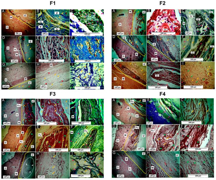 Figure 8
Peer Reviews
No public reviews on file for this paper yet. If you reviewed it on a platform where reviews are public (OpenReview, ICLR, NeurIPS, ICML), you can paste yours below so the community can read it here.
Videos
No videos yet. Explain this paper in a talk, walkthrough, or lecture? Add one.
Taxonomy
TopicsElectrospun Nanofibers in Biomedical Applications · Polymer Nanocomposites and Properties · biodegradable polymer synthesis and properties
Errors in Figures
In the original publication [1], there were two mistakes in Figures 7 and 8 as published.
During the preparation of the original article [1], there was an error in Figure 7. A macroscopic photograph of a biomodel implanted with different scaffolds was unintentionally repeated from our previous work [2].
This mistake arose because, in our research, we routinely employ the same animal biomodels to evaluate multiple materials simultaneously. Specifically, we use a validated subcutaneous pocketing technique that enables the creation of up to ten independent dorsal pockets per animal, each implanted with a distinct material. In some experimental designs, two or three different systems are evaluated in the same biomodel. This methodology enhances resource efficiency, reduces biological variability among animals, and allows comparative evaluation across multiple materials. Importantly, it aligns with the internationally accepted 3R principles (Replacement, Reduction, and Refinement) for ethical animal experimentation.
The figure in question depicts a macroscopic view of the dorsal implantation area. Such images are included only as preliminary visual references of tissue response and healing, serving as complementary context to the histological analyses which remain the primary and valid scientific evidence supporting the study’s conclusions.
The corrected version of Figure 7 is provided below. We confirm that this correction does not alter the validity of the data, the results, or the conclusions of the article. The amendment leaves the core findings and interpretations intact, ensuring that data integrity and scientific outcomes remain unchanged.
Additionally, a histological image was inadvertently duplicated between panels F1B and F4B of Figure 8. This error was unintentional and occurred during the selection and assembly of images for the final figure, due to the large number of images generated for each formulation. Specifically, implants were evaluated at 30, 60, and 90 days, resulting in a total of 209 histological images.
The specific mistake occurred because the image corresponding to panel B of formulation F1 at 30 days was accidentally duplicated in panel B of formulation F4, also at 30 days. Both images were stained using Masson’s trichrome, which explains the strong similarity in coloration and morphological features and contributes to the oversight during the image selection process.
The corrected version of Figure 8 is provided below. It is important to emphasize that this error occurred exclusively during the organization and selection of images. It does not affect data acquisition, histological processing, scientific analysis, or the validity of the results and conclusions presented in the article.
Histological analysis of F1, F2, F3, and F4 membranes. (A–C): 30-day implantations. (D–F): 60-day implantations. (G–I): Implantations at 90 days. Panel F1: (A): Image at 4× HE technique. (B): Image at 10× MT technique. (C): Image at 100× MT technique. (D): Image at 4× HE technique. (E): Image at 40× HE technique. (F): Image at 40× MT technique. (G): Image at 4× HE technique. (H): Image at 10× HE technique. (I): Image at 40× MT. Panel F2: (A): Image at 4× HE technique. (B): Image at 40× HE technique. (C): Image at 10× MT technique. (D): Image at 4× HE technique. (E): Image at 10× HE technique. (F): Image at 40× MT technique. (G): Image at 4× HE technique. (H): Image at 40× HE technique. (I): Image at 40× HE. Panel F3: (A): Image at 4× HE technique. (B): Image at 40× HE technique. (C): Image at 40× MT technique. (D): Image at 4× HE technique. (E): Image at 40× HE technique. (F): Image at 40× MT technique. (G): Image at 4× HE technique. (H): Image at 10× HE technique. (I): Image at 40× MT technique. Panel F4: (A): Image at 4× HE technique. (B): Image at 4× MT technique. (C): Image at 40× HE technique. (D): Image at 4× HE technique. (E): Image at 10× HE technique. (F): Image at 40× HE technique. (G): Image at 4× HE technique. (H): Image at 10× HE technique. (I): Image at 40× HE. D: Dermis. M: Muscle. IZ: Implantation zone. Cc: Connective tissue capsule. Ct: Connective tissue. Fc: Fibrous capsule. H: hypodermis. Red arrow: area with resorbing material. Yellow arrow: Material. Pink arrows: type I collagen fibers. Green arrows: Connective tissue. Red stars: Inflammatory cells. Green stars: Connective tissue. Oval 1: Area with the material in the process of resorption. Oval 2: Zone with less inflammatory activity. Oval 3: Zone with the formation of connective scar tissue. White oval: Histological interest zone. White arrows: material in the process of degradation/resorption. MT: Masson’s trichrome stain. HE: Hematoxylin–Eosin stain.
This correction was approved by the Academic Editor. The original publication has also been updated.
The reference list from the paper itself. Each links out to its DOI / PubMed record.
- 1Castro J.I. Astudillo S. Mina Hernandez J.H. Saavedra M. Zapata P.A. Valencia-Llano C.H. Chaur M.N. Grande-Tovar C.D. Synthesis, Characterization, and Optimization Studies of Polycaprolactone/Polylactic Acid/Titanium Dioxide Nanoparticle/Orange Essential Oil Membranes for Biomedical Applications Polymers 20231513510.3390/polym 1501013536616482 PMC 9823686 · doi ↗ · pubmed ↗
- 2Grande-Tovar C.D. Castro J.I. Valencia Llano C.H. Tenorio D.L. Saavedra M. Zapata P.A. Chaur M.N. Polycaprolactone (PCL)-Polylactic Acid (PLA)-Glycerol (Gly) Composites Incorporated with Zinc Oxide Nanoparticles (Zn O-N Ps) and Tea Tree Essential Oil (TTEO) for Tissue Engineering Applications Pharmaceutics 2023154310.3390/pharmaceutics 1501004336678672 PMC 9864333 · doi ↗ · pubmed ↗