Comparison of Serological Detection Methods for Toxoplasma gondii Antibodies and Seroprevalence in Captive Red Pandas
Chanjuan Yue, Wanjing Yang, Dunwu Qi, Yanshan Zhou, Xueyang Fan, Chao Chen, Yifan Wen, Xiaolan Wang, Mei Yang, Yunli Li, Rong Hou, Songrui Liu

TL;DR
This study compares three antibody tests for Toxoplasma gondii in red pandas and finds that two are reliable for detecting exposure in captive populations.
Contribution
Validated MAT and ELISA as reliable serological tools for T. gondii detection in red pandas, while IHA was ineffective.
Findings
MAT and ELISA showed almost perfect agreement and detected T. gondii antibodies in 35.09% of red panda samples.
IHA failed to detect any positive samples, making it unsuitable for red panda serology.
ELISA confirmed 95.2% sensitivity and 88.9% specificity when compared to MAT.
Abstract
Toxoplasma gondii is a globally distributed parasite that threatens many warm-blooded species, including endangered wildlife such as the red panda. Reliable detection of exposure is essential for monitoring health in captive populations, but the performance of commercial antibody tests can vary across species. This study evaluated the modified agglutination test (MAT), indirect hemagglutination assay (IHA), and enzyme-linked immunosorbent assay (ELISA) for detecting T. gondii antibodies in 57 serum samples from captive red pandas. Results showed that MAT and ELISA had almost perfect agreement and were both effective, whereas IHA failed to detect any positive samples. The combined use of MAT and ELISA determined a seroprevalence of 35.09%, indicating a substantial exposure risk in captivity. These results validate MAT and ELISA as reliable tools for T. gondii surveillance in red pandas…
Genes, proteins, chemicals, diseases, species, mutations and cell lines named across the full text — each resolved to its canonical identifier and authoritative record.
Click any figure to enlarge with its caption.
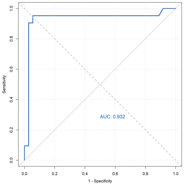 Figure 1
- —Chengdu Science and Technology Program
- —Chengdu Research Base of Giant Panda Breeding
Peer Reviews
No public reviews on file for this paper yet. If you reviewed it on a platform where reviews are public (OpenReview, ICLR, NeurIPS, ICML), you can paste yours below so the community can read it here.
Videos
No videos yet. Explain this paper in a talk, walkthrough, or lecture? Add one.
Taxonomy
TopicsToxoplasma gondii Research Studies · Leptospirosis research and findings · Bartonella species infections research
1. Introduction
The Chinese red panda (Ailurus styani; hereafter referred to as red panda) is an endemic species protected under China’s Class II designation and classified as Endangered by the International Union for Conservation of Nature (IUCN) [1]. Its population has declined by approximately 40% over the past 50 years, primarily due to habitat destruction and fragmentation [2]. Emerging infectious diseases, particularly parasitic infections, now pose significant threats to both in situ and ex situ populations. Over 20 parasite species have been documented in red pandas [3]. Among these, toxoplasmosis—a globally distributed parasitic disease caused by Toxoplasma gondii—represents a parasitic disease of substantial concern for red panda conservation.
Toxoplasma gondii (T. gondii) infects most warm-blooded animals, including red pandas [4]. Infections may cause reproductive failure or acute toxoplasmosis in immunocompromised hosts. Previous studies have reported cases of wildlife mortality attributable to T. gondii infection globally [5,6].
Serological assays are the primary method for detecting T. gondii exposure in wildlife and captive animals [7]. Previous studies have employed the modified agglutination test (MAT), indirect hemagglutination assay (IHA), enzyme-linked immunosorbent assay (ELISA), and immunofluorescence antibody test (IFAT) [8,9,10,11], though the accuracy of these methods varies. MAT and ELISA are widely used in epidemiological studies due to their high sensitivity and applicability across species [4]. Although concurrent MAT and ELISA testing has been reported in captive wildlife, including four red pandas [10], small sample sizes limit robust method comparisons. Therefore, validating reliable serological methods for red pandas remains necessary. To enhance health monitoring for captive red panda conservation, this study aimed to evaluate and compare three commercial serological kits (MAT, IHA, and ELISA) for detecting T. gondii antibodies in captive red pandas in order to identify reliable serosurveillance tools and to determine the seroprevalence in a captive population.
In this study, fifty-seven blood samples were collected from captive red panda individuals in Chengdu, China. The performance of two commercial serological kits (IHA and ELISA) was evaluated against the MAT reference standard for detecting T. gondii antibodies. MAT was adopted as the reference standard for comparison because it is not species-specific and is widely established as a benchmark for evaluating T. gondii antibody detection in wildlife serology. Analytical performance, including sensitivity and specificity, was assessed for each commercial kit relative to MAT. Based on comparative results, optimal serological methods for T. gondii surveillance in red pandas were identified, and the seroprevalence in this captive population is reported.
2. Materials and Methods
2.1. Sample Collection
Fifty-seven blood samples were collected from the cephalic vein of red pandas at the Chengdu Research Base of Giant Panda Breeding (CRBGPB) in Sichuan, China, between November 2015 and July 2019. All blood samples were collected opportunistically during routine health examinations. At the time of sampling, the animals displayed no overt clinical signs suggestive of active parasitic or other infectious diseases. The animal handling procedures were approved by the Institutional Animal Care and Use Committee (IACUC) of the Chengdu Research Base of Giant Panda Breeding (NO. 2019013; Approval Date: 21 November 2019). Serum was separated by centrifugation at 3500 rpm for 10 min and stored at −80 °C.
2.2. Serological Testing
All samples were tested using three commercial kits with three detection methods (Table 1). The MAT kit had a cut-off titer of 1:25, and samples were diluted in twofold serial dilutions from 1:25 to 1:200. The IHA kit had a cut-off titer of 1: 4, and samples were diluted in twofold serial dilutions from 1: 4 to 1: 256. The optical density (OD) values obtained from the ELISA kit were measured using a Multiskan^TM^ Microplate Absorbance Reader (Thermo Scientific, Singapore). All experimental procedures were performed according to the manufacturer’s instructions.
2.3. Seroprevalence Determination
Based on comparative performance results, we selected appropriate commercial kits for T. gondii antibody detection in red panda serum. A dual-method approach was implemented to determine seroprevalence, in which samples were classified as positive only when both methods yielded positive results. Samples with discordant results were retested using the same serum aliquot. Samples exhibiting persistent discordance after retesting were classified as negative.
2.4. Statistical Analysis
All statistical analyses were performed using R version 4.5.0 (R Core Team, 2025) with the following packages: tidyverse (v2.0.0), irr (v0.84.1), pROC (v1.19.0.1), and binom (v1.1.1.1).
Seroprevalence rates with 95% confidence intervals were calculated using the Wilson score method. The McNemar chi-square test with continuity correction was used to compare detection rates between MAT and ELISA (p < 0.05 considered significant). Agreement between methods was assessed using Cohen’s kappa coefficient (κ) with the following interpretations: κ ≤ 0.20 = slight agreement; 0.21–0.40 = fair agreement; 0.41–0.60 = moderate agreement; 0.61–0.80 = substantial agreement; and 0.81–1.00 = almost perfect agreement.
Using MAT as the reference standard, the sensitivity, specificity, positive predictive value (PPV), negative predictive value (NPV), and accuracy of ELISA were calculated. The area under the curve (AUC) was evaluated by receiver operating characteristic (ROC) analysis using the continuous ELISA optical density values. The ROC curve analysis was performed using the raw optical density (OD) values obtained from the ELISA, as the kit manufacturer’s protocol specifies direct use of the measured OD values without conversion.
3. Results
3.1. Detection Rates
According to manufacturer-specified cut-offs, the MAT detected T. gondii antibodies in 21 of 57 red pandas (36.84%; 95% CI: 25.50–49.80), with titers distributed as follows: 1: 50 (n = 2) and 1: 200 (n = 19) (Table 2). The ELISA identified 24 seropositive samples (42.11%; 95% CI: 30.2–55.0). In contrast, the IHA detected no seropositive results (0%) (Table 3).
MAT served as the reference standard for serological comparisons. Among 57 red panda serum samples, IHA yielded no positive results (Table 3), resulting in 100% false negatives (21/21 MAT-positive samples undetected) and 0% false positives. ELISA demonstrated one false-negative result (negative by ELISA but positive by MAT; 4.76% [1/21] of MAT-positive samples) and four false positives (11.11% [4/36] of MAT-negative samples).
3.2. Agreement Analysis
The commercial IHA kit failed to detect T. gondii antibodies in any red panda serum samples and was therefore excluded from further analysis. Cohen’s kappa coefficient (κ) analysis revealed almost perfect agreement between MAT and ELISA for detecting T. gondii exposure in red pandas (κ = 0.817, 95% CI: 0.66–0.97). McNemar’s test demonstrated no significant difference between methods (p = 0.375, χ^2^ = 0.800).
3.3. ROC Analysis
Using MAT as the reference standard, ROC analysis of ELISA yielded an area under the curve (AUC) of 0.932 (95% CI: 0.837–1.000), indicating high diagnostic accuracy (Figure 1). Compared to MAT, ELISA demonstrated 95.20% sensitivity and 88.9% specificity.
3.4. Seroprevalence of T. gondii in Captive Red Pandas
Using a combined MAT/ELISA approach with retesting of discordant samples, the seroprevalence of T. gondii in captive red pandas was determined to be 35.09% (20/57). Five samples (8.77%) remained discordant after retesting and were classified as negative.
4. Discussion
The red panda has been identified as an intermediate host for T. gondii [12]. A fatal case of disseminated toxoplasmosis in this species was documented by Stief et al. (2012) [13], with histopathological examination revealing abundant tissue cysts in the liver and brain, and fewer cysts in other organs. Additionally, Ashley et al. (2020) [14] demonstrated disseminated toxoplasmosis in four red pandas during necropsy through PCR, IHC, and MAT. Given that most wildlife infections remain subclinical [4], serological surveillance provides significant advantages for investigating toxoplasmosis in wildlife due to its non-lethal sampling and operational efficiency.
Serological methods including the MAT, IHA, and ELISA constitute primary diagnostic tools for detecting T. gondii exposure in wildlife [4]. MAT demonstrates particular utility in wildlife serosurveillance due to its simple protocol, high sensitivity, broad species applicability, and minimal equipment requirements [15]. However, interpretive subjectivity necessitates combining MAT with complementary serological methods to enhance detection accuracy [7]. While ELISA requires photometric equipment for result interpretation, its commercial availability, procedural simplicity, and objective output facilitate standardized testing [16].
Our methodological validation demonstrates nearly perfect agreement (κ = 0.817) between MAT and ELISA for T. gondii antibody detection in red pandas, consistent with findings in giant pandas and other wildlife [9,12]. ELISA exhibited high diagnostic performance relative to MAT (sensitivity: 95.2%; specificity: 88.9%; AUC: 0.932), supporting its reliability for serosurveillance. In contrast, IHA failed to detect any seropositive samples, demonstrating complete inadequacy for red panda testing. This aligns with known limitations of IHA, including poor repeatability and reduced sensitivity compared to MAT [13,14]. The observed MAT titer distribution, with a predominance of the 1:200 titer, may reflect a relatively homogeneous exposure intensity or timing within this captive population, though individual immunological variation could also be a factor.
In this study, MAT served as the reference standard for serological comparisons. No significant difference (p > 0.05) was observed between ELISA and MAT for detecting T. gondii antibodies in captive red pandas. Relative to MAT, ELISA demonstrated 95.2% sensitivity and 88.9% specificity, with ROC analysis confirming strong diagnostic agreement (AUC = 0.932). These findings align with previous MAT-ELISA concordance reports in captive giant pandas and other wildlife [10,17].
Furthermore, IHA demonstrates substantial limitations for red panda serosurveillance despite its utility in livestock screening. While MAT and ELISA consistently detected T. gondii antibodies in 35.09% (20/57) of captive red pandas, IHA failed to identify any seropositive samples. This detection failure aligns with documented methodological constraints of IHA, including poor repeatability and erythrocyte instability [18]. Supporting evidence emerges from prior studies. Qin et al. (2007) [9] reported only 19.2% seroprevalence using IHA in 73 red pandas (including CRBGPB seronegatives), while Loeffler et al. (2007) [19] detected zero seropositivity in CRBGPB specimens. These findings collectively validate IHA’s reduced sensitivity relative to MAT [20] and confirm its inadequate performance in wildlife serosurveillance [10].
The seroprevalence of T. gondii antibodies observed in this study (35.09%, 20/57) markedly exceeds rates reported in prior investigations of red pandas, a discrepancy primarily attributable to methodological variations (Table 4). Qin et al. (2007) [9] employed IHA—a method with documented sensitivity limitations—and reported only 19.2% seroprevalence. In contrast, studies using MAT or ELISA consistently yield higher detection rates. Chen et al. (2015) [8] applied MAT to captive red pandas at Fuzhou Zoo, identifying 21.05% seropositivity (4/19), while Yang et al. (2019) [12] detected 12.5% (1/8) in pneumonic individuals using the same method. Zhang et al. (2000) [10] reported 75% seroprevalence (3/4) in Shanghai Zoo red pandas using combined MAT/ELISA, though this small sample size limits generalizability. Notably, captive giant pandas housed under identical environmental conditions at CRBGPB exhibited nearly identical seroprevalence (35.67%, 56/157) when tested via MAT/ELISA [21].
Acquired infection may represent the primary route of T. gondii transmission. Felids serve as definitive hosts for T. gondii, with environmental contamination from oocyst-shedding feces constituting the principal transmission pathway [4]. Although red pandas cohabitate with giant pandas, their broader dietary ecology increases infection risk. In addition to consuming bamboo and prepared foods, captive red pandas engage in opportunistic predation on T. gondii-infected intermediate hosts (rodents and birds). This behavior substantially elevates exposure through tissue cyst ingestion. Consequently, implementing targeted biosecurity measures against stray felids and reservoir rodents is critical for mitigating transmission in captive management programs.
5. Conclusions
Based on the comparative analysis of serological methods for detecting Toxoplasma gondii antibodies in captive red pandas, it is concluded that both the modified agglutination test (MAT) and enzyme-linked immunosorbent assay (ELISA) demonstrate high reliability and strong agreement (κ = 0.817), with no significant difference in detection rates. ELISA showed high sensitivity (95.2%) and specificity (88.9%) relative to MAT. In contrast, the indirect hemagglutination assay (IHA) proved entirely unsuitable, failing to detect any positive samples. The final seroprevalence was determined to be 35.09% using a combined MAT/ELISA approach with retesting of discordant results. The high concordance between MAT and ELISA for detecting T. gondii antibodies in red pandas supports a combined strategy of MAT screening followed by ELISA confirmation in serosurveillance programs. The high seroprevalence underscores the need for integrated management strategies to reduce transmission risks, including control of stray felids and rodents in captive environments.
The reference list from the paper itself. Each links out to its DOI / PubMed record.
- 1Hu Y. Thapa A. Fan H. Ma T. Wu Q. Ma S. Zhang D. Wang B. Li M. Yan L. Genomic evidence for two phylogenetic species and long-term population bottlenecks in red pandas Sci. Adv.20206 eaax 575110.1126/sciadv.aax 575132133395 PMC 7043915 · doi ↗ · pubmed ↗
- 2Wei F. Thapa A. Hu Y. Zhang Z. Red pandas in the wild in China Red Panda 2nd ed. Glatston A.R. Academic Press Beijing, China 2021393411
- 3Liu S. Li Y. Zhang D. Su X. Yue C. Ayala J.E. Yan X. Hou R. Li L. Xie Y. Mortality analysis of captive red panda cubs within Chengdu, China BMC Vet. Res.2022186810.1186/s 12917-022-03170-235144609 PMC 8829990 · doi ↗ · pubmed ↗
- 4Dubey J.P. Toxoplasmosis of Animals and Humans 3rd ed.CRC Press Boca Raton, FL, USA 2022
- 5Hollings T. Jones M. Mooney N. Mc Callum H. Wildlife disease ecology in changing landscapes: Mesopredator release and toxoplasmosis Int. J. Parasitol. Parasites Wildl.2013211011810.1016/j.ijppaw.2013.02.00224533323 PMC 3862529 · doi ↗ · pubmed ↗
- 6Lv Q.B. Zeng A. Xie L.H. Qiu H.Y. Wang C.R. Zhang X.X. Prevalence and risk factors of Toxoplasma gondii infection among five wild rodent species from five provinces of China Vector Borne Zoonotic Dis.20212110510910.1089/vbz.2020.265833290160 · doi ↗ · pubmed ↗
- 7Steinparzer R. Reisp K. Grünberger B. Köfer J. Schmoll F. Sattler T. Comparison of different commercial serological tests for the detection of Toxoplasma gondii antibodies in serum of naturally exposed pigs Zoonoses Public Health 20156211912410.1111/zph.1212224730695 · doi ↗ · pubmed ↗
- 8Chen R.F. Investigation and Genotyping of Toxoplasma gondii for Bred Wild Animals in Fuzhou Master’s Thesis Fujian Agriculture and Forestry University Fuzhou, China 2015