Spectral Precision: The Added Value of Dual-Energy CT for Axillary Lymph Node Characterization in Breast Cancer
Susanna Guerrini, Giulio Bagnacci, Paola Morrone, Cecilia Zampieri, Chiara Esposito, Iacopo Capitoni, Nunzia Di Meglio, Armando Perrella, Francesco Gentili, Alessandro Neri, Donato Casella, Maria Antonietta Mazzei

TL;DR
This study shows that combining dual-energy CT with morphological features improves the non-invasive detection of cancerous lymph nodes in breast cancer patients.
Contribution
The study introduces water concentration from dual-energy CT as a novel complementary diagnostic parameter for lymph node assessment.
Findings
Water concentration in dual-energy CT provides additional diagnostic information beyond iodine concentration.
A model combining morphological features and water concentration achieved an AUC of 0.883 for detecting metastatic lymph nodes.
Morphological features remain the most critical factors in nodal assessment despite the added value of DECT parameters.
Abstract
Accurate assessment of lymph node involvement is crucial for tailoring breast cancer treatment. This study explores an advanced imaging approach that combines dual-energy CT (DECT) with morphological features to distinguish benign from cancer-affected lymph nodes. By analyzing both the physical appearance of nodes and their chemical composition after contrast injection, our model can support non-invasive identification of metastatic nodes. Although iodine concentration remains informative, our findings show that water concentration provides complementary diagnostic information. Morphologic features, however, remain the cornerstone for nodal assessment. This integrated imaging strategy can enhance surgical planning, reduce unnecessary procedures and improve patient outcomes. Future large-scale studies are needed to standardize protocols and confirm these findings across diverse patient…
Genes, proteins, chemicals, diseases, species, mutations and cell lines named across the full text — each resolved to its canonical identifier and authoritative record.
Click any figure to enlarge with its caption.
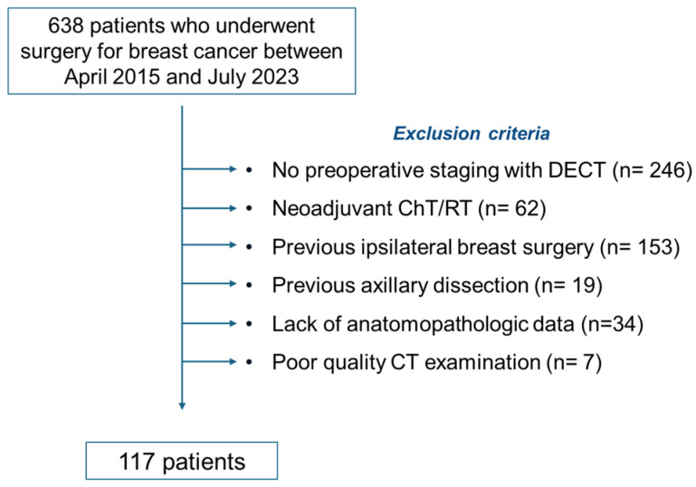 Figure 1
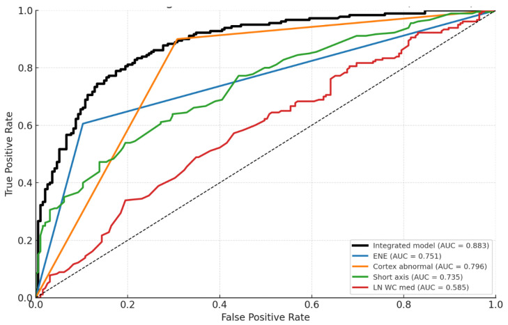 Figure 2
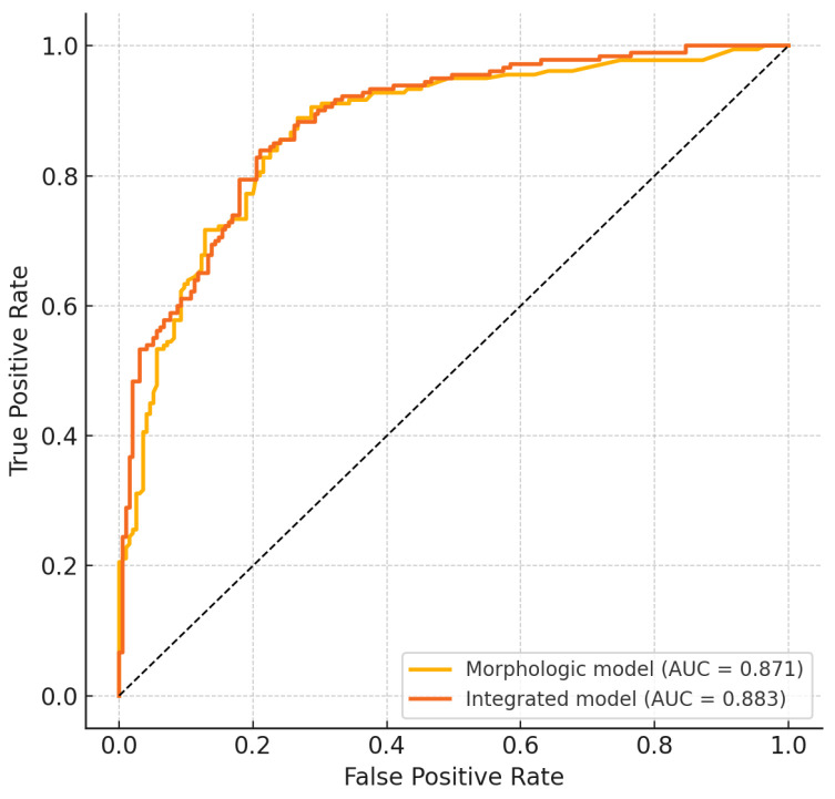 Figure 3
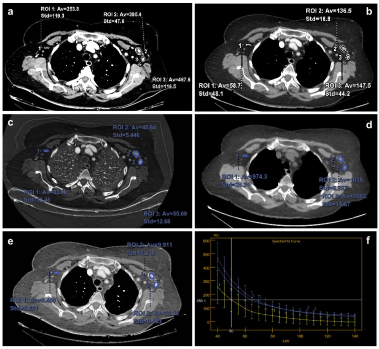 Figure 4
Peer Reviews
No public reviews on file for this paper yet. If you reviewed it on a platform where reviews are public (OpenReview, ICLR, NeurIPS, ICML), you can paste yours below so the community can read it here.
Videos
No videos yet. Explain this paper in a talk, walkthrough, or lecture? Add one.
Taxonomy
TopicsAdvanced X-ray and CT Imaging · Digital Radiography and Breast Imaging · Infrared Thermography in Medicine