Relative and normalized iodine concentrations derived from photon counting computed tomography and their correlation with tumor grade and Ki67 in pancreatic neuroendocrine neoplasia: A pilot study
Marwin‐Jonathan Sähn, Simon Waltermann, Jonas Ottemöller, Raihanatou Diallo‐Danebrock, Julius Henning Niehoff, Marcel Bähr, Berthold Gerdes, Jan Borggrefe, Nehara Begum, Alexey Surov

TL;DR
This study explores using iodine concentration from a new CT technique to predict tumor grade and Ki67 in pancreatic neuroendocrine tumors.
Contribution
The study introduces relative iodine concentration from photon counting CT as a novel, non-invasive biomarker for tumor grade and Ki67 in pancreatic neuroendocrine neoplasms.
Findings
Relative iodine concentration strongly correlates with tumor grade and Ki67 (ρ = 0.54 and 0.53, p = 0.02).
High-grade tumors have significantly higher relative iodine concentration (p = 0.02).
Normalized iodine concentration showed weak and non-significant correlations with tumor grade and Ki67.
Abstract
Neuroendocrine neoplasms are a rare and complex tumor entity, among which pancreatic neuroendocrine neoplasms generally display a more aggressive behavior. Despite a notable stage migration towards lower stages at initial diagnosis, the incidence of pancreatic neuroendocrine neoplasms is rising. In recent publications, iodine concentration derived from dual energy computed tomography was explored as a potential biomarker for pancreatic neuroendocrine neoplasia tumor grade and Ki67. However, methodologies exhibited significant variability, and reported outcomes were ambiguous, ranging from weak correlations to strong predictive performance in complex multivariate analyses. With the advent of photon counting computed tomography and improved technical capabilities, this study revisits the topic and aims to provide evidence for tumor characterization using photon counting computed…
Genes, proteins, chemicals, diseases, species, mutations and cell lines named across the full text — each resolved to its canonical identifier and authoritative record.
Click any figure to enlarge with its caption.
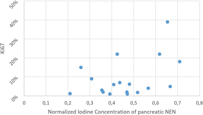 Figure 1
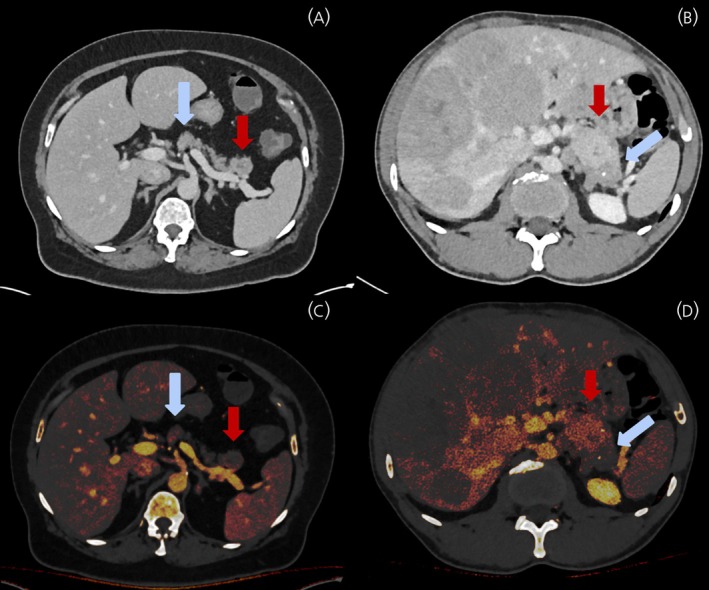 Figure 2
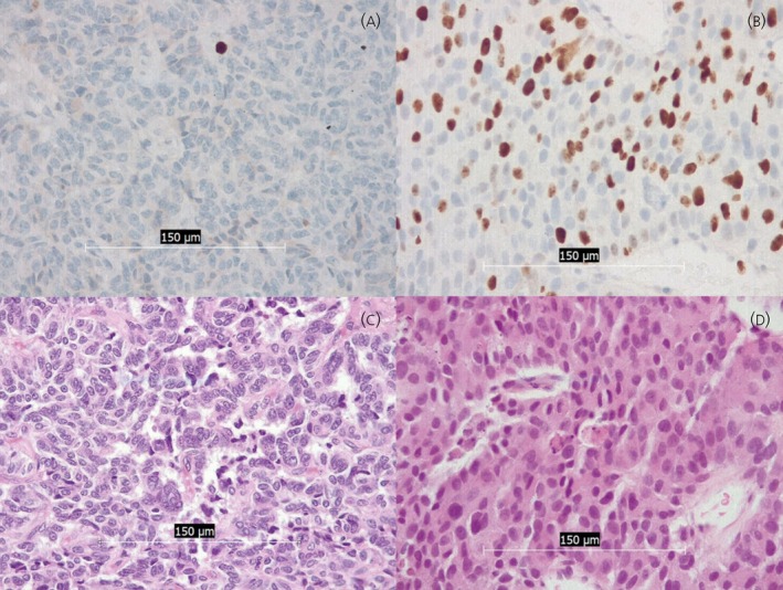 Figure 3
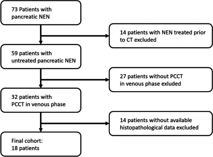 Figure 4
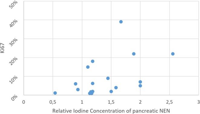 Figure 5
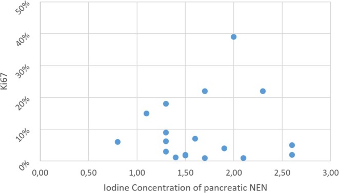 Figure 6
Peer Reviews
No public reviews on file for this paper yet. If you reviewed it on a platform where reviews are public (OpenReview, ICLR, NeurIPS, ICML), you can paste yours below so the community can read it here.
Videos
No videos yet. Explain this paper in a talk, walkthrough, or lecture? Add one.
Taxonomy
TopicsAdvanced X-ray and CT Imaging · Advanced X-ray Imaging Techniques · Advanced Materials Characterization Techniques