Mapping heterogeneous region- and tissue-specific brain ageing patterns using quantitative MRI
Xinjie Chen, Mario Ocampo-Pineda, Po-Jui Lu, Michelle G Jansen, Kwok-Shing Chan, Marcel Zwiers, Joukje M Oosterman, David G Norris, Andre F Marquand, Lester Melie-Garcia, Cristina Granziera, José P Marques

TL;DR
This study uses MRI scans to map how different brain regions and tissues change with age, revealing patterns that could help distinguish normal aging from disease.
Contribution
The study introduces a multiparametric qMRI approach to explore region- and tissue-specific brain aging patterns in healthy adults.
Findings
R1 showed the most robust age modeling, while R2* and susceptibility had greater regional variability.
Peak ages varied across brain regions, showing a posterior-to-anterior gradient in the cortex and an inferior-to-superior gradient in white matter.
The study identified a consistent spatial gradient in aging patterns across cortical grey matter, superficial white matter, and white matter bundles.
Abstract
Brain ageing involves microstructural changes that vary across tissue types and even within regions of those tissues, leading to functional and cognitive alterations. Quantitative MRI (qMRI) offers sensitivity to tissue properties, enabling the identification of differential ageing patterns and distinguishing physiological ageing from pathological changes. In this study, we analysed qMRI data from 293 healthy adults (median age: 52; interquartile range: 36–66; age range: 18–79 years). We applied a multiparametric qMRI approach, including longitudinal relaxation rate (R1), apparent transverse relaxation rate (R2*) and Quantitative Susceptibility Mapping, to model normal ageing effects on qMRI metrics across regions using second-order polynomial regression, adjusting for sex, education and cognition. Peak ages in turning points derived from quadratic fits were extracted to capture…
Genes, proteins, chemicals, diseases, species, mutations and cell lines named across the full text — each resolved to its canonical identifier and authoritative record.
Click any figure to enlarge with its caption.
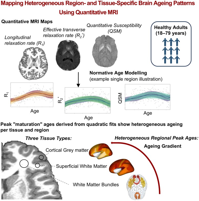 Figure 1
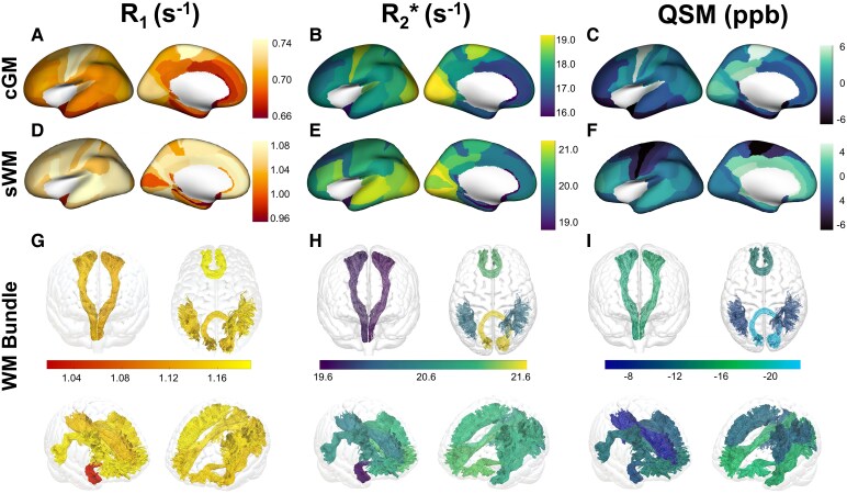 Figure 2
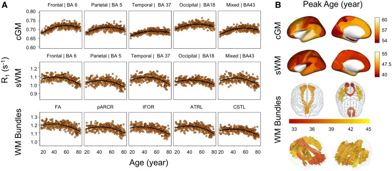 Figure 3
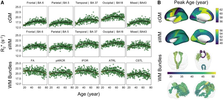 Figure 4
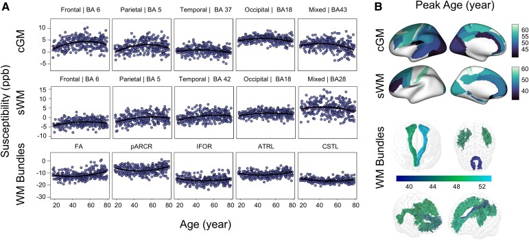 Figure 5
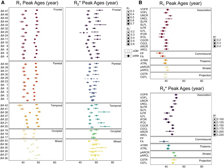 Figure 6
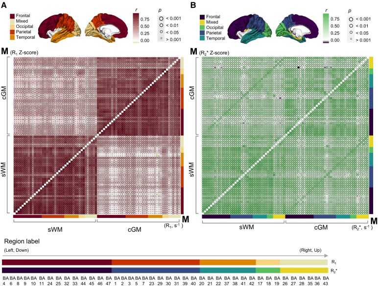 Figure 7
Peer Reviews
No public reviews on file for this paper yet. If you reviewed it on a platform where reviews are public (OpenReview, ICLR, NeurIPS, ICML), you can paste yours below so the community can read it here.
Videos
No videos yet. Explain this paper in a talk, walkthrough, or lecture? Add one.
Taxonomy
TopicsFunctional Brain Connectivity Studies · Advanced Neuroimaging Techniques and Applications · Advanced MRI Techniques and Applications