Towards interpretable prediction of recurrence risk in breast cancer using pathology foundation models
Jakub R. Kaczmarzyk, Sarah C. Van Alsten, Alyssa J. Cozzo, Rajarsi Gupta, Peter K. Koo, Melissa A. Troester, Katherine A. Hoadley, Joel H. Saltz

TL;DR
This paper introduces MAKO, a framework using pathology foundation models to predict breast cancer recurrence risk from histopathology images, offering an accessible alternative to transcriptomic assays.
Contribution
The study introduces MAKO, a novel benchmarking framework for evaluating pathology foundation models in predicting breast cancer recurrence risk.
Findings
Several foundation models outperformed baseline models in predicting ROR-P scores from histopathology images.
CONCH achieved the highest ROC AUC, while H-optimus-0 and Virchow2 showed the best correlation with ROR-P scores.
Pathology models stratified patients by recurrence risk similarly to transcriptomic assays, with tumor regions identified as key for high-risk predictions.
Abstract
Transcriptomic assays such as the PAM50-based ROR-P score guide recurrence risk stratification in non-metastatic, ER-positive, HER2-negative breast cancer but are not universally accessible. Histopathology is routinely available and may offer a scalable alternative. We introduce MAKO, a benchmarking framework evaluating 12 pathology foundation models and two non-pathology baselines for predicting ROR-P scores from H&E-stained whole-slide images using attention-based multiple instance learning. Foundation models, large neural networks pre-trained on millions of pathology images and adaptable to diverse downstream tasks, were trained and validated on the Carolina Breast Cancer Study and externally tested on TCGA BRCA. Several foundation models outperformed baseline models across classification, regression, and survival tasks. CONCH achieved the highest ROC AUC, while H-optimus-0 and…
Genes, proteins, chemicals, diseases, species, mutations and cell lines named across the full text — each resolved to its canonical identifier and authoritative record.
Click any figure to enlarge with its caption.
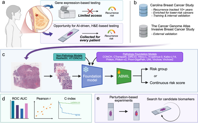 Figure 1
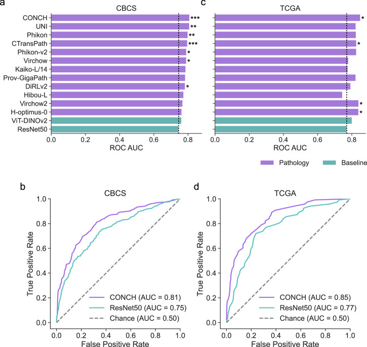 Figure 2
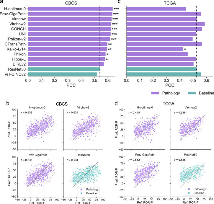 Figure 3
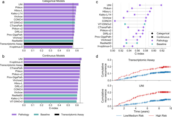 Figure 4
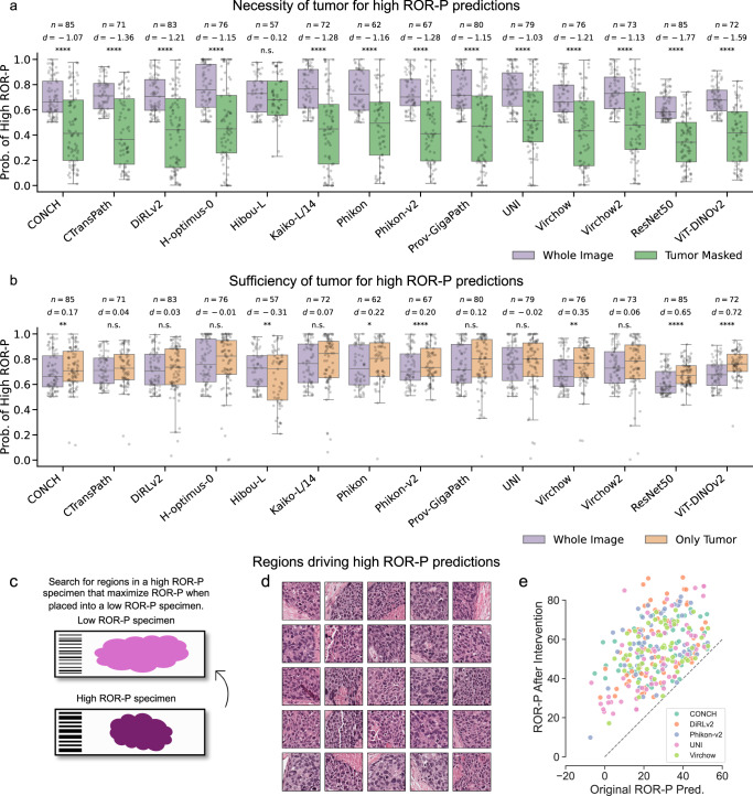 Figure 5
Peer Reviews
No public reviews on file for this paper yet. If you reviewed it on a platform where reviews are public (OpenReview, ICLR, NeurIPS, ICML), you can paste yours below so the community can read it here.
Videos
No videos yet. Explain this paper in a talk, walkthrough, or lecture? Add one.
Taxonomy
TopicsAI in cancer detection · Breast Cancer Treatment Studies · Cell Image Analysis Techniques