Evaluation of Morphologic Dimensions of Humulus Appendix of Pterygoid Plate Using Cone Beam Computed Tomography (CBCT) in Ahvazian Patients, Iran: -
Zahra Saadi, Sanaz Sharifi Shushtari, Alireza Hashemi Ashtiani, Ali Tayebi

TL;DR
This study uses CBCT to evaluate the shape and size of the pterygoid hamulus in Iranian patients, finding age-related changes but no gender differences.
Contribution
The study provides new morphometric data on the pterygoid hamulus using CBCT in an Iranian population.
Findings
Pterygoid hamulus length and width increase with age, then decrease.
Axial and coronal angles increase significantly in specific age groups.
No significant gender differences were observed in morphological dimensions.
Abstract
Today, CBCT has found a special place in dentistry due to the high quality and accuracy of images and providing information, and its use is increasing. With its help, we can examine many parts of the anatomy that are difficult to evaluate. The purpose of this study is to investigate the morphological dimensions of the humulus of the pterygoid appendage using cone beam computed tomography (CBCT). In this retrospective study, the statistical population was the imaging reccords of patients who referred to the radiology department of Ahvaz Dental School for CBCT of the upper jaw, whose values were stored in the NNT software. The size of the humulus (length and width) and its slope in the coronal and axial sections of the images were evaluated by two oral and maxillofacial radiologists. The results were analyzed using SPSS software version 22. Eighty pterygoid hamuli from 38 males and 42…
Genes, proteins, chemicals, diseases, species, mutations and cell lines named across the full text — each resolved to its canonical identifier and authoritative record.
Click any figure to enlarge with its caption.
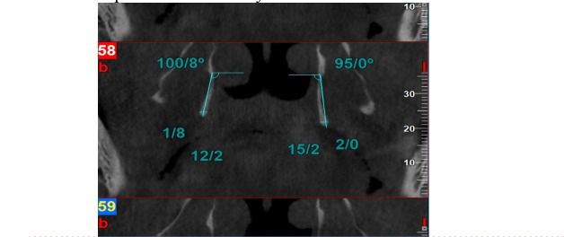 Figure 1
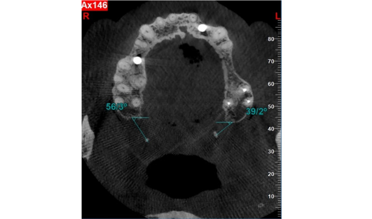 Figure 2
Peer Reviews
No public reviews on file for this paper yet. If you reviewed it on a platform where reviews are public (OpenReview, ICLR, NeurIPS, ICML), you can paste yours below so the community can read it here.
Videos
No videos yet. Explain this paper in a talk, walkthrough, or lecture? Add one.
Taxonomy
TopicsOral and Maxillofacial Pathology · Dental Radiography and Imaging · Oropharyngeal Anatomy and Pathologies