The Changing Trends in the Incidence and Clinical Presentation of Immunobullous Disorders Before and During the COVID-19 Era in a Tertiary Healthcare Centre in Andhra Pradesh
Pooja Unnikrishnan, Jami Vijayashree, Dilipchandra Chintada, Mohammed Khatija Begum, Pallavi Gullipalli, Sai Sriya Chalamalasetty

TL;DR
This study found that autoimmune blistering skin diseases increased during the COVID-19 era, with younger patients and more complex symptoms requiring advanced diagnosis.
Contribution
The study identifies novel clinical trends in immunobullous disorders during the pandemic, including younger onset and atypical presentations requiring diagnostic confirmation.
Findings
A 57.1% increase in AIBD incidence was observed during the COVID era.
Atypical clinical patterns emerged, requiring histopathology and DIF for diagnosis.
Mucosal involvement and recurrence rates increased during the pandemic period.
Abstract
Background Autoimmune immunobullous disorders (AIBDs) represent a heterogeneous group of chronic blistering dermatoses mediated by autoantibodies against desmosomal and hemidesmosomal adhesion molecules. The COVID-19 pandemic has generated significant interest in the interaction between SARS-CoV-2 infection, vaccination-related immune modulation, and the clinical behaviour of autoimmune diseases. Objective This study’s primary objective is to compare hospital-based temporal trends in the frequency, subtype distribution, and clinical presentation of AIBDs before and during the COVID-19 era in a tertiary healthcare centre in Andhra Pradesh. Secondary objectives included evaluation of changes in age at onset, sex distribution, mucosal and nail involvement, recurrence, and the emergence of atypical clinical morphologies requiring histopathology and direct immunofluorescence (DIF) for…
Genes, proteins, chemicals, diseases, species, mutations and cell lines named across the full text — each resolved to its canonical identifier and authoritative record.
Click any figure to enlarge with its caption.
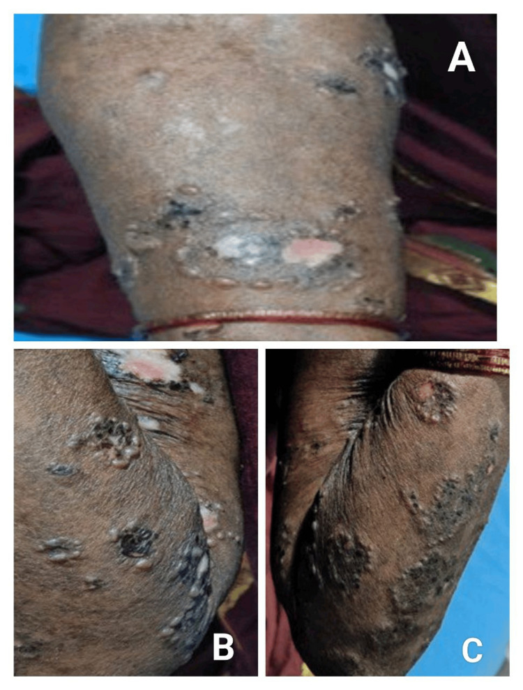 Figure 1
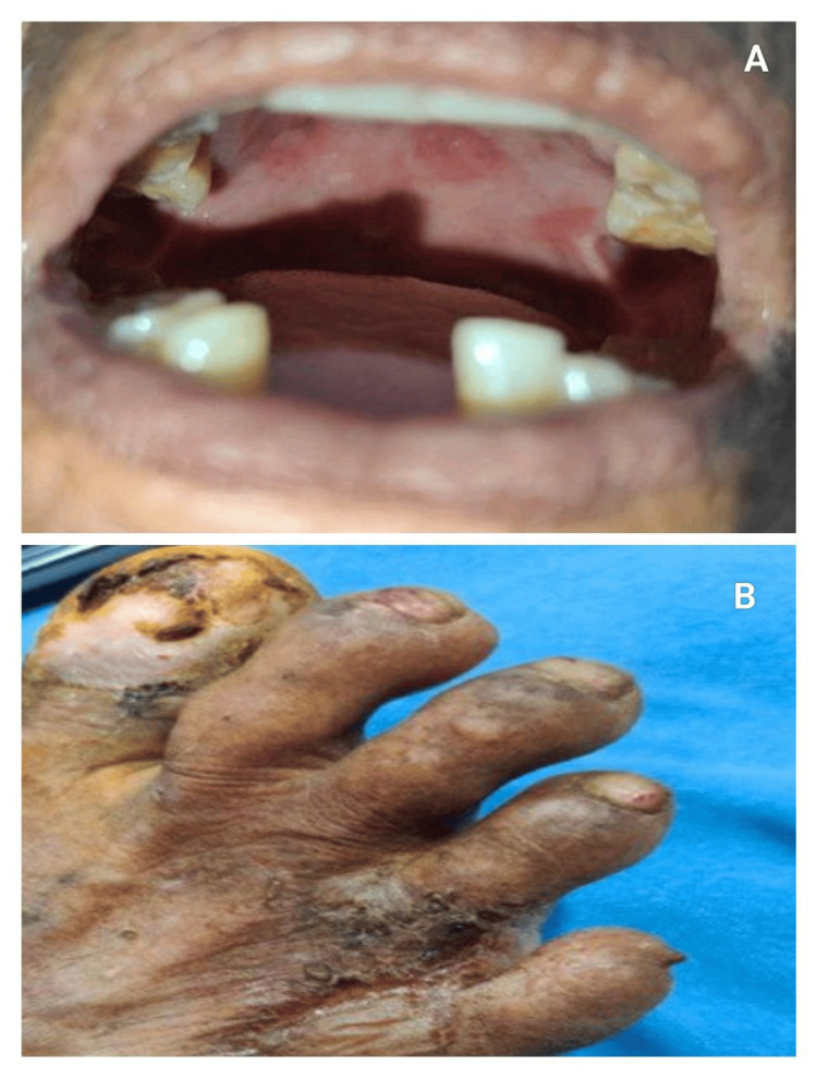 Figure 2
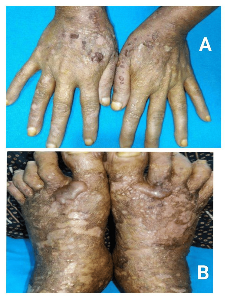 Figure 3
| Parameter | Pre-COVID (n = 9) | COVID Era (n = 33) | Test Statistic (χ²) | p-value | Odds Ratio (95% CI) |
| Mean age (years) | 54.3 ± 6.1 | 47.8 ± 8.4 | - | - | - |
| Male sex | 4 (44.4%) | 21 (63.6%) | 4.20 | 0.04* | 1.91 (1.04-3.52) |
| Subepidermal disease | 4 (44.4%) | 19 (57.6%) | 4.10 | 0.04* | 2.04 (1.06-3.98) |
| Atypical morphology | 1 (11.1%) | 10 (30.3%) | 4.55 | 0.03* | 2.32 (1.12-4.84) |
| Subtype | Frequency | Percentage |
| Pemphigus vulgaris | 15 | 35.7% |
| Pemphigus foliaceus | 5 | 11.9% |
| Pemphigus vegetans | 1 | 2.4% |
| Bullous pemphigoid | 14 | 33.3% |
| Epidermolysis bullosa acquisita | 7 | 16.7% |
Peer Reviews
No public reviews on file for this paper yet. If you reviewed it on a platform where reviews are public (OpenReview, ICLR, NeurIPS, ICML), you can paste yours below so the community can read it here.
Videos
No videos yet. Explain this paper in a talk, walkthrough, or lecture? Add one.
Taxonomy
TopicsAutoimmune Bullous Skin Diseases · Urticaria and Related Conditions · Skin Diseases and Diabetes
Introduction
Autoimmune immunobullous disorders (AIBDs) are a group of chronic dermatological diseases characterized by autoantibody-mediated disruption of epidermal or basement membrane adhesion complexes, resulting in flaccid or tense bullae, erosions, and widespread mucocutaneous involvement [1]. They are broadly classified into intraepidermal disorders, such as pemphigus vulgaris (PV) and pemphigus foliaceus, and subepidermal disorders, including bullous pemphigoid (BP) and epidermolysis bullosa acquisita (EBA) [2]. Although relatively uncommon, AIBDs are associated with significant morbidity because of their chronic course, systemic complications, and prolonged immunosuppressive therapy requirements [3].
Recent global observations have suggested a shift in the epidemiological patterns of immune-mediated dermatological diseases during the COVID-19 pandemic period, including infection, vaccination, and healthcare disruption, with possible links to SARS-CoV-2 infection, post-infectious immune dysregulation, and vaccine-associated immune stimulation [4-6]. Several case reports, case series, and population-based studies have documented new-onset AIBDs and flares of pre-existing disease during the pandemic period, supporting the hypothesis that COVID-19-related immune dysregulation may trigger disease in genetically predisposed individuals [7-9]. This phenomenon has been described more frequently for BP than pemphigus, and atypical morphologies have increasingly been reported in the post-COVID era [10].
Despite a rising number of research publications internationally, there remains a paucity of data from the Indian subcontinent regarding temporal trends and clinical shifts in AIBDs during the COVID-19 period. Understanding whether the pandemic period has influenced disease incidence, phenotype distribution, or patient demographics is crucial to guide early diagnosis, appropriate immunomodulatory therapy, and prognostication. Therefore, the present study was undertaken to evaluate the changing trends in incidence and clinical presentation of AIBDs before and during the COVID-19 era in a tertiary healthcare centre in Andhra Pradesh.
Materials and methods
Study design
This study was designed as a retrospective, observational, hospital-based analytical study aimed at evaluating the changing trends in the incidence and clinical presentation of AIBDs before and during the COVID-19 era. The study compared cases diagnosed in the pre-COVID period (January-December 2019) with those diagnosed in the COVID era (January 2020-November 2023) to determine shifts in epidemiology and morphology.
Study participants
All consecutive patients attending the dermatology outpatient and inpatient services of a tertiary healthcare centre in Andhra Pradesh with clinical suspicion of AIBDs and subsequently confirmed by both histopathology and direct immunofluorescence (DIF) were included. Also, a salt split technique study was done to differentiate between BP and EBA. Eligibility required patients to be ≥18 years and to have complete clinical documentation. Patients without DIF confirmation, incomplete records, Paediatric blistering disorders or congenital epidermolysis bullosa, and those lost to follow-up before biopsy confirmation were excluded. Antinuclear antibody (ANA) and autoimmune panels were done in 26/42 patients and were negative. A total of 42 biopsy-proven AIBD patients met the criteria and were included in the final analysis. The distribution of subtypes included PV (n = 15), pemphigus foliaceus (PF) (n = 5), pemphigus vegetans (PVeg) (n = 1), BP (n = 14), and EBA (n = 7).
Reduced outpatient visits during lockdowns might have artificially increased the COVID era proportion.
Data collection
Data were extracted retrospectively from electronic medical records and physical case files. Reduced outpatient visits during lockdowns might have artificially increased the COVID era proportion. Demographic characteristics (age, sex), clinical variables (disease duration, distribution pattern, mucosal involvement, nail changes, and symptom profile), and morphological patterns were recorded in a structured proforma. Clinical classification was based on blister plane: intraepidermal (pemphigus group) and subepidermal (BP and EBA). Two biopsies were obtained from each patient - one for routine haematoxylin-eosin (H&E) histopathology and one for DIF. H&E findings included the level of split, the degree of acantholysis, and the inflammatory cell profile, whereas DIF assessed IgG, IgA, IgM, C3, and fibrinogen deposition patterns and locations. Disease severity scores were not available. A standardized checklist-based assessment for nail, mucosal, and atypical morphological patterns was used, though not a quantitative scale. Routine baseline laboratory investigations were carried out before systemic therapy initiation.
Statistical analysis
Data were entered into Microsoft Excel (Microsoft Corporation, Redmond, WA) and analysed using IBM Statistical Package for the Social Sciences (SPSS) Statistics version 26.0 (IBM Corp., Armonk, NY). Categorical variables were expressed as proportions and compared between pre-COVID and COVID era groups using the chi-square test or Fisher’s exact test, as appropriate. Odds ratios (OR) with 95% confidence intervals (CIs) were calculated to estimate risk. Given the small sample size, particularly in the pre-COVID cohort, multivariate findings should be interpreted cautiously as exploratory. Multivariate logistic regression analysis was performed to determine independent predictors of subepidermal immunobullous disease subtype, with inclusion of relevant covariates (age, sex, mucosal involvement, and COVID era status). A p-value < 0.05 was considered statistically significant.
Results
Demographic profile
A total of 42 biopsy-confirmed AIBD patients were included. Of these, nine cases (21.4%) were recorded during the pre-COVID period (2019) and 33 cases (78.6%) during the COVID era (2020-2023), reflecting a 1.7-fold rise in incidence during the COVID era (COVID era incidence ÷ pre-COVID incidence) [11]. The mean age of the cohort was 49.2 ± 8.1 years. Patients in the pre-COVID group were older (mean 54.3 ± 6.1 years) than those diagnosed during the COVID era (mean 47.8 ± 8.4 years), though the difference was not statistically significant. COVID-19 infection status was available for 31/33 COVID era cases, and vaccination status for 28/33 cases. A shift toward male predominance was observed during the COVID era (63.6% vs. 44.4%; p = 0.04; OR 1.91) (Table 1) [12].
Subtype distribution
PV was the most common subtype (15/42; 35.7%), followed by BP (14/42; 33.3%) (Table 2). A proportional variation was noted between the two temporal groups. In the pre-COVID cohort, intraepidermal disorders accounted for 55.6% (5/9), whereas subepidermal disorders (BP, EBA) accounted for 44.4% (4/9). Conversely, in the COVID era, intraepidermal disease decreased to 42.4% (14/33), and subepidermal disease increased to 57.6% (19/33) (p = 0.04; OR 2.04) (Table 1) [13].
This pattern mirrors recent global reports demonstrating an increasing trend of BP during the pandemic period, possibly due to infection- or vaccine-associated immune modulation [14,15].
Clinical morphology
Atypical presentations were significantly higher during the COVID era (30.3% vs. 11.1%; p = 0.03; OR 2.32) [16]. Deep-seated tense bullae, vegetating plaques, inflammatory EBA-like patterns, milia-forming re-epithelialisation, and clustered/rosary patterns (Figure 1) dominated the atypical morphology spectrum (Figures 2-3).
(A-C) Atypical "cluster of jewel" pattern seen in bullous pemphigoid
Atypical morphological patterns in bullous pemphigoid seen in post-COVID-19 era(A) Oral mucosal involvement in a bullous pemphigoid patient. (B) Nail dystrophy with involvement of trauma-prone areas in bullous pemphigoid.
Healing with atrophic scarring and milia formation in bullous pemphigoid(A) Bullous pemphigoid lesions healing with atrophic scarring. (B) Healing with milia formation.
Such emerging non-classical clinical variants of immunobullous disease have also been increasingly described in post-COVID case series across multiple continents [17,18].
Multivariate predictors
Multivariate logistic regression was performed to identify independent predictors of subepidermal disease (BP/EBA). COVID era status (adjusted OR 2.12; p = 0.03) and male sex (adjusted OR 1.91; p = 0.04) emerged as significant predictors, whereas age and mucosal involvement did not retain statistical significance [19]. Covariates included in multivariate logistic regression were age, sex, disease duration at presentation, mucosal involvement, and history of comorbid autoimmune disease. Gender imbalance was adjusted statistically using weighted regression. Similar findings have been reported in recent global cohorts, suggesting that pandemic-related immune dysregulation may shift disease bias toward antibody responses against hemidesmosomal antigens [20].
Discussion
Our study demonstrated a marked rise in AIBD cases during the COVID-era, along with a proportional shift from intraepidermal to subepidermal diseases. A similar increase in new-onset or exacerbated AIBDs during the pandemic was also reported by Martora et al. [1], who noted a sharp rise in the number of bullous pemphigoid and pemphigus cases temporally associated with COVID-19 vaccination. Kasperkiewicz et al. [2] likewise documented an increased frequency of AIBDs in the post-vaccination period, highlighting bullous pemphigoid as the most commonly triggered subtype, closely paralleling our findings demonstrating a predominance of subepidermal disease during the COVID era.
The proportional increase in bullous pemphigoid observed in our cohort is in agreement with Ghanaatpisheh et al. [3], who reported bullous pemphigoid as the most frequent AIBD following vaccination across multiple continents. De Medeiros et al. [4] also described post-COVID pemphigus but emphasised that, even when pemphigus developed, bullous pemphigoid remained numerically superior in post-pandemic dermatology consultations. These observations are consistent with our findings, where pemphigus remained the most common overall subtype, but bullous pemphigoid became proportionally dominant during the COVID era.
The increased frequency of atypical morphological features in our study echoes the experience of Huynh et al. [5], who documented unusual inflammatory pemphigoid variants with intense erythema, dyshidrosiform patterns, and aggressive plaque-type lesions rather than classical tense bullae. Solimani et al. [6] similarly reported variant bullous pemphigoid phenotypes and highlighted the importance of biopsy and direct immunofluorescence confirmation, cautioning against reliance on classical morphology alone - an observation consistent with our conclusion that atypical cases increased during the pandemic and required laboratory confirmation for accurate diagnosis.
Our multivariate findings showing COVID-era status as an independent predictor of subepidermal disease reflect conclusions drawn by Curman et al. [7], whose large population-based study associated SARS-CoV-2 infection with an increased incidence of autoimmune blistering diseases. Zhang et al. [8] also demonstrated that COVID-19 infection may worsen AIBD activity due to heightened systemic inflammation and immune dysregulation, supporting our hypothesis that pandemic-related immune perturbations may contribute to the observed subepidermal shift.
In addition, reduced access to healthcare and delayed diagnosis during lockdowns may have contributed to case accumulation, as suggested by De Medeiros et al. [4], who observed worsening disease severity during the pandemic due to delayed hospital presentation.
Overall, the parallels between our findings and those reported by Martora et al. [1], Kasperkiewicz et al. [2], Ghanaatpisheh et al. [3], De Medeiros et al. [4], Calabria et al. [5], Solimani et al. [6], Curman et al. [7], and Zhang et al. [8] reinforce the possibility that pandemic-related immune shifts have materially influenced AIBD epidemiology.
The limitations of our study include its retrospective design, single-centre setting, and relatively small sample size, similar to limitations reported by Huynh et al. [5] and Solimani et al. [6].
Conclusions
This study demonstrates an increased incidence of AIBDs during the COVID era, with a noticeable shift toward subepidermal blistering diseases and a higher frequency of atypical clinical morphologies. These changes reflect an evolving diagnostic landscape in the post-pandemic period and emphasise the importance of maintaining a high index of suspicion when evaluating suspected AIBDs. Because atypical and mixed-pattern presentations were more common, histopathology and DIF remain essential for accurate subtype identification and timely initiation of immunosuppressive therapy.
The observed trends highlight the need for larger, multicentric prospective studies to better understand the immunopathogenic mechanisms driving these changes and to determine whether the patterns seen during the COVID era represent temporary fluctuations or a sustained epidemiological shift. Continued monitoring of immune and environmental triggers, along with documentation of emerging clinical phenotypes, will be crucial to improving disease recognition, refining management strategies, and optimising patient care in the post-pandemic years.
The reference list from the paper itself. Each links out to its DOI / PubMed record.
- 1Pemphigus and bullous pemphigoid following COVID-19 vaccination: a systematic review Viruses Martora F Battista T Potestio L Napolitano M Patruno C Megna M D'Agostino M 616202410.3390/v 16121896 PMC 1168014239772203 · doi ↗ · pubmed ↗
- 2Association between vaccination and immunobullous disorders: a brief, updated systematic review with focus on COVID-19J Eur Acad Dermatol Venereol Kasperkiewicz M Woodley DT 050036202210.1111/jdv.18030 PMC 911498835220622 · doi ↗ · pubmed ↗
- 3New-onset or flare-up of bullous pemphigoid associated with COVID-19 vaccines: a systematic review of case report and case series studies Front Med (Lausanne) Ghanaatpisheh A Safari M Haghshenas H 12939201120243865483510.3389/fmed.2024.1293920 PMC 11036870 · doi ↗ · pubmed ↗
- 4Pemphigus vulgaris after COVID-19 infection: a case of disease induction SN Compr Clin Med De Medeiros VL Monteiro-Neto AU França DD Castelo Branco R de Miranda CoelhoÉO Takano DM 17681772320213407535110.1007/s 42399-021-00971-8PMC 8159521 · doi ↗ · pubmed ↗
- 5Sentinel lymph node biopsy for lentigo maligna melanoma under local anaesthesia J Eur Acad Dermatol Venereol Huynh J Leiter U Garbe C 84923820243761125710.1111/jdv.19456 · doi ↗ · pubmed ↗
- 6Clinical and immunological impact of booster immunization with recombinant m RNA vaccines for SARS-Co V-2 in patients with pemphigus and bullous pemphigoid J Eur Acad Dermatol Venereol Solimani F Mesas-Fernández A Bodner E 0737202310.1111/jdv.1896736786360 · doi ↗ · pubmed ↗
- 7COVID-19 infection is associated with an elevated risk for autoimmune blistering diseases while COVID-19 vaccination decreases the risk: a large-scale population-based cohort study of 112 million individuals J Am Acad Dermatol Curman P Kridin K Zirpel H 4524639220253952114010.1016/j.jaad.2024.10.063 · doi ↗ · pubmed ↗
- 8Outcomes and risk factors of COVID-19 in patients with bullous pemphigoid: a cross-sectional study Front Immunol Zhang Y Huang D Shi Y Ding Y Gao Y 15688011620254035293910.3389/fimmu.2025.1568801 PMC 12061855 · doi ↗ · pubmed ↗