Clinical application of confocal laser endomicroscopy in neurosurgery: a scoping review
Yuan Xu, Carlos E. Calderon-Valero, Thomas J. On, Francesco Restelli, Francesco Acerbi, Jürgen Schlegel, Evgenii Belykh, Mark C. Preul

TL;DR
This review explores how confocal laser endomicroscopy (CLE) can help neurosurgeons by providing real-time imaging during brain tumor surgeries, potentially replacing traditional methods.
Contribution
The study provides a comprehensive scoping review of clinical applications and performance of CLE in neurosurgery, highlighting its potential as an optical biopsy tool.
Findings
CLE demonstrated diagnostic accuracy comparable to frozen section pathology with high sensitivity and specificity.
CLE interpretation is faster than frozen section and can be integrated with fluorescence-guided surgery and telepathology.
Most studies on CLE in neurosurgery are recent, but more interventional trials are needed to confirm its effectiveness.
Abstract
Confocal laser endomicroscopy (CLE) is an emerging intraoperative “optical biopsy” tool that enables real-time, in vivo, cellular-resolution visualization of brain tumor histoarchitecture. It offers the potential to complement frozen section pathology by providing rapid intraoperative feedback. We conducted a scoping review of prospective clinical studies to characterize CLE platforms, fluorophores, operative applications, and diagnostic performance in neurosurgical patients. This review followed PRISMA-ScR guidelines. A systematic search of PubMed, Scopus, and Embase was performed. Eligible studies were prospective clinical studies of intraoperative CLE imaging in neurosurgical patients. Two independent reviewers screened and extracted data on study design, CLE system, fluorophore use, pathology types, diagnostic performance, and workflow characteristics. From 379 initial records, 19…
Genes, proteins, chemicals, diseases, species, mutations and cell lines named across the full text — each resolved to its canonical identifier and authoritative record.
Click any figure to enlarge with its caption.
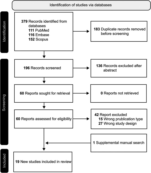 Figure 1
 Figure 2
 Figure 3
 Figure 4
 Figure 5
Peer Reviews
No public reviews on file for this paper yet. If you reviewed it on a platform where reviews are public (OpenReview, ICLR, NeurIPS, ICML), you can paste yours below so the community can read it here.
Videos
No videos yet. Explain this paper in a talk, walkthrough, or lecture? Add one.
Taxonomy
TopicsEsophageal Cancer Research and Treatment · Optical Imaging and Spectroscopy Techniques · Cerebrospinal fluid and hydrocephalus