Circulating Extracellular Vesicle Protein Biomarkers for the Early Detection of High-Grade Serous Ovarian Cancer
Sagar Rayamajhi, Jared Sipes, Bidii Ngala, Amrita Mitra, Meizhang Li, Camille V. Trinidad, Wei Cui, Mohammod Mahmudur Rahman, Foyez Ahmmed, Leonidas E. Bantis, Mihaela E. Sardiu, Dennis W. Province, Harsh B. Pathak, Andrew K. Godwin

TL;DR
Researchers identified proteins in tiny cell-derived particles that can detect early-stage ovarian cancer, offering a promising non-invasive diagnostic tool.
Contribution
The study introduces a novel combinatorial protein panel for early detection of high-grade serous ovarian cancer using extracellular vesicle proteomics.
Findings
A four-protein panel (MUC1, MYL6, TTYH3, GSTP1) achieved 90% sensitivity and 95% specificity for early-stage HGSOC detection.
Circulating sEV proteins were found to reflect precursor lesions in the fallopian tube, enabling early disease detection.
MUC1 levels in sEVs showed strong diagnostic potential with an AUC of 0.84 for early-stage HGSOC versus controls.
Abstract
Small extracellular vesicles (sEVs), lipid-bilayer delimited particles (50–200 nm) released by cells, are emerging as a promising class of liquid biopsy biomarkers for elusive cancers, such as high-grade serous ovarian cancer (HGSOC). HGSOC originates from the fallopian tube (FT), progressing from p53 signatures to a precursor lesion known as serous tubal intraepithelial carcinoma (STIC). We hypothesize that sEVs contribute to ovarian cancer pathogenesis, carry cargo reflective of their site of origin, and serve as diagnostic biomarkers for early detection. To test this, we established a case–control cohort using archival plasma samples from 30 HGSOC patients (10 early stage [ES] and 20 late stage [LS]) and 40 healthy controls (HC). sEVs were enriched by size-exclusion chromatography and profiled by LC–MS/MS. Across all samples, 1078 EV-associated proteins (exoproteins) were identified,…
Genes, proteins, chemicals, diseases, species, mutations and cell lines named across the full text — each resolved to its canonical identifier and authoritative record.
Click any figure to enlarge with its caption.
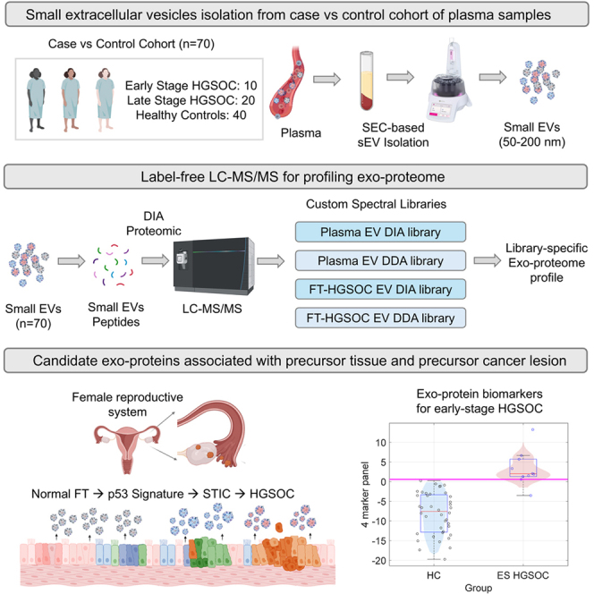 Figure 1
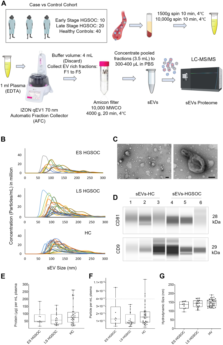 Figure 2
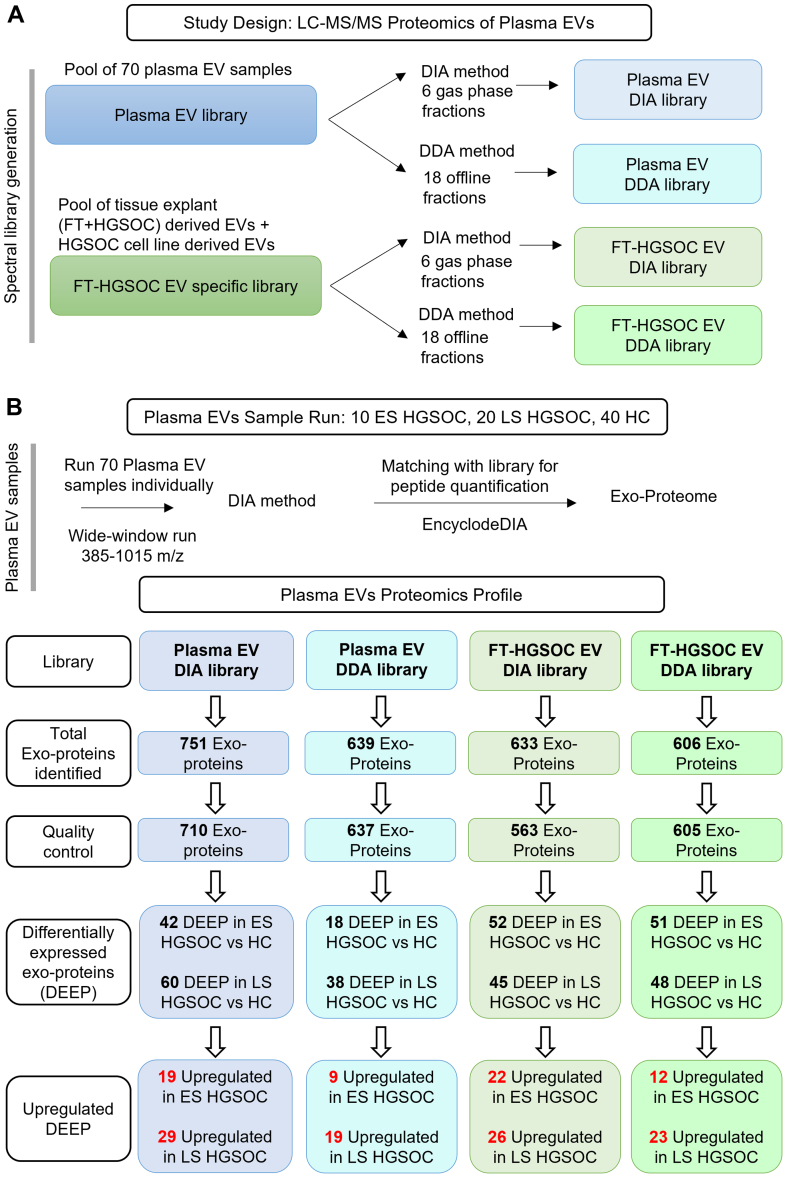 Figure 3
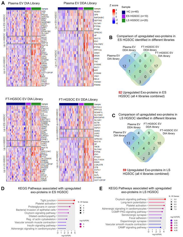 Figure 4
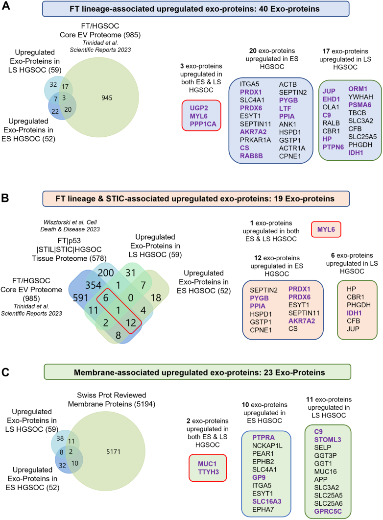 Figure 5
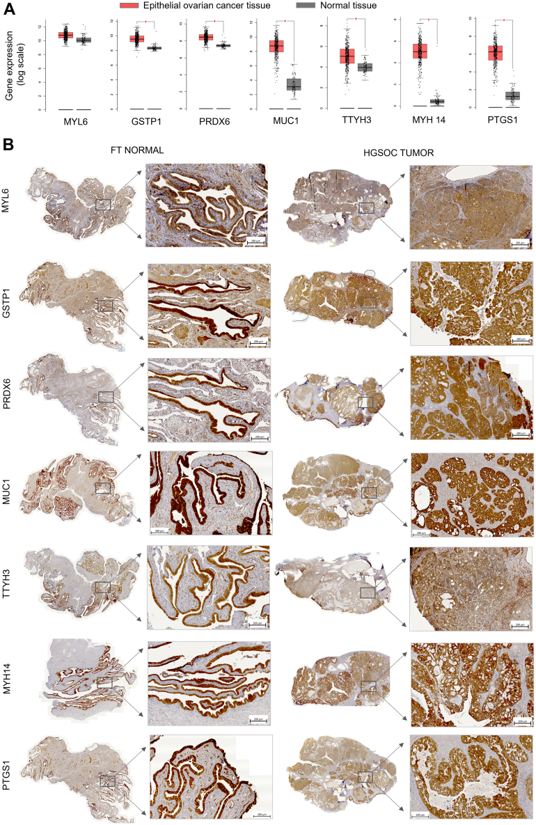 Figure 6
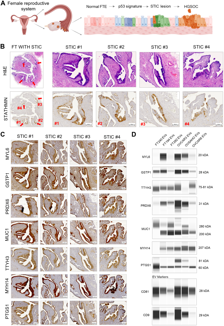 Figure 7
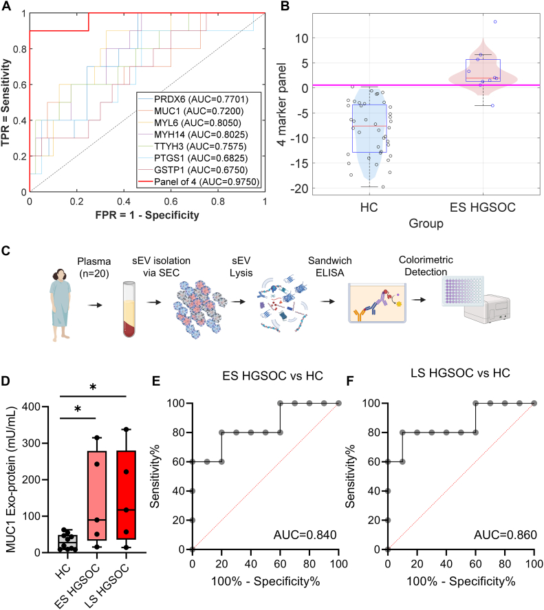 Figure 8
Peer Reviews
No public reviews on file for this paper yet. If you reviewed it on a platform where reviews are public (OpenReview, ICLR, NeurIPS, ICML), you can paste yours below so the community can read it here.
Videos
No videos yet. Explain this paper in a talk, walkthrough, or lecture? Add one.
Taxonomy
TopicsExtracellular vesicles in disease · Ovarian cancer diagnosis and treatment · Ferroptosis and cancer prognosis