Pigmented Spitz nevus of the lip
João Teixeira, Mariana Pedroso, José Carlos Cardoso, Inês Coutinho, Hugo Schönenberger de Oliveira

Abstract
Genes, proteins, chemicals, diseases, species, mutations and cell lines named across the full text — each resolved to its canonical identifier and authoritative record.
Click any figure to enlarge with its caption.
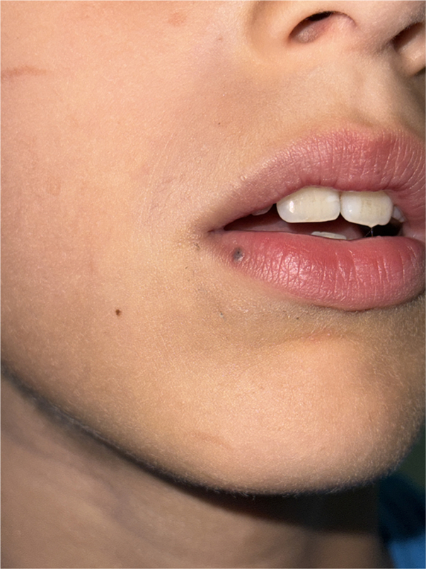 Figure 1
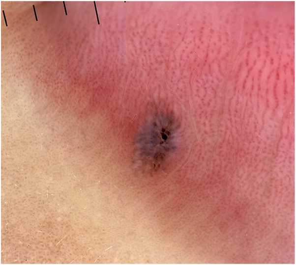 Figure 2
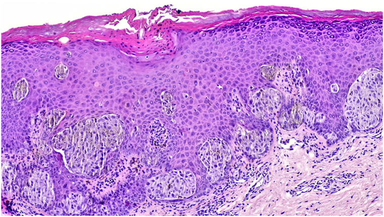 Figure 3
Peer Reviews
No public reviews on file for this paper yet. If you reviewed it on a platform where reviews are public (OpenReview, ICLR, NeurIPS, ICML), you can paste yours below so the community can read it here.
Videos
No videos yet. Explain this paper in a talk, walkthrough, or lecture? Add one.
Taxonomy
TopicsGenetic and rare skin diseases. · Cutaneous Melanoma Detection and Management · melanin and skin pigmentation
Clinical presentation
A 7-year-old boy presented with a solitary, asymptomatic pigmented papule on the lower vermilion lip that had been present for several months. Clinical examination revealed a 3-mm, well-circumscribed blue-gray papule with a smooth surface (Fig 1).Fig 1. Clinical picture showing a 3-mm, well-circumscribed bluish papule with a smooth surface on the lower vermilion lip of a 7-year-old boy.
Dermoscopic appearance
Dermoscopy showed a structureless blue-gray background with central brown-black dots and globules, blue globules radiating peripherally, and peripheral shiny white structures (Fig 2).Fig 2. Polarized contact dermoscopy (Heine Delta One, 10×) demonstrating a structureless blue-gray background with central brown-black and blue globules radiating peripherally. The overall architecture is symmetrical and globular, producing a starburst-like appearance.
Histologic diagnosis
Excisional biopsy revealed a symmetrical, well-circumscribed junctional melanocytic proliferation composed of epithelioid and spindle melanocytes arranged in nests at the dermoepidermal junction without significant cytologic atypia (Fig 3). These findings confirmed a junctional pigmented Spitz nevus.Key messageSpitz nevi are uncommon melanocytic proliferations, representing about 1% of melanocytic nevi, typically arising in children and adolescents.1 They are classified as nonpigmented, appearing as pink to reddish papules, and pigmented Spitz nevi, presenting as brown to black macules or papules. Common sites include the extremities, face, and trunk.Dermoscopically, pigmented Spitz nevi often display the starburst pattern, with radial streaks or pseudopods at the periphery and homogeneous central pigmentation.1 Other features include symmetrical streaks, regularly distributed dotted vessels, and homogeneous pigmentation. In this case, the globular pattern corresponded to the large junctional nests, and the grayish color was likely attributable to the overlying acanthotic epithelium. These features are consistent with those described in pigmented Spitz nevi at other cutaneous sites, although the lack of follicular structures and the prominent vascularity of the lip semimucosa may attenuate the globular pattern. Of note, the dermoscopic pattern in this case, although previously described in cutaneous pigmented Spitz nevi, has not, to our knowledge, been documented on the vermilion.In fact, in mucosal locations such as the lip, Spitz nevi are exceedingly rare.2 In this context, traumatized angioma, venous lake, and melanotic labial macule should be considered in the differential diagnosis, particularly when typical dermoscopic features are absent, making histopathologic confirmation essential for accurate diagnosis.Fig 3. Histopathology (hematoxylin-eosin stain, 200×) showing epithelioid and spindle melanocytes at the dermoepidermal junction with no significant cytologic atypia, with clefts surrounding the nests, consistent with a junctional pigmented Spitz nevus. Focal pigmented parakeratosis was also noted.
Conflicts of interest
None disclosed.
The reference list from the paper itself. Each links out to its DOI / PubMed record.
- 1Requena C.Requena L.Kutzner H.Spitz nevus: a clinicopathologic study of 349 cases Am J Dermatopathol 31220091071161931879510.1097/DAD.0b 013e 3181934218 · doi ↗ · pubmed ↗
- 2Tosios K.Nikitakis N.G.Sklavounou A.Junctional spitz tumor (nevus) of the upper lip Head Neck Pathol 73201334034410.1007/s 12105-013-0510-8PMC 412691724293157 · doi ↗ · pubmed ↗