Diagnosis of Bilateral Quadriceps Tendon Rupture Using Point-of-Care Ultrasound
Edward Guo, Akaysha Duran, Alexander Kuc, Alfred B. Cheng

TL;DR
A 32-year-old man with bilateral knee pain was diagnosed with a rare quadriceps tendon rupture using point-of-care ultrasound, enabling timely surgery.
Contribution
Demonstrates the effectiveness of point-of-care ultrasound in diagnosing rare bilateral quadriceps tendon ruptures in emergency settings.
Findings
Point-of-care ultrasound confirmed bilateral quadriceps tendon rupture in a timely manner.
The patient underwent successful surgical repair the day after diagnosis.
Ultrasound is a viable alternative to MRI in emergency settings for this rare injury.
Abstract
A healthy 32-year-old man presented to the emergency department with bilateral knee pain after landing from a jump. He was unable to extend his knees and had pain to palpation superior to the patella. Bilateral quadriceps tendon rupture was confirmed using point-of-care ultrasound, and the patient underwent operative repair the next day. Bilateral quadriceps tendon rupture is exceedingly rare, which often leads to misdiagnosis. Magnetic resonance imaging is the gold standard diagnostic imaging study but has multiple disadvantages, especially in emergency settings. Point-of-care ultrasound is an excellent tool to screen for this injury and prevent morbidity from delay in surgical repair.
Genes, proteins, chemicals, diseases, species, mutations and cell lines named across the full text — each resolved to its canonical identifier and authoritative record.
Click any figure to enlarge with its caption.
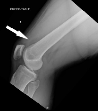 Figure 1
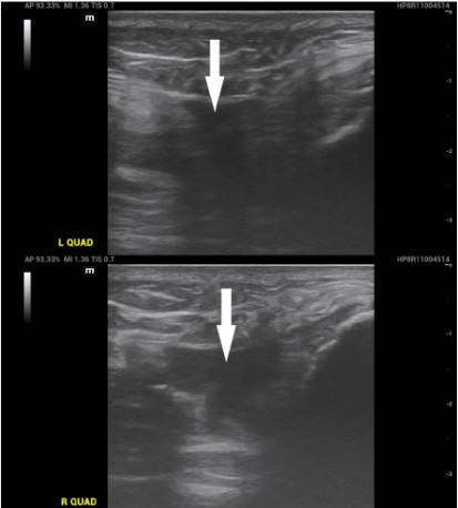 Figure 2
Peer Reviews
No public reviews on file for this paper yet. If you reviewed it on a platform where reviews are public (OpenReview, ICLR, NeurIPS, ICML), you can paste yours below so the community can read it here.
Videos
No videos yet. Explain this paper in a talk, walkthrough, or lecture? Add one.
Taxonomy
TopicsTendon Structure and Treatment · Musculoskeletal synovial abnormalities and treatments · Ultrasound and Hyperthermia Applications
CASE PRESENTATION
A 32-year-old man with no past medical history presented to the emergency department (ED) for bilateral knee pain. He had been jumping on a trampoline when he landed in a squat position, felt pain in both knees, and was then unable to bear weight. He had an athletic build and body mass index of 32.1 (reference range 18.5–24.9) kilograms per square meter. Physical examination was significant for bogginess and tenderness to palpation to the distal femur bilaterally with inability to extend either knee. The patient denied medication or drug use and had no previous orthopedic surgeries.
Plain radiographs of the bilateral knees demonstrated trace amounts of bilateral suprapatellar joint effusions with no fracture or dislocation (Image 1). Point-of-care ultrasound (POCUS) of the suprapatellar regions revealed discontinuity of the bilateral quadriceps tendons with adjacent hematomas (Image 2). Dynamic POCUS of the quadriceps tendons while the patient attempted to extend at the knee further supported a diagnosis of bilateral quadriceps tendon rupture (Video). No further diagnostic imaging studies were obtained. The patient underwent operative repair the following day and was confirmed to have complete rupture of the bilateral quadriceps tendons.
DISCUSSION
Bilateral quadriceps tendon rupture is rare with just over 100 reported cases in the literature. It is commonly misdiagnosed at initial presentation due to its rarity and the inability to compare the affected limb to the unaffected limb.1 Risk factors include chronic renal disease, diabetes mellitus, obesity, and steroid use.2 Complete quadriceps tendon rupture is typically caused by forceful contraction of the quadriceps muscles with the knee in a flexed position while regaining balance such as in the case of our patient. Orthopedic consultation is indicated as patients suffer ambulatory dysfunction from injury to the extensor mechanism of both lower extremities. Delay in surgical repair for complete rupture is correlated with quadriceps retraction, muscle atrophy, and decreased functional outcomes.3
CPC-EM Capsule What do we already know about this clinical entity? Quadriceps tendon rupture is uncommon; bilateral cases are rare and frequently missed, delaying surgical repair and worsening patient outcomes. What is the major impact of the image(s)? Video of left and right suprapatellar regions with the patient attempting to extend his knees demonstrates bilateral quadriceps tendon rupture. How might this improve emergency medicine practice? Dynamic point-of-care ultrasound may improve recognition of tendon rupture and expedite a rapid, accurate diagnosis.
Plain radiographs may show indirect signs of quadriceps tendon rupture but are rarely diagnostic. Magnetic resonance imaging (MRI) of the knee is the gold standard imaging study but has several limitations including cost, time, and availability. Physical examination paired with radiology-performed ultrasonography has been used to diagnose quadriceps tendon rupture with sensitivities reported as high as 100%.4 Thus, ED POCUS is an excellent screening modality to assess for bilateral rupture and prevent delay in diagnosis, treatment, and potential morbidity. It was particularly valuable in our case due to the inability to compare findings to an unaffected limb. Furthermore, to our knowledge, no prior peer-reviewed case report has included annotated, dynamic POCUS images visualizing quadriceps tendon rupture.
Our images provide novel educational value, demonstrating a practical, real-time imaging technique to improve recognition of this rare injury. Advantages of POCUS include universal availability and rapid utility, making it an ideal screening tool for quadriceps tendon rupture in the ED that may also be diagnostic. Its major limitation is operator dependence. In cases with any doubt of the diagnosis, an MRI should be obtained given its superior specificity.4,5
The reference list from the paper itself. Each links out to its DOI / PubMed record.
- 1Abduljabbar FH Aljurayyan A Ghalimah B Bilateral simultaneous quadriceps tendon rupture in a 24-year-old obese patient: a case report and review of the literature Case Rep Orthop 2016201647131372784075710.1155/2016/4713137 PMC 5093266 · doi ↗ · pubmed ↗
- 2Camarda LD’Arienzo A Morello S Bilateral ruptures of the extensor mechanism of the knee: a systematic review J Orthop 20171444454532881934210.1016/j.jor.2017.07.008PMC 5548366 · doi ↗ · pubmed ↗
- 3Hak DJ Sanchez A Trobisch P Quadriceps tendon injuries Orthopedics 201033140462005295310.3928/01477447-20091124-20 · doi ↗ · pubmed ↗
- 4Perfitt JS Petrie MJ Blundell CM Acute quadriceps tendon rupture: a pragmatic approach to diagnostic imaging Eur J Orthop Surg Traumatol 2014247123712412399608010.1007/s 00590-013-1307-x · doi ↗ · pubmed ↗
- 5La Rocco BG Zlupko G Sierzenski P Ultrasound diagnosis of quadriceps tendon rupture J Emerg Med 20083532932951797682310.1016/j.jemermed.2007.05.015 · doi ↗ · pubmed ↗