Clinical outcomes of the EyeWatch system: three-year results
Constance Liegl, Sarah Hundertmark, Frank G. Holz, Karl Mercieca

TL;DR
A three-year study shows the EyeWatch system effectively lowers eye pressure in glaucoma patients, with lasting results and fewer medications needed.
Contribution
This is the first study to report three-year clinical outcomes of the EyeWatch system for glaucoma management.
Findings
Eye pressure dropped from 34.6 mmHg pre-surgery to 13.6 mmHg after three years.
All patients maintained eye pressure below 21 mmHg without needing frequent device adjustments after the first year.
MRI exposure caused temporary eye pressure spikes, highlighting the need for monitoring in magnetic environments.
Abstract
The EyeWatch™ System (EWS) is an adjustable glaucoma drainage device that allows non-invasive postoperative intraocular pressure (IOP) control.Early studies have shown promising short- and mid-term results with EWS in managing refractory glaucoma. The EyeWatch™ System (EWS) is an adjustable glaucoma drainage device that allows non-invasive postoperative intraocular pressure (IOP) control. Early studies have shown promising short- and mid-term results with EWS in managing refractory glaucoma. This study presents the first three-year clinical outcomes of EWS implantation, showing a sustained IOP reduction from a preoperative mean of 34.6 mmHg to 13.6 mmHg, with all patients achieving IOP levels below 21 mmHg and a significant decrease in medication burden.Most device adjustments occurred within the first postoperative year, after which IOP remained stable without further intervention.A…
Genes, proteins, chemicals, diseases, species, mutations and cell lines named across the full text — each resolved to its canonical identifier and authoritative record.
Click any figure to enlarge with its caption.
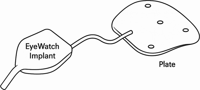 Figure 1
- —Rheinische Friedrich-Wilhelms-Universität Bonn (1040)
Peer Reviews
No public reviews on file for this paper yet. If you reviewed it on a platform where reviews are public (OpenReview, ICLR, NeurIPS, ICML), you can paste yours below so the community can read it here.
Videos
No videos yet. Explain this paper in a talk, walkthrough, or lecture? Add one.
Taxonomy
TopicsGlaucoma and retinal disorders · Ocular Surface and Contact Lens · Ophthalmology and Visual Impairment Studies
Brief communications
The eyeWatch™ System (EWS; Rheon Medical, Lausanne, Switzerland) offers an innovative solution for managing refractory glaucoma (Fig. 1). The EWS, used with a glaucoma drainage device (GDD), regulates aqueous flow via a magnetically adjustable deformable tube, allowing postoperative adjustment of intraocular pressure (IOP) without surgical intervention. This study presents three-year outcomes in five eyes undergoing EWS implantation (with 200 mm^2^ plate) at the University Hospital of Bonn.Fig. 1. Schematic picture of the EWS with EyePlate
All patients (mean age 66.2 years) presented with advanced glaucoma (2 primary open-angle glaucoma, 3 secondary glaucomas). Preoperative mean IOP was 34.6 mmHg (range: 23–45 mmHg) on 2.8 topical agents. At 12, 24, and 36 months, mean IOP declined to 12.2, 13.4, and 13.6 mmHg, respectively. The need for IOP-lowering agents decreased significantly, with all patients off acetazolamide postoperatively. Visual acuity remained stable or improved in most patients (Table 1).Table 1. Demographic and clinical characteristics of patients who underwent EWS surgeryPatient 1Patient 2Patient 3Patient 4Patient 5Sex Male/FemaleMFFMFEthnicityCaucasianCaucasianCaucasianCaucasianCaucasianAge6676695248Glaucoma typeSecondary glaucoma due to panuveitisPOAGPOAGTraumatic secondary glaucomaNeovascular glaucoma due to pDRPPrevious vitrectomyNoNoYesNoyesPrevious glaucoma surgeryNoYesNoYesnoWhich one?MP-CPCXEN, trabectomeFollow-up time3736383636AnesthesiaGALocalGAGAGA# of pressure-lowering eye drops24341acetazolamidenononoyesyesLens statusPseudophakicPseudophakicPseudophakicPseudophakicPhakicBCVA (logMAR)1.60.10.40.41.6IOP preoperative4045252340Maximum IOP without eye drops4148314140
EWS adjustments using the eyeWatch Pen™ (EWP) were performed in four of five cases, with 12 total adjustments in the first year. IOP decreased from a pre-adjustment mean of 21.3 mmHg to 8.6 mmHg after modification. No further adjustments were necessary after the first year.
Postoperative complications were limited. One patient required surgical revision due to tube exposure and inadequate IOP control, attributed to prior trauma and multiple surgeries. Minor complications included self-resolving hyphema (2 eyes) and a vitreous hemorrhage (1 eye). Notably, one case of transient IOP elevation followed an MRI scan; this was corrected by EWS adjustment. No vision-threatening events occurred.
Compared to Roy et al.‘s 2-year results (IOP 11.5 mmHg, 1.0 medications; 42 eyes), our cohort had higher baseline IOP but achieved comparable reductions (IOP 13.4 mmHg, 0.6 medications at 24 months) [1]. Roy et al. also compared EWS with Ahmed Glaucoma Valves (AGV) in a smaller study, showing a reduction from 27.3 mmHg to 12.8 mmHg in the EWS group at 12 months (12 AGV, 9 EWS combined with Baerveldt plate) [2]. Detorakis et al. described two patients with IOP reductions from 45 to 12 mmHg and 36 to 15 mmHg at one year; one patient did not respond despite valve opening [3]. The ability to modulate IOP non-invasively distinguishes EWS from traditional GDD such as Ahmed or Baerveldt implants, which rely on fixed resistance or surgical revisions [4]. Roy et al. reported 23 EWS adjustments in 8/9 patients during year one, aligning with our experience [5].
MRI compatibility is a unique consideration with the EWS [6]. In our cohort, one IOP spike occurred after cranial MRI. This phenomenon has been described in the literature and highlights the need for IOP monitoring post-MRI. EWS position was likely affected by magnetic interaction but was reversible.
This study confirms the EWS as an effective option for long-term IOP control with a low complication rate. Its non-invasive adjustability provides a significant advantage in early postoperative management. Limitations include the small sample size, lack of endothelial data, and retrospective design. Longer-term and comparative studies are warranted.