Protective Effects of Myrtus communis Essential Oil Against Bisphenol A‐Induced Metabolic Dysfunction‐Associated Fatty Liver Disease in Wistar Rats
Mhimdi Mariem, Selmi Slimen, Amira Zammali, Manel Hraoui, Soumaya Wahabi, Dallacqua Stefano, Sebai Hichem

TL;DR
This study shows that Myrtus communis essential oil can protect against liver damage caused by BPA in rats, similar to vitamin E.
Contribution
The study demonstrates the protective potential of Myrtus communis essential oil against BPA-induced MASLD in a rat model.
Findings
BPA exposure increased liver enzymes, lipid levels, and oxidative stress in rats.
EOMC reduced triglycerides and oxidative stress markers in a dose-dependent manner.
Liver histology showed EOMC significantly attenuated BPA-induced steatosis.
Abstract
Endocrine disruptors such as bisphenol A (BPA) are increasingly associated with metabolic disorders, including metabolic dysfunction‐associated fatty liver disease (MASLD). This preclinical study investigates the effect of BPA on liver function and lipid metabolism in Wistar rats and evaluated the protective potential of Myrtus communis essential oil (EOMC) and vitamin E (Vit E). MASLD was induced in Wistar rats by oral administration of BPA (100 mg/kg/day) for 30 days. Rats were co‐treated with EOMC at doses of 50,100, or 200 mg/kg body weight, or Vit E at 100 mg/kg. Liver and kidney function markers, lipid profiles, oxidative stress parameters, and organ histology were assessed. BPA exposure significantly increased plasma liver enzymes (AST: 195,76 ± 4,47 U/L, ALT: 91,05 ± 0,58 U/L) and lipid levels (triglycerides: 1,30 ± 0,13 g/L) compared to controls, and elevated oxidative stress…
Genes, proteins, chemicals, diseases, species, mutations and cell lines named across the full text — each resolved to its canonical identifier and authoritative record.
Click any figure to enlarge with its caption.
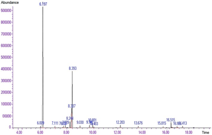 FIGURE 1
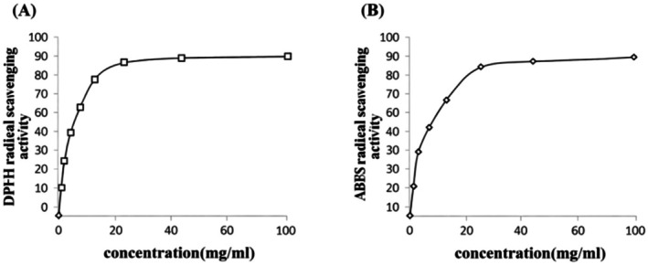 FIGURE 2
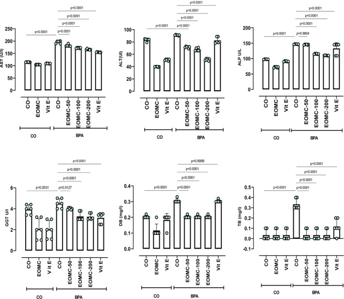 FIGURE 3
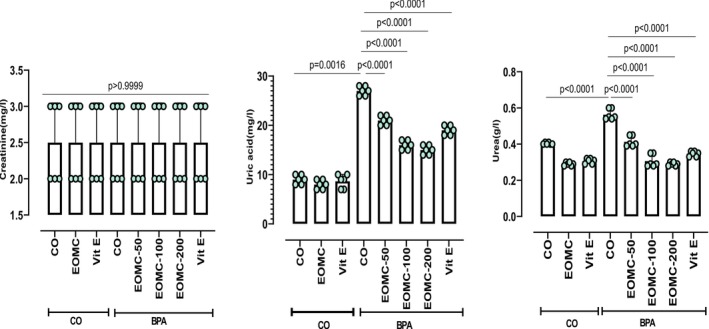 FIGURE 4
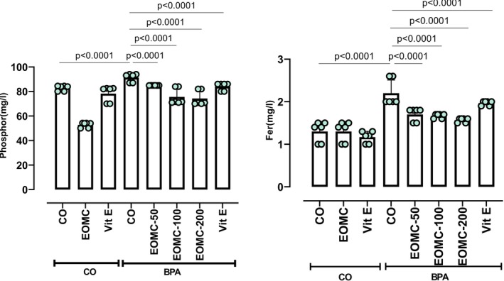 FIGURE 5
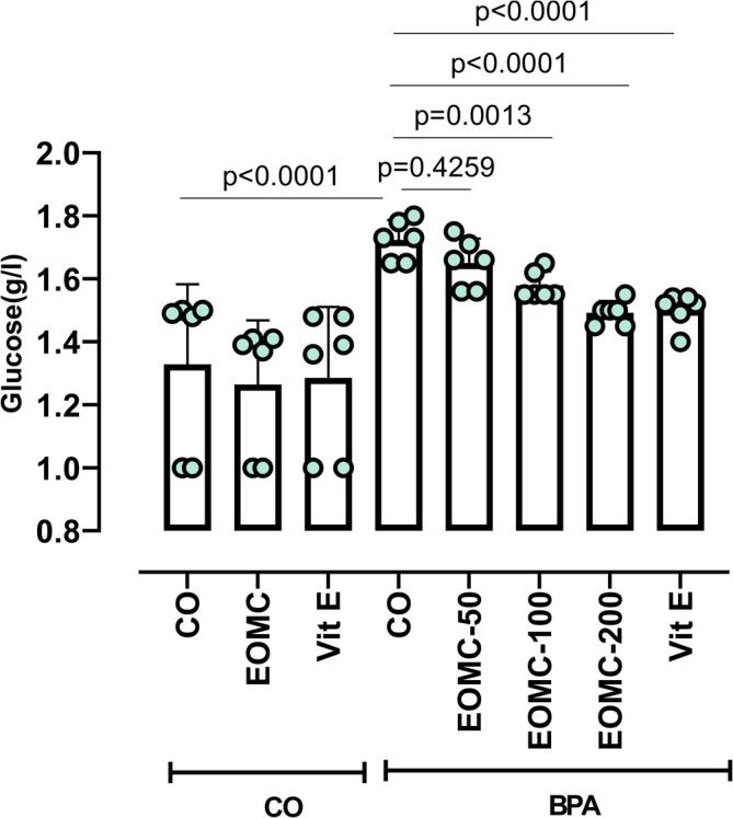 FIGURE 6
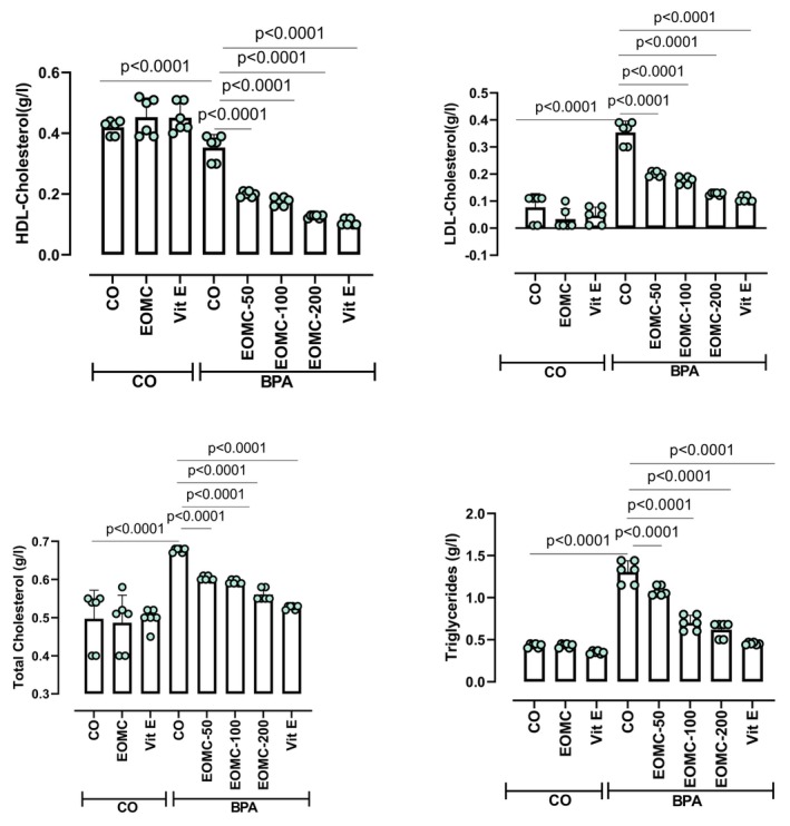 FIGURE 7
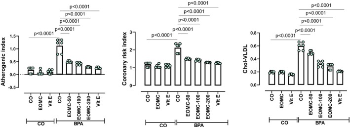 FIGURE 8
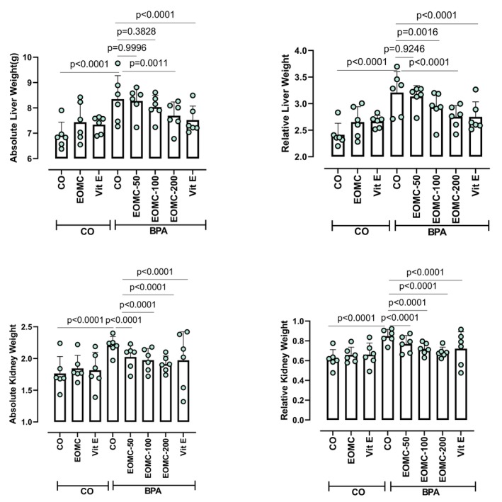 FIGURE 9
 FIGURE 10
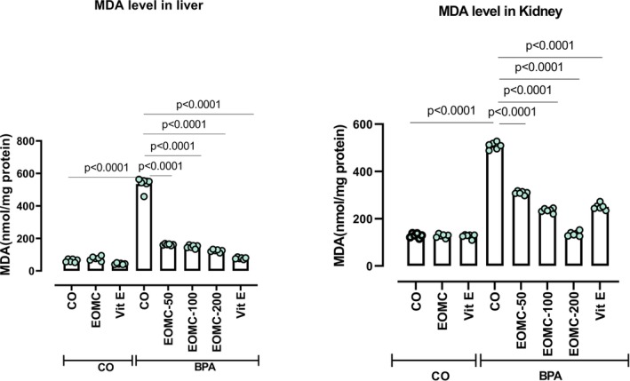 FIGURE 11
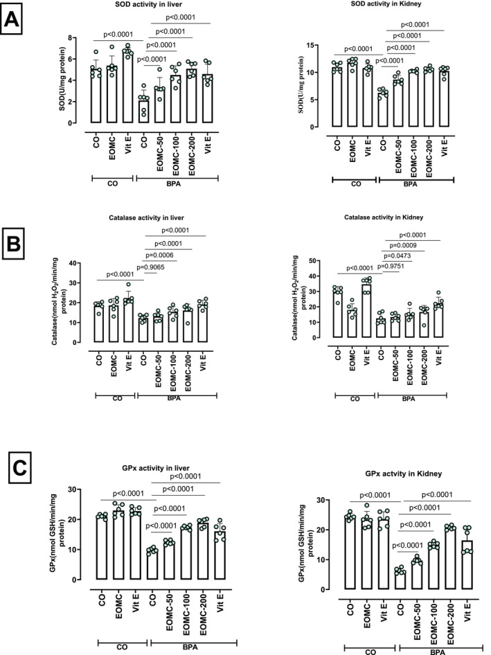 FIGURE 12
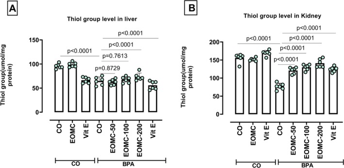 FIGURE 13
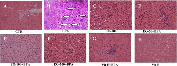 FIGURE 14
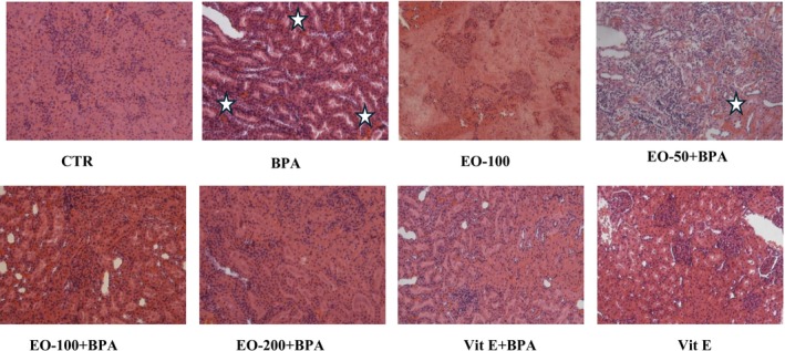 FIGURE 15
| Groups | Treatments |
|---|---|
| G1 | Vehicle control: 0.4 mL/kg/day of corn oil |
| G2 | BPA (100 mg/kg) |
| G3 | EOMC (100 mg/kg) |
| G4 | EOMC (50 mg/kg) + BPA |
| G5 | EOMC (100 mg/kg) + BPA |
| G6 | EOMC (200 mg/kg) + BPA |
| G7 | Vitamin E (100 mg/kg) + BPA |
| G8 | Vitamin E (100 mg/kg) |
| Peak | Compounds | Relative content (%) | RT (min) |
|---|---|---|---|
| 1 | Alpha. ‐Thujene | 0.618 | 6.009 |
| 2 | Alpha. ‐Pinene | 59.749 | 6.197 |
| 3 | Beta. ‐Pinene | 0.410 | 7.111 |
| 4 | Butanoic acid, 2‐methyl‐, 2‐methylpropyl ester | 0.260 | 7.674 |
| 5 | l‐Phellandrene | 0.450 | 7.739 |
| 6 | Delta.3‐Carene | 0.768 | 7.879 |
| 7 | p‐Cymene | 2.297 | 8.214 |
| 8 | dl‐Limonene | 7.020 | 8.337 |
| 9 | 1,8‐Cineole | 18.651 | 8.393 |
| 10 | gamma‐Terpinene | 0.808 | 9.030 |
| 11 | Alpha‐Terpinolene | 1.164 | 9.746 |
| 12 | Linalool | 1.666 | 10.001 |
| 13 | Butanoic acid, 2‐methyl‐, 2‐methylbutyl ester | 0.435 | 10.093 |
| 14 | Alpha. Terpineol | 1.241 | 12.203 |
| 15 | Linalyl acetate | 0.624 | 13.676 |
| 16 | α‐Terpinenyl acetate | 0.447 | 15.815 |
| 17 | Geranyl acetate | 1.911 | 16.515 |
| 18 | Methyleugenol | 0.804 | 16.996 |
| 19 | Caryophyllene | 0.676 | 17.413 |
| Bacterial Strains | Inhibition diameter (mm) | |
|---|---|---|
| EOMC (20 μL/disc) | Gentamicin (10 IU/disc) | |
| Gram (+) | ||
|
| (−) | 18 |
|
| (−) | 36 |
|
| 17.66 ± 2.56 | 22 |
|
| (−) | |
| Gram (−) | ||
|
| 12 ± 0 | 30 |
|
| (−) | 18 |
|
| (−) | 20 |
| Bacterial strains | EOMC | ||
|---|---|---|---|
| CMI (mg/ml) | CMB (mg/ml) | CMB/CMI | |
| Gram (+) | |||
|
| 25 | 200 | 8 |
|
| 6.25 | 200 | 32 |
|
| 12.5 | 200 | 16 |
|
| 1.562 | 200 | 128.04 |
| Gram (−) | |||
|
| 3.125 | 200 | 64 |
|
| 12.5 | 200 | 10 |
|
| 25 | 200 | 8 |
- —Ministry of Higher Education and Scientific Research10.13039/100010450
Peer Reviews
No public reviews on file for this paper yet. If you reviewed it on a platform where reviews are public (OpenReview, ICLR, NeurIPS, ICML), you can paste yours below so the community can read it here.
Videos
No videos yet. Explain this paper in a talk, walkthrough, or lecture? Add one.
Taxonomy
TopicsEffects and risks of endocrine disrupting chemicals · Phytochemistry and biological activity of medicinal plants · Saffron Plant Research Studies
Introduction
1
Metabolic dysfunction associated steatotic liver disease (MASLD), recently adopted to replace the term non‐alcoholic fatty liver disease (NAFLD), represents the hepatic manifestation of metabolic dysfunction and is now the most prevalent chronic liver disorder worldwide (Machado 2023, Syed‐Abdul 2023). MASLD affects approximately 40% of adults and is closely associated with obesity, insulin resistance, dyslipidemia, and type 2 diabetes mellitus (Mentsiou Nikolaou et al. 2024). The disease spectrum ranges from simple steatosis to steatohepatitis, fibrosis, and cirrhosis, making MASLD a major nutritional and public health challenge (AbdalHussin et al. 2025).
In the past decade, environmental factors have received a well‐deserved increase in attention due to the parallel rise of both metabolic pathologies and the production or usage of endocrine‐disruptive chemicals (Lința et al. 2024) particularly bisphenol A (BPA), which is a widely used synthetic compound found in polycarbonate plastics and epoxy resins, leading to chronic exposure in humans and animals. BPA exhibits estrogenic activity and has been shown to induce oxidative stress, inflammation, and mitochondrial dysfunction in several organs, especially the liver (Alotaibi et al. 2024). After oral ingestion, BPA is metabolized in the liver into BPA‐G and BPA‐S, which are primarily excreted via bile, urine, and feces, with a half‐life of less than 12 h (Inoue et al. 2001; Mao et al. 2023). However, repeated exposure to moderate or high doses of BPA can overwhelm detoxification pathways, resulting in excessive generation of reactive oxygen species (ROS), lipid peroxidation, and hepatic injury (Hassan et al. 2012; Kobayashi et al. 2020).
Oxidative stress plays a central role in BPA‐induced hepatotoxicity and MASLD progression by enhancing lipid peroxidation, altering antioxidant defenses, and promoting inflammatory responses (Mentsiou Nikolaou et al. 2024; Tang et al. 2024; Yuan et al. 2025). Accordingly, nutritional strategies aimed at reinforcing antioxidant capacity have gained considerable interest. Vitamin E has been extensively investigated as a dietary antioxidant and has demonstrated beneficial effects on liver oxidative status and histological features in MASLD (Gheonea et al. 2025; Vrentzos et al. 2025). However, the search for natural, food‐derived bioactive compounds with hepatoprotective properties remains an important research focus.
Essential oils are increasingly recognized as valuable sources of natural (Bhavaniramya et al. 2019). Myrtus communis L., a Mediterranean aromatic plant traditionally used in food preservation and folk medicine, is rich in monoterpenes such as 1,8‐cineole, α‐pinene, linalool, and α‐terpineol (Özkan and Güray 2009; Hennia et al. 2019). These compounds exhibit strong antioxidant and anti‐inflammatory activities (Al‐Harrasi et al. 2022), and it was selected for this study due to their high content of bioactive monoterpenes, making them a promising candidate for hepatoprotection. Despite these promising properties, the protective effect of Myrtus communis essential oil against environmentally induced hepatic steatosis, particularly BPA‐related MASLD, has not yet been thoroughly investigated.
This study, therefore, aimed to investigate the potential of EOMC to prevent BPA‐induced MASLD in a rat model, comparing its efficacy to that of Vit E, by assessing metabolic profiles, oxidative stress status, and hepatic histopathology.
Materials and Methods
2
Reagents and Chemicals
2.1
2,2‐Diphenyl‐1‐picrylhydrazyl (CAS 1898‐66‐4 | Sigma‐Aldrich); 2,2′‐Azino‐bis(3‐ethylbenzothiazoline‐6‐sulfonic acid) (CAS 30931‐67‐0 | Sigma‐Aldrich); Gentamicin sulfate salt (CAS 1405‐41‐0 | Sigma‐Aldrich); Bisphenol A (CAS 80‐05‐7 | Sigma‐Aldrich); Butylated hydroxytoluene (CAS 128‐37‐0 | Sigma‐Aldrich); Trichloroacetic acid (CAS 76‐03‐9 | Sigma‐Aldrich); 2‐thio‐barbituric acid (CAS 84030‐12‐6 | Sigma‐Aldrich); 5,5′‐dithiobis(2‐nitrobenzoic acid) (CAS 69‐78‐3 | Sigma‐Aldrich); Vitamin E was obtained in the form of commercially available capsules from a pharmacy.
Essential Oil Extraction and Analysis
2.2
Myrtle leaves were harvested in March in the Hammam Bourguiba region (in the north‐west of Tunisia) and identified by the botanist Chokri Hafsi.
The essential oil was extracted by hydrodistillation using a Clevenger‐type apparatus for 3 h. The distillate was dried over anhydrous sodium sulfate and stored at 4°C until use.
The essential oil yield was calculated as (Mohamadi et al. 2021):
To ensure reproducibility, the extraction was performed in three independent replicates.
GC–MS Analysis
2.2.1
The extracted essential oil of myrtle leaves was analyzed using gas chromatography coupled with mass spectrometry using Trace GC ULTRA/Polaris Q (GC‐MS, Thermo Electron) by the method of Abidi et al., with slight modifications (Abidi et al. 2018).
In Vitro Antioxidant and Antimicrobial Tests
2.3
Antioxidant Assays (DPPH and ABTS)
2.3.1
Antioxidant capacity of EOMC was assessed using DPPH and ABTS assays in triplicate, following (Abramovič et al. 2018; Wahabi et al. 2023), with slight modifications.
Antimicrobial Activity
2.3.2
The antibacterial activity of essential oil from Myrtus Communis was assessed against Gram‐positive bacteria Staphylococcus aureus ATCC 29213, Enterococcus faecalis ATCC29212, Bacillus cereus (clinical isolate) and Listeria monocytogenes and Gram‐negative bacteria ( Escherichia coli ATCC 25983, Salmonella enteritidis ATCC 13076, and Pseudomonas aeruginosa ATCC 27853), using two methods: the disk diffusion method and liquid microdilution method (Dakhli et al. 2025). Gentamicin (10 μg/disc) served as a positive control.
Animal Study Design
2.4
All animal procedures were approved by the Biomedical Ethics Committee (CEBM, Pasteur Institute of Tunis, approval no. JORT472001, 15/04/2024) and complied with NIH and ARRIVE guidelines. Healthy male Wistar rats (170–180 g) were housed under standard pet shop conditions (22°C ± 0.5°C, and 12 h/12 h light/dark cycle), with access to food (standard pellet diet‐ Badr Utique‐TN) and water ad libitum. After 2 weeks of acclimatization, rats were randomly assigned to 8 groups (n = 6). The sample size (n = 6 per group) was determined based on commonly used group sizes in similar preclinical studies of hepatoprotection to ensure adequate power for detecting significant biochemical and histological differences. The animals received oral treatment for 4 weeks. To evaluate the preventive potential of EOMC, rats received EOMC or vitamin E for 7 days before and concurrently with BPA administration for the subsequent 30 days. In the following Table 1:
Vitamin E‐stripped corn oil was prepared by treating commercial corn oil with activated alumina (aluminum oxide) to remove tocopherols, following the method described by Rokosik et al. (2019).
Bisphenol A (100 mg/kg) was selected based on previous studies demonstrating its ability to reliably induce hepatic steatosis and oxidative stress in reflecting a toxicologically relevant exposure in rats (Gurmeet et al. 2014; Kamel and Ahmed 2018).
Note: From the 7th day of treatment, bisphenol A is introduced into groups 4, 5, 6, and 7.
Tissue Collection and Biochemical Analysis
2.5
After 4 weeks, rats were fasted overnight and euthanized. Blood, liver, and kidneys were collected for biochemical and histological analyses. Additionally, white adipose tissues, including epididymal (EAT) and subcutaneous (SAT) adipose tissues, were meticulously gathered and weighed.
Plasma biomarkers such as liver enzymes, lipids, and biomarkers of kidney function were measured using certified Biomaghreb kits and a SELECTRA PRO XL analyzer.
Atherogenic index (AI), Coronary risk index (CRI), and the cholesterol‐VLDL were calculated using the following formulas (Kazemi et al. 2018):
- AI = TC‐HDL‐C/HDL
- CRI = TC/HDL‐C
- Chol‐VLDL (Borschovetska and Marchenko 2019) = Chol‐VLDL = TG / 5 × 2,29
Oxidative Stress and Antioxidant Enzyme Assays
2.6
Liver and kidney homogenates were prepared for the assessment of MDA, SOD, CAT, GPx, and thiol groups using standard protocols (Aebi 1974; Flohé and Günzler 1984; Kakkar et al. 1984; Draper and Hadley 1990; Hu 1994).
Histopathology
2.7
Liver and kidney tissues were fixed, paraffin‐embedded, sectioned (5 μm), and stained with hematoxylin–eosin following (Hajji et al. 2020).
Statistical Analysis
2.8
All results were presented as mean ± SD. Statistical analysis was performed using one‐way analysis of variance (ANOVA) with GraphPad Prism statistical software, Version 9.0.2 (GraphPad Software Inc., La Jolla, CA, USA), to compare between groups. When significant differences were found, post hoc comparisons were performed using Tukey's HSD to correct for multiple testing. P‐values less than 0.05 were considered statistically significant.
Results
3
Composition of EOMC
3.1
In this study, the yield of essential oil was 1% in Myrtus Communis leaves. The composition of EOMC with the retention times of the compounds are presented in Table 2.
TABLE 2: Chemical composition of the essential oil of Myrtus communis L. leaves as determined by GC–MS analysis.
Based on the findings from GC–MS chromatographic analysis (Figure 1), the most abundant constituents present in this oil were alpha‐pinene (59.749%), 1.8‐cineole (18.651%), and d‐Limonene (7.020%).
GC–MS chromatogram of the essential oil extracted from the leaves of Myrtus communis L., showing the retention times of the identified volatile compounds.
In Vitro Antioxidant Activity of EOMC
3.2
EOMC exhibited a significant in vitro antioxidant capacity, as demonstrated by the different assays employed, such as DPPH (Figure 2A) and ABTs (Figure 2B). The oil showed a dose‐dependent free radical scavenging activity, confirming its ability to counteract oxidative stress in vitro.
Dose response of the antioxidant capacity of EOMC against the 2,2‐diphenyl‐1 picrylhydrazyl radical (DPPH) (A) and the 2,2‐azino‐bis (3‐ethylbenzothiazoline‐6‐sulfonic acid) radical (ABTS) (B).
Antimicrobial Activity of EOMC
3.3
The antimicrobial activity of EOMC varied according to the assay method used.
Disk Diffusion Assay
3.3.1
Using the disk diffusion method, EOMC exhibited inhibitory activity against Bacillus cereus , with an inhibition zone diameter of 17.66 mm, and against Escherichia coli ATCC, with an inhibition zone diameter of 12 mm. No inhibition zones were observed for the other tested bacterial strains (Table 3).
TABLE 3: Antibacterial activity of Myrtus communis L. essential oil (EOMC) evaluated by the disk diffusion method against Gram‐positive and Gram‐negative bacterial strains. Inhibition zones are expressed as mean ± SD (mm).
Broth Microdilution Assay
3.3.2
In contrast, the liquid microdilution method showed that EOMC exerted bacteriostatic activity against all tested Gram‐positive and Gram‐negative bacterial strains (Table 4).
TABLE 4: Antibacterial activity of Myrtus communis L. essential oil (EOMC) determined by the broth microdilution method against Gram‐positive and Gram‐negative bacterial strains.
The difference in activity between the disk diffusion and broth microdilution assays may be attributable to the limited diffusion of the hydrophobic essential oil components in the agar matrix, a limitation overcome in the liquid broth medium.
Effects of EOMC and Vit E on Liver and Renal Function Plasma
3.4
BPA exposure severely impaired liver function, as evidenced by significant increases in plasma AST, ALT, ALP, DB, and GGT (Figure 3). Co‐treatment with EOMC, particularly at 100 and 200 mg/kg, effectively reversed these changes. However, TB remained unchanged between the groups.
Effects of EOMC and Vit E on plasma liver and renal function parameters in BPA‐exposed rats (n = 6 per group). CO: Corn oil negative groups, EOMC: Essential oil of myrtus comminus, Vit E: Vitamin E, BPA: Bisphenol A, EOMC‐BPA: Essential oil of myrtus comminus at (50,100, and 200 mg/kg) with bisphenol A, Vit E‐BPA: Vitamin E with bisphenol A; AST: Aspartate aminotransferase; ALT: Alanine aminotransferase; ALP: Alkaline phosphatase; GGT: Gamma‐glutamyl transferase; DB: Direct bilirubin; TB: Total bilirubin. Values are the mean ± SD. Significance: p values of the compared conditions are indicated.
BPA exposure also profoundly affected plasma markers of renal function, as shown by significant increases in plasma uric acid and urea (Figure 4). As expected, co‐treatment with EOMC at different concentrations 50,100 and 200 mg/kg, ameliorated these changes. while creatinine levels remained unaffected.
Effect of EOMC and Vit E on levels of creatinine, uric acid, and urea in plasma in rats exposed to BPA. Number of rats: n = 6/group. CO: Corn oil negative groups, EOMC: Essential oil of myrtus comminus, Vit E: Vitamin E, BPA: Bisphenol A, EOMC‐BPA: Essential oil of myrtus comminus at (50,100, and 200 mg/kg) with bisphenol A, Vit E‐BPA: Vitamin E with bisphenol A. Values are the mean ± SD. Significance: p values of the compared conditions are indicated.
Effect of EOMC and Vit E on Mineral Levels in Plasma
3.5
BPA exposure significantly altered plasma mineral homeostasis, as evidenced by a marked decrease in plasma phosphorus levels and a significant increase in plasma iron concentrations in BPA‐treated rats (Figure 5). Treatment with EOMC at doses of 50,100, and 200 mg/kg, as well as vitamin E, effectively attenuated these alterations.
Effect of EOMC and Vit E on mineral levels in plasma in rats exposed to BPA. Number of rats: n = 6/group. CO: Corn oil negative groups, EOMC: Essential oil of myrtus comminus, Vit E: Vitamin E, BPA: Bisphenol A, EOMC‐BPA: Essential oil of myrtus comminus at (50,100, and 200 mg/kg) with bisphenol A, Vit E‐BPA: Vitamin E with bisphenol A. Values are the mean ± SD. Significance: p values of the compared conditions are indicated.
Effect of EOMC and Vit E on Glucose in Plasma
3.6
BPA exposure stringently exhibited significantly higher plasma glucose concentrations (Figure 6) compared with the control group. EOMC at different concentrations 50,100, and 200 mg/kg effectively modulated this alteration.
Effect of EOMC and Vit E on plasma glucose in rats exposed to BPA. Number of rats: n = 6/group. CO: Corn oil negative groups, EOMC: Essential oil of myrtus comminus, Vit E: Vitamin E, BPA: Bisphenol A, EOMC‐BPA: Essential oil of myrtus comminus at (50,100, and 200 mg/kg) with bisphenol A, Vit E‐BPA: Vitamin E with bisphenol A. Values are the mean ± SD. Significance: p values of the compared conditions are indicated.
Effect of EOMC and Vit E on Lipid Profile
3.7
BPA exposure markedly disrupted the plasma lipid profile (triglycerides, total cholesterol, LDL‐C, and HDL‐C) (Figure 7). EOMC at different concentrations 50,100, and 200 mg/kg effectively attenuated these changes.
Effect of EOMC and Vit E on serum lipid profile (HDL‐C, LDL‐C, total cholesterol, and triglycerides) in rats exposed to BPA. Number of rats: n = 6/group. CO: Corn oil negative groups, EOMC: Essential oil of myrtus comminus, Vit E: Vitamin E, BPA: Bisphenol A, EOMC‐BPA: Essential oil of myrtus comminus at (50,100, and 200 mg/kg) with bisphenol A, Vit E‐BPA: Vitamin E with bisphenol A, HDL: High‐density lipoprotein; LDL: Low‐density lipoprotein. Values are the mean ± SD. Significance: p values of the compared conditions are indicated.
BPA exposure led to a significant increase in the atherogenic index (AI), cardiac risk index (CRI), and very low‐density lipoprotein cholesterol (VLDL‐C) levels (Figure 8). As expected, EOMC at different doses and Vit E effectively decreased AI, CRI, and VLDL‐C levels in BPA‐intoxicated Wistar rats.
Effects of EOMC and vit E on the atherogenic index (AI), Coronary risk index (CRI), and very low‐density lipoprotein cholesterol (VLDL‐C) in rats exposed to BPA. Number of rats: n = 6/group. CO: Corn oil negative groups, EOMC: Essential oil of myrtus comminus, Vit E: Vitamin E, BPA: Bisphenol A, EOMC‐BPA: Essential oil of myrtus comminus at (50,100, and 200 mg/kg) with bisphenol A, Vit E‐BPA: Vitamin E with bisphenol A, AI: Atherogenic index; CRI: Coronary risk index; VLDL‐C: Very‐low‐density lipoprotein cholesterol. Values are the mean ± SD. Significance: p values of the compared conditions are indicated.
Effect of EOMC and Vit E on Weight Organs and Adipose Tissue
3.8
At the end of the experiment, on the day of sacrifice, the weights of the liver (Figure 9), epididymal, and subcutaneous fat tissues (Figure 10) were measured. Rats exposed to BPA exhibited significantly higher liver and fat tissue weights compared with the control group. As anticipated, administration of EOMC at different concentrations 50,100, and 200 mg/kg, and Vit E significantly reduced liver, epididymal, and subcutaneous fat weights compared with the BPA‐treated group (Figures 9 and 10).
Effect of EOMC and Vit E on absolute and relative weight of organs in rats exposed to BPA. Number of rats: n = 6/group. CO: Corn oil negative groups, EOMC: Essential oil of myrtus comminus, Vit E: Vitamin E, BPA: Bisphenol A, EOMC‐BPA: Essential oil of myrtus comminus at (50,100, and 200 mg/kg) with bisphenol A, Vit E‐BPA: Vitamin E with bisphenol A. Values are the mean ± SD. Significance: p values of the compared conditions are indicated.
Effect of EOMC and Vit E on subcutaneous and epididymal adipose tissue in rats exposed to BPA. Number of rats: n = 6/group. CO: Corn oil negative groups, EOMC: Essential oil of myrtus comminus, Vit E: Vitamin E, BPA: Bisphenol A, EOMC‐BPA: Essential oil of myrtus comminus at (50,100, and 200 mg/kg) with bisphenol A, Vit E‐BPA: Vitamin E with bisphenol A. Values are the mean ± SD. Significance: p values of the compared conditions are indicated.
Effect of EOMC and Vit E on Liver and Kidneys on Lipid Peroxidation
3.9
Rats treated with BPA exhibited significantly higher malondialdehyde (MDA) levels in the liver and kidneys (Figure 11). Consistent with expectations, treatment with EOMC Specifically at 100 and 200 mg/kg, and vit E significantly decreased MDA levels with greater reductions observed at higher doses compared with the BPA‐treated group.
Effect of EOMC and Vit E on malondialdehyde (MDA) in liver and kidney in rats exposed to BPA. Number of rats: n = 6/group. CO: Corn oil negative groups, EOMC: Essential oil of myrtus comminus, Vit E: Vitamin E, BPA: Bisphenol A, EOMC‐BPA: Essential oil of myrtus comminus at (50,100, and 200 mg/kg) with bisphenol A, Vit E‐BPA: Vitamin E with bisphenol A. Values are the mean ± SD. Significance: p values of the compared conditions are indicated.
Effect of EOMC and Vit E on the Activity of Antioxidant Enzymes
3.10
As shown in Figure 12, BPA‐treated rats exhibited a significant reduction in the activity of superoxide dismutase (SOD) (Figure 12A), catalase (CAT) (Figure 12B), and glutathione peroxidase (GPx) (Figure 12C). Co‐treatment with EOMC was more pronounced at 100 and 200 mg/kg, and Vit E effectively reversed these changes.
Effect of EOMC and Vit E on the activity of antioxidant enzymes (SOD, CAT, and GPx) in rats exposed to BPA. Number of rats: n = 6/group. CO: Corn oil negative groups, EOMC: Essential oil of myrtus comminus, Vit E: Vitamin E, BPA: Bisphenol A, EOMC‐BPA: Essential oil of myrtus comminus at (50,100, and 200 mg/kg) with bisphenol A, Vit E‐BPA: Vitamin E with bisphenol A. Values are the mean ± SD. Significance: p values of the compared conditions are indicated.
Effect of EOMC and Vit Eon the Variation in the Level of Thiol Groups
3.11
Rats treated with BPA (100 mg/kg) exhibited a marked decrease in thiol group levels in the liver (Figure 13A) and kidneys (Figure 13B). Co‐treatment with EOMC at all tested doses (50,100, and 200 mg/kg) as well as with vit E significantly restored thiol group levels, with greater reductions observed at higher doses compared with the BPA‐treated group.
Effect of EOMC and Vit Eon the variation in the level of thiol groups in rats exposed to BPA. Number of rats: n = 6/group. CO: Corn oil negative groups, EOMC: Essential oil of myrtus comminus, Vit E: Vitamin E, BPA: Bisphenol A, EOMC‐BPA: Essential oil of myrtus comminus at (50,100, and 200 mg/kg) with bisphenol A, Vit E‐BPA: Vitamin E with bisphenol A. Values are the mean ± SD. Significance: p values of the compared conditions are indicated.
Histopathological Changes in Liver and Kidney Tissues
3.12
Histopathological analysis revealed significant liver (Figure 14) and kidney (Figure 15) damage in the BPA‐exposed group, characterized by hepatocyte swelling, sinusoidal congestion, necrosis, inflammatory infiltration, tubular necrosis, and glomerular alterations, indicative of severe oxidative stress. Co‐treatment with EOMC was associated with a progressive reduction in the severity of these histological alterations, with more preserved tissue architecture observed at higher doses. Similarly, rats co‐treated with vitamin E displayed fewer histopathological alterations in both liver and kidney tissues compared with the BPA‐treated group.
Histopathological changes in liver tissues following bisphenol a exposure and protective effects of myrtle essential oil and vitamin E. Representative liver tissue sections stained with hematoxylin and eosin (H&E), magnification 400×. (A) Control group showing normal liver histology with intact hepatocytes and sinusoids. (B) BPA‐treated group displayed marked hepatocyte swelling, sinusoidal congestion (white arrow), necrosis, and inflammatory cell infiltration (star), indicative of severe liver damage. (C, D, E) Groups treated with BPA and Myrtle essential oil at 50,100, and 200 mg/kg, respectively, demonstrated a dose‐dependent improvement: Reduced necrosis and inflammation (stars), with near‐complete restoration of normal architecture at 200 mg/kg. (F) BPA + Vitamin E‐treated group showed significant protection with reduced inflammation and preserved hepatocyte structure. (G) Positive control (Vitamin E only), exhibiting normal liver morphology. (H) Negative control (Myrtle essential oil only) also displays intact histological features.
Histopathological changes in kidney tissues following bisphenol a exposure and protective effects of myrtle essential oil and vitamin E. Representative kidney tissue sections stained with hematoxylin and eosin (H&E), magnification 200×. (A) Control group showing normal kidney histology with intact glomeruli and tubules. (B) BPA‐treated group displayed severe tubular necrosis, glomerular alterations, and interstitial inflammation (stars), indicative of significant nephrotoxicity. (C, D, E) Groups treated with BPA and Myrtle essential oil at 50,100, and 200 mg/kg, respectively, demonstrated a dose‐dependent protective effect: Reduced tubular damage and inflammation, with near‐complete restoration of normal kidney morphology at 200 mg/kg. (F) BPA + Vitamin E‐treated group showed marked recovery with reduced necrosis and improved glomerular and tubular structure. (G) Positive control (Vitamin E only), exhibiting normal renal morphology. (H) Negative control (Myrtle essential oil only) also displays intact renal architecture.
Discussion
4
During this study, we demonstrated that EOMC exerts a protective effect against hepatic steatosis associated with BPA‐MASLD in rats. Exposure to BPA resulted in biochemical, oxidative, and histological alterations consistent with hepatic steatosis and liver dysfunction, while co‐administration of myrtle oil significantly attenuated these disturbances. These results suggest that EOMC can limit the development of MASLD‐like features in this experimental model. Importantly, this protection appears to be multifactorial, involving improvements in lipid metabolism, attenuation of oxidative stress, and preservation of hepatic histoarchitecture.
One of the central features of BPA has been associated with marked disruption of plasma lipid homeostasis (Fang et al. 2022), reflecting a dyslipidemic profile characteristic of MASLD development (Mosca et al. 2024; Romeo et al. 2025). The attenuation of these parameters following preventive treatment with EOMC and Vit E suggests an improvement in lipid metabolism and a potential reduction of BPA‐induced lipotoxicity. This protective effect was more pronounced at high doses of EOMC, demonstrating a dose‐dependent relationship. These results are consistent with previous studies showing that BPA promotes lipid accumulation and contributes to hepatic steatosis (Lin et al. 2017; Figueiredo et al. 2020). Furthermore, similar lipid‐lowering and hepatoprotective effects of EOMC have been described in experimental models of metabolic and oxidative liver injury, suggesting a protective role against xenobiotic‐induced dyslipidemia (Odeh et al. 2022).
Oxidative stress is a well‐established contributor to MASLD progression, playing a central role in hepatocellular injury and disease progression (Mignini et al. 2024; Jiang et al. 2025; Miller et al. 2025). BPA exposure is known to promote reactive oxygen species generation and disrupt antioxidant defenses in hepatic tissue (Bindhumol et al. 2003; Wang et al. 2019). In the current study, BPA‐induced oxidative imbalance was evidenced by increased lipid peroxidation and altered antioxidant defenses, whereas EOMC administration was associated with partial restoration of redox balance. This antioxidant effect may be attributed to the bioactive constituents of EOMC, which have been reported to possess free radical scavenging and antioxidant properties in both in vitro and in vivo models (Mimica‐Dukić et al. 2010; Giampieri et al. 2020). These effects could be linked to the chemical composition of EOMC, particularly its chemotype, such as alpha pinene, which may modulate oxidation processes in experimental models (Rahimi et al. 2023, 2025). Also, alpha pinene is an inhibitor of cytochrome P450 (Zehetner et al. 2019), as several studies suggest that the generation of free radicals by CYP2E1 contributes to MASLD (Hardwick and Garcia 2024), especially given that CYP2E1 becomes more toxic in the presence of BPA (Zaulet et al. 2017). Although such mechanisms were not directly assessed in this study, the observed attenuation of oxidative damage supports the hypothesis that reduction of oxidative stress may contribute to the hepatoprotective effects of EOMC in this experimental model.
Histological analysis showed that EOMC administration attenuated BPA‐induced structural alterations in the liver, suggesting that the observed protection is not limited to biochemical or antioxidant changes but may also involve preservation of liver architecture. These observations are like previous studies on experimental models of MASLD that have shown that the combination of lipid improvement and reduced oxidative stress contributes to the histological protection of the liver (Eweda et al. 2020; Salau et al. 2025).
It should be acknowledged that the current study has some limitations. Indeed, no formal sample size calculation was performed before the experiment; the number of animals used was determined based on previous similar studies and practical considerations. Also, this work was conducted in a single experimental setting with male Wistar rats, which may limit the generalizability of the results to other models, including females or other species. Moreover, the exclusive use of males introduces a sex bias, and the selected BPA dose may not fully reflect environmental exposures. Additionally, multiple comparisons increase the risk of type I errors. Future studies should consider including both sexes, testing different animal models, and isolating the main active compounds of Myrtus communis essential oil to better understand its protective effects.
Conclusion
5
In summary, EOMC, like Vit E, may effectively regulate fatty liver and related disorders by improving plasma lipid levels, reducing oxidative stress, and supporting hepatic structural integrity. This study underscores the detrimental effects of BPA on liver and kidney tissues, leading to oxidative stress and significant structural damage, as confirmed by histopathological analyses. Co‐treatment with EOMC has a dose‐dependent protective effect, restoring normal histology at 200 mg/kg, likely due to its strong antioxidative properties. Vit E also has beneficial effects in reducing oxidative damage. Although these results suggest a potential protective role of EOMC against BPA‐induced toxicity, further studies are warranted, including the use of chronic low‐dose BPA exposures that better reflect real environmental conditions and the isolation of the main active compounds (α‐pinene and 1,8‐cineole) to elucidate the underlying molecular mechanisms.
Author Contributions
Mariem Mhimdi, Slimen Selmi, Amira Zammali, Manel Hraoui: Investigation: Roles/Writing – original draft; Conceptualization; Supervision; Methodology, Slimen Selmi, Stefano Dallacquaand Hichem Sebai: Project administration; Software; Resources, Writing – review and editing.
Funding
This research did not receive any specific grant from funding agencies in the public, commercial, or non‐profit sectors. The study was supported by the Higher Institute of Biotechnology of Beja, University of Jendouba, Tunisia, through the provision of laboratory facilities and materials.
Ethics Statement
All animal procedures were approved by the biomedical ethics committee of the Pasteur Institute of Tunis (approval no. JORT472001, April 15, 2024) and were conducted in accordance with the National Institutes of Health (NIH) guidelines and the ARRIVE guidelines for animal research.
Conflicts of Interest
The authors declare no conflicts of interest.
The reference list from the paper itself. Each links out to its DOI / PubMed record.
- 1Abdal Hussin, E. A. , Z. Abd Hamid , M. H. Md Idris , M. H. Omar , and I. S. Taib . 2025. “Bisphenol F and Steatotic Liver Disease: Resolving the PXR Paradox Through Stress Pathway Mechanisms.” Biomedicine 14, no. 1: 30.10.3390/biomedicines 14010030 PMC 1283925141595567 · doi ↗ · pubmed ↗
- 2Abidi, A. , E. Sebai , M. Dhibi , et al. 2018. “Chemical Analyses and Anthelmintic Effects of Artemisia campestris Essential Oil.” Veterinary Parasitology 263: 59–65.30389026 10.1016/j.vetpar.2018.10.003 · doi ↗ · pubmed ↗
- 3Abramovič, H. , B. Grobin , N. S. Ulrih , et al. 2018. “Relevance and Standardization of in Vitro Antioxidant Assays: ABTS, DPPH, and Folin–Ciocalteu.” Journal of Chemistry 2018, no. 1: 4608405.
- 4Aebi, H. 1974. “Catalase.” In Methods of Enzymatic Analysis, 673–684. Elsevier.
- 5Al‐Harrasi, A. , S. Bhatia , P. B. Sharma , et al. 2022. “Anti‐Inflammatory, Antioxidant, and Immunomodulatory Effects of E Os.” In Role of Essential Oils in the Management of COVID‐19, 239–255. CRC Press.
- 6Alotaibi, K. S. , M. Elobeid , P. Virk , et al. 2024. “Plausible Effect of Hesperetin and Nano‐Hesperetin Against Bisphenol‐A Induced Hepatoxicity in a Rat Model.” Arabian Journal of Chemistry 17, no. 2: 105563.
- 7Bhavaniramya, S. , A. Vishnupriya , M. Saleh Al‐Aboody , et al. 2019. “Role of Essential Oils in Food Safety: Antimicrobial and Antioxidant Applications.” Grain & Oil Science and Technology 2, no. 2: 49–55.
- 8Bindhumol, V. , K. C. Chitra , and P. P. Mathur . 2003. “Bisphenol A Induces Reactive Oxygen Species Generation in the Liver of Male Rats.” Toxicology 188, no. 2–3: 117–124.12767684 10.1016/s 0300-483x(03)00056-8 · doi ↗ · pubmed ↗