Cystic Lung and Hepatic Lesions in Hydatid Disease
Danilo de Oliveira Santana Ramos, André Vaz, Vinícius Cardoso Serra

Abstract
Genes, proteins, chemicals, diseases, species, mutations and cell lines named across the full text — each resolved to its canonical identifier and authoritative record.
Click any figure to enlarge with its caption.
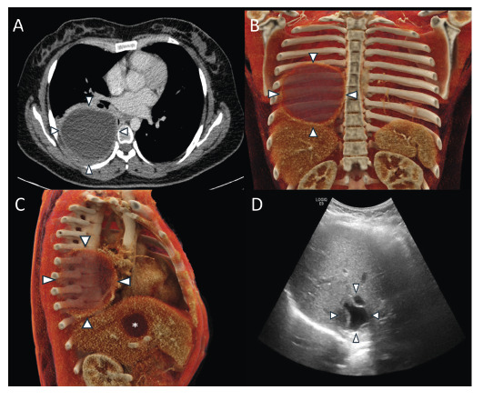 Figure 1
Peer Reviews
No public reviews on file for this paper yet. If you reviewed it on a platform where reviews are public (OpenReview, ICLR, NeurIPS, ICML), you can paste yours below so the community can read it here.
Videos
No videos yet. Explain this paper in a talk, walkthrough, or lecture? Add one.
Taxonomy
TopicsParasitic infections in humans and animals · Amoebic Infections and Treatments · Congenital Anomalies and Fetal Surgery
A 24-year-old woman from Bolivia, residing in Brazil for the past 10 years, presented with intermittent back pain and occasional nonproductive cough. She reported no fever, night sweats, or hemoptysis but described an unintentional weight loss of 3 kg over the preceding months. Multimodal imaging (Figure 1) revealed large well-defined cystic lesions in the right lung and liver. Based on the patient’s clinical and epidemiologic backgrounds and radiologic findings, hydatid disease was suspected and subsequently confirmed by the detection of serum immunoglobulin G antibodies against Echinococcus species. The pulmonary cyst was removed via elective thoracotomy, and histopathological analysis confirmed the diagnosis. Hepatic cyst was treated by percutaneous drainage. The patient recovered uneventfully and remained asymptomatic at 1-year follow-up.
FIGURE 1: (A) Axial contrast-enhanced chest CT shows a large, well-defined cystic lesion in the right lower lobe of the lung (white arrowheads). (B,C) Cinematic volume-rendered reconstructions redemonstrate the lung (white arrowheads) and liver (asterisk) cystic lesions. (D) Abdominal ultrasound showing a well-defined cystic lesion in the right hepatic lobe containing a detached, irregular laminated membrane floating within its contents, consistent with the “water lily sign.”
Hydatid disease, also known as echinococcosis, is a parasitic infection caused by the Echinococcus granulosus tapeworm, which is endemic in parts of South America, Africa, and Asia1 ^,^ 2. Although the disease can be multisystemic, the liver and lungs are the most frequently involved organs2 ^,^ 3. Definitive treatment generally involves surgical resection because of the low complication and recurrence rates. Antiparasitic medications such as albendazole are often used as supplementary therapy, and percutaneous methods may be suitable for selecting hepatic cases. Imaging is crucial for early diagnosis, as it helps prevent complications such as rupture, secondary infection, or spread, and assists in timely organ-preserving interventions, especially in patients from endemic areas, where hydatid disease should remain on the list of differential diagnoses.
The reference list from the paper itself. Each links out to its DOI / PubMed record.
- 1Morar R Feldman C Pulmonary echinococcosis Eur Respir J 2003216106910771279750410.1183/09031936.03.00108403 · doi ↗ · pubmed ↗
- 2Zalaquett E Menias C Garrido F Vargas M Olivares JF Campos D Imaging of hydatid disease with a focus on extrahepatic involvement Radiographics 20173739019232849380110.1148/rg.2017160172 · doi ↗ · pubmed ↗
- 3Durhan G Tan AA Düzgün SA Akkaya S Arıyürek OM Radiological manifestations of thoracic hydatid cysts: pulmonary and extrapulmonary findings Insights Imaging 20201111161163317529510.1186/s 13244-020-00916-0PMC 7658283 · doi ↗ · pubmed ↗