Multifocal Extrapulmonary Tuberculosis Presenting with a Masticatory Space Abscess
Ragıp Afşın Alay, Elif Gözgeç, Alperen Aksakal, Handan Alay

Abstract
Genes, proteins, chemicals, diseases, species, mutations and cell lines named across the full text — each resolved to its canonical identifier and authoritative record.
Click any figure to enlarge with its caption.
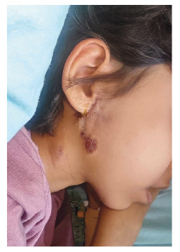 Figure 1
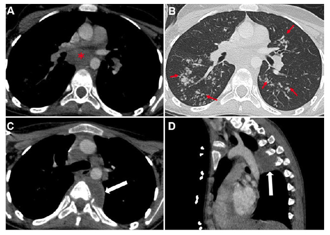 Figure 2
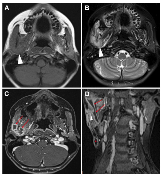 Figure 3
Peer Reviews
No public reviews on file for this paper yet. If you reviewed it on a platform where reviews are public (OpenReview, ICLR, NeurIPS, ICML), you can paste yours below so the community can read it here.
Videos
No videos yet. Explain this paper in a talk, walkthrough, or lecture? Add one.
Taxonomy
TopicsInfectious Diseases and Tuberculosis · Mycobacterium research and diagnosis · IgG4-Related and Inflammatory Diseases
A 14-year-old girl with autism presented with persistent right cervical swelling for eight months. She had undergone multiple abscess drainages and prolonged antibiotic therapy without improvement. Cultures and serologic tests for Francisella tularensis, EBV, CMV, and Brucella were negative. Tuberculosis testing (purified protein derivative [PPD] 9 mm, QuantiFERON negative, gastric aspirate smear, and polymerase chain reaction [PCR] negative) was initially inconclusive. An excisional lymph node biopsy revealed no caseating necrosis and was negative for acid-fast bacilli. Family history revealed maternal pulmonary tuberculosis eight years earlier, without household prophylaxis.
On admission, she reported night sweats, a 5-kg weight loss, and persistent lymphadenopathy. Physical examination showed cervical lymph nodes with a chronic draining fistula (Figure 1). Chest computed tomography (CT) and neck magnetic resonance imaging (MRI) demonstrated necrotic mediastinal lymphadenopathy, parenchymal consolidations, paravertebral rib destruction, and a masticatory space abscess (Figures 2 and 3). Endobronchial ultrasound-guided bronchoalveolar lavage culture yielded Mycobacterium tuberculosis. The patient was treated with rifampicin, pyrazinamide, streptomycin, and linezolid due to isoniazid resistance, resulting in clinical improvement.
FİGURE 1:External draining fistula and scar in the right auricular-submandibular region.
FİGURE 2:Axial non-contrast chest CT, soft tissue window (A), demonstrates necrotic lymphadenopathy (asterisks) in the mediastinum and bud-like branch patterns, as well as patchy consolidated areas (arrows) in the bilateral parenchyma on the parenchymal window (B). Axial (C) and sagittal (D) non-contrast chest CT images show a hypodense lesion causing rib destruction in the left paravertebral region (arrow).
FİGURE 3:Axial MR images show a hypointense appearance on the T1-weighted image (A) and a hyperintense appearance (arrow) on the T2-weighted image (B) in the right masticatory space. Post-contrast T1-weighted axial (C) and coronal (D) sections demonstrate a peripherally contrast-enhancing abscess loculation (red arrows) and a fistula tract (arrowhead) extending to the skin. A central necrotic LAP formation (asterisks) is noted inferior to the lesion.
Extrapulmonary tuberculosis remains challenging to diagnose because clinical samples obtained from relatively inaccessible sites are often paucibacillary, reducing the sensitivity of conventional diagnostic tests1. Persistent cervical lymphadenitis with a draining fistula should prompt repeat tuberculosis evaluation, even when initial PPD, QuantiFERON, culture, and PCR results are negative2. Imaging (MRI/CT) is crucial to delineate abscess tracts, lymph node necrosis, and bone involvement, thereby guiding both diagnosis and management3.
The reference list from the paper itself. Each links out to its DOI / PubMed record.
- 1Lee JY Diagnosis and treatment of extrapulmonary tuberculosis Tuberc Respir Dis Seoul 2015782475510.4046/trd.2015.78.2.47PMC 438890025861336 · doi ↗ · pubmed ↗
- 2Fontanilla JM Barnes A Fordham von Reyn C Current diagnosis and management of peripheral tuberculous lymphadenitis Clin Infect Dis 20115365555622186519210.1093/cid/cir 454 · doi ↗ · pubmed ↗
- 3Pattamapaspong N Kanthawang T Peh WCG Hammami N Bouaziz MC Ladeb MF Imaging of thoracic tuberculosis: pulmonary and extrapulmonary BJR Open 202461 tzae 031tzae 0313936390810.1093/bjro/tzae 031PMC 11449295 · doi ↗ · pubmed ↗