Primary Alveolar Echinococcosis of the Kidney
Hicham Esselmani, Rachid Hnini, Eliane Silva, Mustapha Najimi, Mohamed Merzouki

Abstract
Genes, proteins, chemicals, diseases, species, mutations and cell lines named across the full text — each resolved to its canonical identifier and authoritative record.
Click any figure to enlarge with its caption.
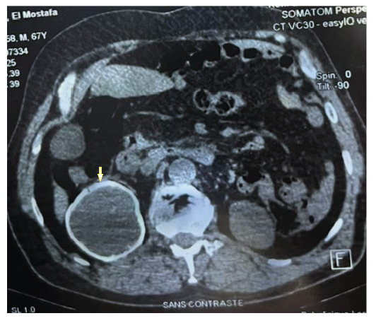 Figure 1
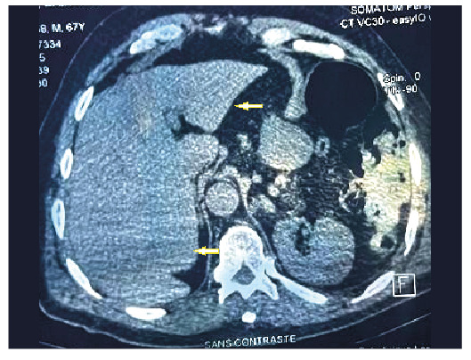 Figure 2
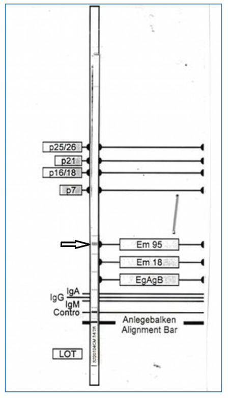 Figure 3
Peer Reviews
No public reviews on file for this paper yet. If you reviewed it on a platform where reviews are public (OpenReview, ICLR, NeurIPS, ICML), you can paste yours below so the community can read it here.
Videos
No videos yet. Explain this paper in a talk, walkthrough, or lecture? Add one.
Taxonomy
TopicsParasitic infections in humans and animals · Urinary and Genital Oncology Studies · Tuberous Sclerosis Complex Research
Alveolar echinococcosis (AE), which is caused by the larval stage of** ** Echinococcus multilocularis , is a rare and aggressive parasitic disease1. Over 95% of cases involve the liver, with primary extrahepatic AE being exceptionally uncommon2. Here, we present a rare case of primary renal AE in a 67-year-old asymptomatic Moroccan man.
The lesion was incidentally detected on thoraco-abdomino-pelvic computed tomography (CT). Abdominal CT revealed a calcified cystic mass in the right kidney, associated with renal atrophy (Figure 1). The liver and other abdominal organs showed no signs of lesions (Figure 2). Serology using an indirect hemagglutination test forE. granulosus yielded a negative result. However, an Anti-Echinococcus EUROLINE-Western Blot IgG assay tested positive for antibodies against theE. multilocularis-specific antigen Em95 (Figure 3), confirming the diagnosis of AE.
FIGURE 1:Abdominal tomography showing a calcified cyst in the right kidney with atrophy.
FIGURE 2:Abdominal tomography showing the liver without cystic or solid lesions.
FIGURE 3:Western blot analysis showing the presence of antibodies against *E. multilocularis-*specific antigen Em95.
To our knowledge, this represents the first case of primary renal AE reported in Morocco and the second described worldwide3. Isolated renal AE is a clinical mimic that can easily be misdiagnosed as a renal tumor based solely on imaging4. Maintaining a high index of suspicion is essential in non-endemic regions and cases with atypical presentations. Specific immunoblotting is a valuable noninvasive tool for distinguishing AE from other cystic renal lesions and for guiding appropriate management, which typically involves a combination of surgery and long-term albendazole therapy4.
The reference list from the paper itself. Each links out to its DOI / PubMed record.
- 1Kodama Y Fujita N Shimizu T Endo H Nambu T Sato N Alveolar echinococcosis: MR findings in the liver Radiology 20032281721771275045910.1148/radiol.2281020323 · doi ↗ · pubmed ↗
- 2Piarroux M Piarroux R Giorgi R Knapp J Bardonnet K Sudre B Clinical features and evolution of alveolar echinococcosis in France from 1982 to 2007: results of a survey in 387 patients J Hepatol 201155102510332135444810.1016/j.jhep.2011.02.018 · doi ↗ · pubmed ↗
- 3Türker Köksal I Tefekli A Kiliçaslan I Erdemir F Kadioğlu T Esen T Hydatid disease of the kidney caused by Echinococcus multilocularis Urol Int 2001673103121174113410.1159/000051009 · doi ↗ · pubmed ↗
- 4Meinel TR Gottstein B Geib V Keel MJ Biral R Mohaupt M Vertebral alveolar echinococcosis-a case report, systematic analysis, and review of the literature Lancet Infect Dis 2018183 e 87-e 982880762810.1016/S 1473-3099(17)30335-3 · doi ↗ · pubmed ↗