Assembly and glycosylation of Helicobacter pylori sheathed flagella
Rajeev Kumar, Shoichi Tachiyama, Huaxin Yu, Samira Heydari, Jiaqi Guo, Jack M Botting, Wangbiao Guo, Timothy R Hoover, Jun Liu

TL;DR
This study reveals how Helicobacter pylori uses glycosylated flagella to move through stomach mucus and colonize the gastric epithelium.
Contribution
The paper presents near-atomic structures of H. pylori flagella and identifies glycosylation's role in filament stability and motility.
Findings
FlaA and FlaB flagellins form a conserved core with variable surface domains.
Pseudaminic acid glycans stabilize the flagellar filament and create a negatively charged surface.
The flagellar filament rotates independently of the membranous sheath to drive motility.
Abstract
The bacterial flagellum is a complex nanomachine essential for motility, colonization, and invasion in diverse species. Helicobacter pylori has evolved elaborate sheathed flagella that enable migration through the highly viscous gastric mucus layer to reach its colonization niche on the gastric epithelium, yet the molecular basis for these unique adaptations has remained elusive. Here, we use in situ single-particle cryo-electron microscopy to determine near-atomic structures of the flagellar filament within the membranous sheath of H. pylori. The major flagellin FlaA constitutes the bulk of the filament, whereas the minor flagellin FlaB contributes critically to the hook-proximal region. Both FlaA and FlaB form a conserved core surrounded by variable surface-exposed domains. Our structures further reveal that pseudaminic acid glycans decorate these domains, where they mediate inter-…
Genes, proteins, chemicals, diseases, species, mutations and cell lines named across the full text — each resolved to its canonical identifier and authoritative record.
Click any figure to enlarge with its caption.
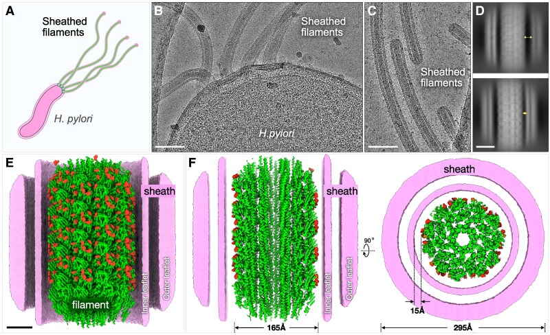 Fig. 1
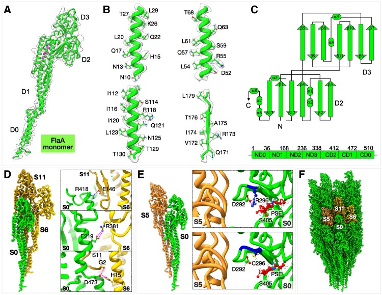 Fig. 2
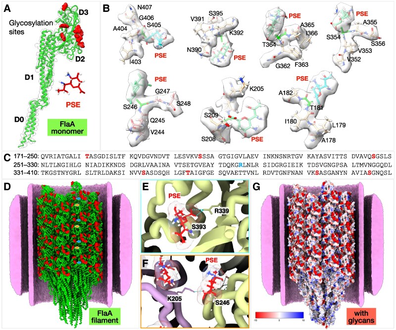 Fig. 3
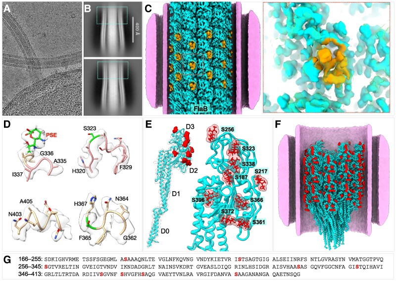 Fig. 4
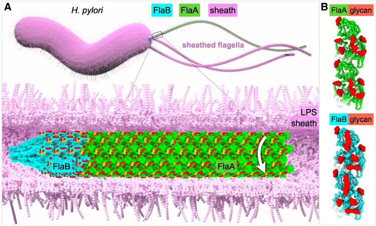 Fig. 5
- —National Institute of Allergy and Infectious Diseases10.13039/100000060
- —National Institutes of Health10.13039/100000002
Peer Reviews
No public reviews on file for this paper yet. If you reviewed it on a platform where reviews are public (OpenReview, ICLR, NeurIPS, ICML), you can paste yours below so the community can read it here.
Videos
No videos yet. Explain this paper in a talk, walkthrough, or lecture? Add one.
Taxonomy
TopicsGlycosylation and Glycoproteins Research · Bacterial Genetics and Biotechnology · Lipid Membrane Structure and Behavior
Introduction
Helicobacter pylori is a gastric bacterium that colonizes approximately half the world's population (1, 2). Having coexisted with humans for at least 50,000 years (3), H. pylori has evolved remarkable strategies to evade the immune system and colonize its preferred niche, the gastric mucosa. Although H. pylori colonization does not cause symptoms in most people, it can trigger chronic stomach inflammation and significantly increase the risk of developing peptic ulcer disease, chronic gastritis, and gastric cancer (4–6). The ability of H. pylori to traverse the gastric mucous layer and colonize the underlying epithelial cell surface is promoted by its characteristic helical shape (7, 8), urease production (9, 10), and robust motility in viscous media (10–12). Helicobacter pylori motility is driven by a bundle of unipolar flagella, each of which is enclosed by a membranous sheath that is continuous with the outer membrane. Although the role of the sheath has yet to be determined experimentally, possible functions include protecting the flagella from dissociating in the gastric acidic environment, facilitating adherence, and avoiding surveillance by the host innate immune system (13).
The bacterial flagellum has been extensively characterized in model organisms such as Escherichia coli and Salmonella enterica. It is composed of three principal parts: the rotary motor, the hook, and the filament (14). The motor is embedded in the cell envelope and is responsible for flagellar assembly and rotation. At least 20 different proteins are involved in building the core components of the motor, including the MS-ring, C-ring, LP-rings, rod, export apparatus, and torque-generating stator units. The hook functions as a universal joint, transmitting torque from the rod to the filament (15, 16). The rotating filament serves as a propeller to drive bacterial motility.
Although the core components of bacterial flagella are broadly conserved, the sheathed flagella of H. pylori have evolved distinctive adaptations that enable motility within and colonization of the gastric mucosa. The H. pylori genome encodes numerous motor accessory proteins that are structurally and functionally distinct from those of enteric bacteria and are essential for motility (17–20). Furthermore, the H. pylori flagellar filament differs from that of enteric species in that it is assembled from two flagellins, FlaA and FlaB, which do not activate Toll-like receptor 5, contributing to immune evasion and persistent colonization (21–23). FlaA constitutes the majority of the filament, whereas the minor flagellin FlaB localizes at the filament base near the hook (24). Consistent with their distinct roles, deletion of flaA results in truncated filaments and severely impaired motility, whereas flaB mutants produce full-length filaments with moderately reduced motility (25). Intriguingly, both FlaA and FlaB undergo extensive O-glycosylation with 5,7-diacetamido-3,5,7,9-tetradeoxy-L-glycero-L-manno-nonulosonic acid (Pse5Ac7Ac), a nine-carbon pseudaminic acid derivative structurally related to the sialic acid Neu5Ac found on human cell surfaces. Disruption of flagellin glycosylation abolishes filament assembly and motility in H. pylori (26) ). Although the precise functional role of glycosylation remains incompletely understood, it has been proposed to stabilize inter-subunit interactions during filament assembly (27).
Here, we deploy in situ single-particle cryo-electron microscopy (cryo-EM) to determine near-atomic structures of the sheathed flagellar filament in H. pylori, revealing how two flagellins, FlaA and FlaB, assemble the filament through highly conserved domains that form in the central core of the filament and variable domains located on the surface of the filament. We further identify residues within the variable domains of FlaA and FlaB that are glycosylated, as well as protein–ligand interactions that likely promote filament assembly and stability.
Results
High-resolution in situ structures of the sheathed flagellar filament
FlaA is the main component of the filament, whereas FlaB is a minor flagellin found near the hook (24). To elucidate the unique adaptations of the sheathed flagellum in H. pylori (Fig. 1A), we employed an in situ single-particle cryo-EM approach to determine the filament structures directly from frozen-hydrated bacteria, without filament isolation or purification (Fig. 1). Using cryoSPARC (28), filaments were first automatically selected and processed to generate 2D class averages (Figs. 1D and S1). The majority of 2D classes display an intact bilayer membranous sheath surrounding the filament, with filaments lacking the sheath also observed (Figs. 1D and S1A). The spacing between the filament and sheath varies from ∼1 to 4 nm for the majority of 2D classes (Figs. 1D and S1A). Filaments enclosed by the sheath were selected for subsequent helical reconstruction and refinement, yielding a 2.65 Å resolution structure of the sheathed filament (Figs. 1E, F, S1B–D, and S2A–C). To determine the filament structures near the filament tip and the hook, we manually selected filament segments and determined their in situ structures at 3.19 and 3.22 Å resolution, respectively (Fig. S2). All three filament structures exhibit nearly identical helical parameters: a helical twist of 65.40° and a rise of 4.68 Å (Table S1).
In situ cryo-EM structure of the sheathed flagellar filament at near-atomic resolution. A) A schematic model of a H. pylori cell with multiple sheathed flagella. B) A typical cryo-EM micrograph collected from a cell pole containing multiple sheathed flagella. Scale bar is 100 nm. C) A typical cryo-EM micrograph collected from the filament tips. D) 2D class averages of the flagellar filaments show variable distance (1–4 nm) between the flagellar filament and the membranous sheath. Scale bar is 10 nm. E) A surface rendering of the flagellar filament structure at 2.65 Å resolution with a helical symmetry of twist 65.40° and rise 4.68 Å. A cut-through view of the membranous sheath enables a better view of the filament, the inner and outer leaflets of the membranous sheath. The filament is colored in green and glycans are colored in red. Scale bar is 4 nm. F) Left panel: A cross-sectional view of the filament shows the filament core. The diameter of the filament is 165 Å. The averaged diameter of the membranous sheath is 295 Å. Right panel: A top view of the sheathed flagella shows 11 protofilaments and the surrounding membranous sheath.
Consistent with flagellar filaments of S. enterica and other bacteria (29, 30), the H. pylori flagellar filament forms an 11-protofilament tubular structure composed of helical arrays of flagellin subunits (Fig. 1E–G). The filament diameter is ∼165 Å, and the central lumen measures ∼28 Å in diameter (Figs. 1F, S1, and S2). Including the surrounding membranous sheath, the total diameter of the sheathed flagellum reaches ∼295 Å (Fig. 1F). Importantly, our in situ structures at near-atomic resolution afforded us the opportunity to analyze unique features of the sheathed flagellar filament in intact bacteria.
The in situ structure of the major flagellin FlaA
Two flagellins, FlaA and FlaB, share 58% amino acid identity and 75% similarity across their entire lengths (24) (Fig. S3A). AlphaFold-predicted flagellin structures show a canonical feature where a single-flagellin peptide folds to form a helical coiled-coil structure and beta sheets and can be classified into the typical four domains: D0, D1, D2, and D3 (Fig. S3B) (31, 32). Domains D0 and D1 are highly conserved, whereas the surface-exposed domains are more divergent (Fig. S3C). To test which flagellin is present in our maps near the tip and middle regions of the filament, we fit the predicted FlaA and FlaB models into our cryo-EM density maps, followed by manual model building in Coot (33) and refinement in Phenix (34). Our data showed that the refined FlaA model fit well into the densities corresponding to the tip and central regions of the filament, while the FlaB model did not (Fig. S4A and B). Specifically, residues Arg-23, Ser-26, Arg-66, Thr-237, and Arg-294 in FlaB did not fit the density appropriately, whereas the corresponding FlaA residues—Asn-23, Lys-26, Ala-66, Arg-227, and Glu-284—aligned well with the map (Fig. S4A and B).
Our cryo-EM structures confirm that FlaA consists of four major domains: D0, D1, D2, and D3 (Fig. 2A–C). The D0 domain is composed of ND0 (residues 1–36) and CD0 (residues 472–510), and D1 includes ND1 (residues 37–167) and CD1 (residues 412–472) (Fig. 2C). Assembly of FlaA subunits within the filament revealed that Arg-381 and Glu-219 form bifurcated hydrogen bonds, while Glu-146 and Arg-418 engage in inter-chain hydrogen bonding with neighboring protomers (Fig. 2D). These interactions form an extensive hydrogen-bond network that is also found in other bacterial filaments and is critical for filament assembly and structural stability (Fig. 2D) (30, 35).
Structure of major flagellin FlaA from H. pylori. A) Refined model of the FlaA monomer fitted into the cryo-EM density map. B) Representative density fittings for the D0 and D2 domains of FlaA. C) Schematic representation of the D2 and D3 domains (top) and linear arrangement of the four FlaA domains (bottom). Domains D2 and D3 adopt immunoglobulin-like folds. D) Interactions between adjacent FlaA monomers (S0, S6, and S11). The right panels highlight key residues involved in hydrogen-bond interactions; left, side view of the filament model; right, top view. E) Point mutation in domain D3 of H. pylori B128 FlaA (Cys-296→Arg-296, blue). The top panel shows Arg-296 forming a hydrogen bond with Asp-292, whereas no such interaction occurs in the wild-type Cys-296 (bottom). F) Side view of the FlaA filament. Three adjacent FlaA monomers (S5, S6, and S11) are colored in orange.
The in situ structure of FlaA further reveals that residue 296 is an arginine (Figs. 2E and S5A), which is different from a cysteine in the original sequence of H. pylori B128 FlaA (36). DNA sequencing of flaA from the H. pylori B128 strain used in this study confirmed that codon 296 encodes arginine (CGC) rather than cysteine (TGC) (Fig. S5A). Comparative analysis of FlaA sequences from different H. pylori strains—including 26695, J99, SS1, G27, and ATCC 43504—showed that all possess arginine at position 296 (Fig. S5B). Notably, the H. pylori 7.13 strain, a mouse-adapted derivative of B128, also contains Arg-296 and exhibits a significantly larger swim halo in soft-agar assays than its parental strain (36). Although it remains unclear whether the Cys-to-Arg substitution contributes to enhanced motility of H. pylori 7.13 compared with its parental strain, our in situ structure indicates that Arg-296 forms a hydrogen bond with Asp-292 (Fig. 2E), potentially contributing to filament stability and function.
Distinct flagellin surface glycosylation sites in H. pylori
Post-translational modifications of bacterial flagellins, particularly glycosylation, are critical for motility in many bacterial species (35, 37). Mass-spectrometric analysis of FlaA from H. pylori strain 1061 previously identified glycosylation by pseudaminic acid (Pse5Ac7Ac) at seven serine and threonine residues within the D2 and D3 domains (26). Consistent with these findings, fitting the refined FlaA model into the in situ cryo-EM density map derived from middle regions of the filament revealed additional densities at seven sites near specific serine and threonine residues on the surface of the variable D2 and D3 domains (Fig. 3A and B). Five of these glycosylated residues—Thr-181, Ser-246, Ser-354, Thr-364, and Ser-405—match those identified by mass spectrometry (Fig. S6A and B) (26). The remaining two glycosylated residues, Ser-207 and Ser-393, are adjacent to previously reported sites (Ser-208 and Ser-395; Figs. 3B and S6B). This discrepancy likely does not result from sequence heterogeneity, as the amino acid sequences surrounding Ser-207 and Ser-393 are identical between FlaA from H. pylori strains B128 and 1061 (used in the mass-spectrometric analysis; Fig. S6B).
The FlaA filament surface is extensively decorated with glycans. A) The FlaA monomer contains seven glycosylation sites located on domains D2 and D3. B) Model of Pse5Ac7Ac fitted into glycan densities, showing O-linked glycosylation via serine and threonine residues. C) Sequence of the FlaA D2 and D3 domains, with glycosylation sites colored in red and Arg-296 in blue. D) Ribbon model of the FlaA filament showing extensive glycan densities on the filament surface (red blobs). Representative image showing formation of H-bond by Pse5Ac7Ac within the same protofilament (E) and with the neighboring protofilament (F). G) Electrostatic surface representations reveal an overall negative surface charge of the H. pylori flagellar filament, primarily due to glycosylation.
The in situ structure further revealed that several Pse5Ac7Ac glycans form hydrogen bonds with neighboring amino acid side chains, suggesting a stabilizing role in filament architecture (Fig. S6A). For example, the glycan attached to Ser-393 in D2 forms hydrogen bonds with Asn-407, Asn-398, and Arg-339, while the glycan at Ser-246 in D3 forms a hydrogen bond with Lys-205 in D2 of an adjacent protofilament (Fig. S6C and D). These interactions likely contribute to filament stability and inter-subunit packing. Importantly, the glycan decorations render an overall negative surface charge to the filament (Fig. 3F). Two of the major phospholipids in H. pylori, cardiolipin and phosphatidylglycerol (38), are anionic phospholipids, and the negatively charged surface of the filament imparted by the glycan decorations may electrostatically repel these negatively charged phospholipids within the inner leaflet in the membranous sheath.
FlaB structure and distribution in H. pylori
We further determined a cryo-EM structure derived from the filaments near the hook region and built a FlaB model (Figs. 4A–F and S2G–I). The overall domain organization of FlaB resembles that of FlaA, comprising four domains (D0–D3; Fig. 4B). Although the D0 and D1 domains of FlaA and FlaB share high structural similarity, distinct differences were observed within the D2 and D3 domains, which constitute the variable, surface-exposed regions (Fig. 4E).
Structure of FlaB and its distinct glycosylation sites. A) Cryo-EM image from the cell tip showing the FlaB filament near the hook region. B) Representative 2D class averages from near-hook regions. The blue boxes are the FlaB filament regions. C) Surface rendering of the FlaB filament structure. The orange region shows a distinct loop in FlaB. D) Representative density fittings of the D2 and D3 domains of FlaB with glycans. E) Cryo-EM structure reveals nine glycosylation sites located on domains D2 and D3 of FlaB. F) Ribbon model of the FlaB filament showing extensive glycan densities on the filament surface (red blobs). G) Sequence of FlaB D2–D3 domains, with glycans colored in red.
Notably, the D2 and D3 domains of FlaB contain clear densities corresponding to nine O-linked glycosylation sites modified by Pse5Ac7Ac (Figs. 4E and S7). Earlier work identified ten Pse5Ac7Ac-modified sites in FlaB by mass spectrometry (26) though the precise positions could not be determined due to the predominance of FlaA in those samples. The glycosylated tryptic peptides and number of attached Pse5Ac7Ac units, however, were quantified. Except for one discrepancy, the number of glycosylation sites resolved in our in situ FlaB structure closely matches that inferred from the mass-spectrometric data. The difference involves the peptide spanning Met-173 to Lys-200, which was previously reported to contain two Pse5Ac7Ac units (26) but in our cryo-EM map only a single glycosylation site (Ser-187) was observed (Fig. 4E).
Four of the FlaB glycosylation sites lie within regions of greater sequence homology to FlaA than to the rest of the D2 and D3 domains (Fig. S7A–C). While FlaA and FlaB share 43% identity and 66% similarity across the entire D2/D3 domains, the regions surrounding their common glycosylation sites exhibit 58% shared identity and 84% similarity (Fig. S7A). Structural alignment of the D2 and D3 domains of FlaA and FlaB yielded an RMSD of 0.513 Å, indicating strong structural conservation despite sequence divergence (Fig. S7D).
Overall, our near-atomic in situ cryo-EM structure of FlaB defines its domain architecture and pinpoints glycosylation sites, highlighting the power of in situ single-particle cryo-EM in resolving post-translational modifications in complex assemblies.
Discussion
Using in situ single-particle cryo-EM, we determined near-atomic structures of the sheathed flagellar filament, revealing molecular adaptations that underlie the unique motility and persistent infection of H. pylori. FlaA forms the bulk of the filament, while FlaB localizes proximally near the hook, consistent with previous observations (24). Both flagellins share conserved D0/D1 domains and variable D2/D3 domains (Figs. 2 and 4). The D0/D1 domains form the filament core and the central channel for unfolded subunit secretion (39), whereas the D2/D3 domains comprise the sheath-proximal outer surface (Figs. 3A and 4A).
Our in situ structures identify these glycosylation sites (Figs. 3–5) and reveal Pse5Ac7Ac-mediated hydrogen bonding that stabilizes the filament. In FlaA, Ser-207 and Ser-393 are glycosylated, differing slightly from the predicted Ser-208 and Ser-395; this discrepancy likely reflects peptide overlap during mass-spectrometric analysis. In FlaB, nine glycosylation sites were observed, compared with 10 previously predicted (26). This difference may stem from strain-specific variation in deglycosylation by Cds6 (HP0518), an L,d-carboxypeptidase implicated in peptidoglycan trimming and cell-shape control (40, 41). Flagellins from a cds6 mutant are hyper-glycosylated, containing roughly 3-fold more Pse5Ac7Ac than those from wild type (37). These results suggest that FlaA and FlaB are glycosylated in the cytoplasm before export, after which Cds6 removes excess Pse5Ac7Ac units. Strain-dependent Cds6 activity may therefore explain variations reported for the number of Pse5Ac7Ac units associated with FlaB in the previous mass-spectrometric analysis (25) and the in situ structure of FlaB.
Model of assembly and rotation of the sheathed flagellar filament. A) Top: Model of an H. pylori cell possessing multiple sheathed flagella, each comprising a motor, hook, and filament. The filament consists primarily of the major flagellin FlaA, with the minor flagellin FlaB located at the hook-proximal region. Bottom: Atomic models of the FlaB and FlaA filaments, both extensively covered by glycans. The white arrow indicates that the filament could rotate independently within the surrounding membranous sheath. B) Comparison of distinct surface glycans decorating FlaA and FlaB.
We hypothesize that flagellin glycosylation directly contributes to filament–sheath dynamics. Decoration of FlaA and FlaB with Pse5Ac7Ac introduces a dense array of negative charges on the filament surface, which likely repels the negatively charged phospholipid head groups of the inner leaflet of the membranous sheath (Fig. 5). This electrostatic repulsion supports a model in which the flagellar filament rotates independently from the surrounding membranous sheath (42). Consistent with this model, 2D class averages reveal that the sheath is not tightly bound to the filament but separated by a variable gap (Figs. 1D and S1A). Comparable electrostatic interactions were recently observed in the Vibrio cholerae sheathed flagellum (43), although the filament–sheath spacing is substantially narrower than in H. pylori (Fig. S8). Interestingly, unlike H. pylori, V. cholerae flagellins are not glycosylated, but the filament surface is enriched in acidic residues (PDB 9N8B), suggesting that many bacterial species have evolved distinct strategies to generate a negatively charged, hydrophilic flagellar filament surface for optimal rotation against the surrounding membranous sheath.
An intriguing question is why does H. pylori employ two flagellin species to assemble the flagellar filament? H. pylori FlaB is dispensable for the formation of functional flagellar filaments, although the H. pylori flaB mutant displays reduced motility compared with wild type (25). It is possible that the higher number of glycosylation sites in FlaB than in FlaA increases the stability of the filament, allowing H. pylori to better navigate viscous environments. A recent study that generated a cryo-EM structure of the S. enterica FlgK–FlgL hook–filament junction proposed that the hook–filament junction protects the filament from mechanical stress emanating from the flexible hook (44). The portion of the H. pylori flagellar filament that is formed by FlaB may be more resistant to any mechanical stress not buffered by the hook–filament junction and thereby help stabilize the filament. Further analysis of H. pylori FlaB and its relationships with FlaA and hook–filament junction proteins will be needed to understand the specific roles of FlaB in the assembly and function of the flagellar filament, as well as the role of glycosylation in FlaB function.
In summary, our in situ near-atomic structures of the H. pylori flagellar filament reveal a highly specialized motility apparatus. Similar to many flagellar filaments from different bacterial species, the H. pylori filament is stabilized by a conserved, canonical hydrogen-bonding network and inter-domain contacts. Unique to H. pylori, extensive Pse5Ac7Ac glycosylation reinforces filament integrity, modulates electrostatic properties, and supports a model of independent filament rotation within a flexible sheath. Together, these unique properties of the H. pylori flagellar filament likely equip the bacterium to endure the harsh gastric environment and sustain persistent colonization.
Materials and methods
Bacterial culture preparation
Helicobacter pylori B128 was grown on tryptic soy agar (TSA) plate with 30 μg/mL of kanamycin and 5% heat-inactivated horse serum at 37 °C in 10% CO_2_ atmosphere for 2 days. Then, the strain was inoculated and grown in Brucella broth with the antibiotics and 10% heat-inactivated fetal bovine serum at 37 °C with shaking in a reduced O_2_ condition using CampyGen 2.5 L for the overnight. The next day, the overnight bacterial culture was inoculated into a fresh medium and grown for 4 h. The bacterial culture was centrifuged at 1,500 × g for 10 min, and bacterial pellets were resuspended into phosphate-buffered saline at pH 7.4 and adjusted to OD_600_ of 0.8.
Cryo-EM sample preparation
To prepare the sample for the single-particle cryo-EM experiment, 5 μL of the bacterial sample was deposited on fresh glow-discharged cryo-EM grids (Quantifoil, Cu, 200 mesh). Grids were blotted for 6–8 s under 90% humidity and rapidly plunge-frozen in a liquid propane–ethane mixture using a GP2 plunger (Leica).
Single-particle cryo-EM data collection
The grids of frozen-hydrated specimens were loaded into a Titan Krios electron microscope (Thermo Fisher Scientific) equipped with a field emission gun, K3 summit detector, and GIF BioQuantum imaging filter (Gatan). SerialEM (45) was used to collect data from bacterial flagellar filaments at magnification of 81,000× with a physical pixel size of 1.068 Å and defocus ranging from −1.0 to −2.0 μm. In total, 50,562 movies were collected in dose fraction mode with a dose of 73 e⁻/Å^2^ distributed over 40 frames per movie.
Cryo-EM image processing and 3D structure reconstruction
All data processing was performed using CryoSPARC (28). Beam-induced image drift was corrected using patch motion correction, and the contrast transfer function (CTF) was estimated with patch CTF estimation. Filament tracer was utilized to select 1,070,759 particles, initially extracted from 4× binned images (4.272 Å/pixel), followed by 2D classification to remove nonfilament particles. Selected particles were then re-extracted at the original pixel size (1.068 Å/pixel) and used for ab initio reconstruction to create an initial model for iterative heterogeneous refinement, further eliminating low-quality particles. These particles underwent iterative nonuniform refinement without imposed symmetry, followed by local refinement, achieving a 3.26 Å resolution reconstruction. A symmetry search determined helical parameters with a rise of 4.675 Å and a twist of 65.45°. Using 228,410 high-quality particles, helical refinement produced a 3D reconstruction at 2.80 Å resolution without cyclic symmetry, and final nonuniform refinement yielded a map at 2.65 Å resolution (later identified as FlaA through modeling). From the initial 2D classification, a filament-like class with a varying diameter was noted and attributed to the filament–hook junction. The volume alignment tool shifted particle centers to the near-pole flagellar filament after determining a 3D structure through homogeneous refinement. The resulting 60,029 particles were re-extracted at the original pixel size (1.068 Å/pixel), and subsequent homogeneous and helical refinements resolved the structure at 3.22 Å resolution without cyclic symmetry (later identified as FlaB through modeling), sharing the same helical parameters as FlaA with a rise of 4.675 Å and a twist of 65.45°.
A total of 2,830 particles from the flagellar tip were manually picked and extracted from 2× binned images (2.136 Å/pixel) for ab initio reconstruction to generate initial models. Nonuniform refinement was performed, followed by symmetry expansion using parameters determined for FlaA and FlaB, yielding 30,715 particles. The FlaA density map was imported, low-pass filtered to 10 Å and used as the reference volume for multiple rounds of local refinement. Particle centers were shifted along the flagellar tip at 48.06 Å intervals using Volume Alignment Tools. The resulting 275,229 particles were re-extracted at the original pixel size (1.068 Å/pixel) and subjected to additional rounds of local refinement, achieving a final resolution of 3.19 Å.
Model building and refinement
Helicobacter pylori flagella consist of two flagellins, FlaA and FlaB (24). We retrieved the FlaA and FlaB flagellin sequences of H. pylori B128 from JGI IMG database (46) (IMG ID 2877310586 and 2877311207 for FlaA and FlaB, respectively). We used these sequences to generate AlphaFold models (31, 32). The AlphaFold monomer structures for FlaA were initially docked into the cryo-EM density map using UCSF ChimeraX (47). This docked model was manually fitted in Coot (33) for the mismatched residue and then real space refined using Phenix (34). This step was repeated until the best-fitted model was generated and the model quality was validated by Molprobity (48). A similar approach was used to build an atomic model for flagellin FlaB. Although the overall structures of FlaA and FlaB are similar, the difference lies in the surface-exposed loop region (Fig. S7D). This minor loop difference was prominent enough to distinguish between FlaA and FlaB. Our next step was to build the complete filament structure fragment for each density map. Hence, we docked the refined FlaA and FlaB monomer model against the cryo-EM density maps in a stepwise manner (i.e. one at a time), followed by real space refinement of flagellin structures in Phenix and model building in Coot. The final filament structure was achieved by real space refinement in Phenix (34), and the model was validated in Molprobity (48). Similar protocol was followed for each map corresponding to the tip area, the middle area, and the near-hook area. The refinement statistics are provided in Table S1. UCSF ChimeraX (47) was used to visualize the cryo-EM maps and the atomic models.
Sequencing of B128 flaA
Genomic DNA was prepared from the B128 strain using the QIAamp DNA kit (Qiagen). PCR primer pair 5′-ATGGCTTTTCAGGTCAATACAAATATCAATGC-3′ (flaA-F) and 5′-CTAAGTTAAAAGCCTTAAGATATTTTGTTGAACGG-3′ (flaA-R) together with Q5 high-fidelity DNA polymerase (NEB) were used to amplify the entire coding region of flaA from the H. pylori B128 gDNA. The resulting amplicon was submitted to the Keck DNA Sequencing Facility (Yale University) for Sanger sequencing using the flaA-F and flaA-R primers.
Supplementary Material
pgag011_Supplementary_Data
The reference list from the paper itself. Each links out to its DOI / PubMed record.
- 1Amieva MR, El-Omar EM. 2008. Host-bacterial interactions in Helicobacter pylori infection. Gastroenterology. 134:306–323.18166359 10.1053/j.gastro.2007.11.009 · doi ↗ · pubmed ↗
- 2Atherton JC, Blaser MJ. 2009. Coadaptation of Helicobacter pylori and humans: ancient history, modern implications. J Clin Invest. 119:2475–2487.19729845 10.1172/JCI 38605 PMC 2735910 · doi ↗ · pubmed ↗
- 3Linz B, et al 2007. An African origin for the intimate association between humans and Helicobacter pylori. Nature. 445:915–918.17287725 10.1038/nature 05562 PMC 1847463 · doi ↗ · pubmed ↗
- 4Blaser MJ . 1993. Helicobacter pylori: microbiology of a ‘slow’ bacterial infection. Trends Microbiol. 1:255–260.8162405 10.1016/0966-842x(93)90047-u · doi ↗ · pubmed ↗
- 5Cover TL, Blaser MJ. 1992. Helicobacter pylori and gastroduodenal disease. Annu Rev Med. 43:135–145.1580578 10.1146/annurev.me.43.020192.001031 · doi ↗ · pubmed ↗
- 6Kuipers EJ . 1997. Helicobacter pylori and the risk and management of associated diseases: gastritis, ulcer disease, atrophic gastritis and gastric cancer. Aliment Pharmacol Ther. 11 Suppl 1:71–88.9146793 10.1046/j.1365-2036.11.s 1.5.x · doi ↗ · pubmed ↗
- 7Bonis M, Ecobichon C, Guadagnini S, Prévost MC, Boneca IG. 2010. A M 23B family metallopeptidase of Helicobacter pylori required for cell shape, pole formation and virulence. Mol Microbiol. 78:809–819.20815828 10.1111/j.1365-2958.2010.07383.x · doi ↗ · pubmed ↗
- 8Sycuro LK, et al 2010. Peptidoglycan crosslinking relaxation promotes Helicobacter pylori's helical shape and stomach colonization. Cell. 141:822–833.20510929 10.1016/j.cell.2010.03.046PMC 2920535 · doi ↗ · pubmed ↗