CT Diagnosis of Tibial Post Fracture in a Posterior-Stabilized Total Knee Arthroplasty
Alexander Cotza, Filip Robijns, Jan Vandevenne

TL;DR
This paper presents a case where CT scans helped diagnose a rare tibial post fracture in a knee replacement that was missed on regular X-rays.
Contribution
The study highlights the use of CT attenuation measurements to identify polyethylene fractures in knee implants.
Findings
CT demonstrated a displaced polyethylene fragment with low attenuation values.
Intraoperative confirmation and polyethylene exchange led to a good clinical outcome.
Abstract
Fracture of the polyethylene tibial post is a rare complication of posterior-stabilized total knee arthroplasty and is frequently missed on conventional radiography due to the radiolucent nature of polyethylene. We report a case of post-traumatic instability in which computed tomography demonstrated a displaced polyethylene fragment with characteristic low attenuation values. The diagnosis was confirmed intraoperatively, and isolated polyethylene exchange resulted in a good clinical outcome. Teaching point: CT attenuation measurements can aid in identifying displaced polyethylene fragments and facilitate the diagnosis of tibial post fracture in posterior-stabilized knee arthroplasty.
Genes, proteins, chemicals, diseases, species, mutations and cell lines named across the full text — each resolved to its canonical identifier and authoritative record.
Click any figure to enlarge with its caption.
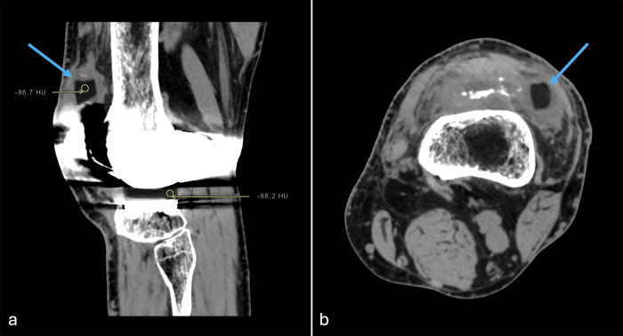 Figure 1
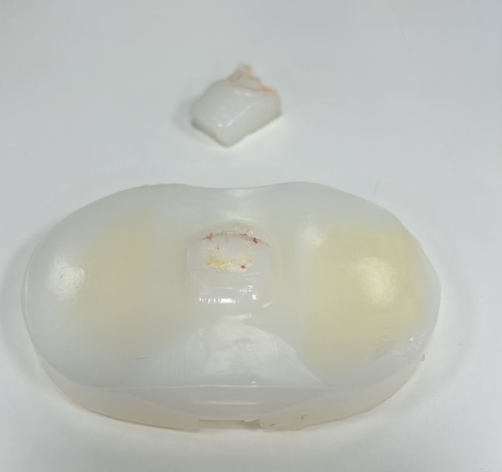 Figure 2
Peer Reviews
No public reviews on file for this paper yet. If you reviewed it on a platform where reviews are public (OpenReview, ICLR, NeurIPS, ICML), you can paste yours below so the community can read it here.
Videos
No videos yet. Explain this paper in a talk, walkthrough, or lecture? Add one.
Taxonomy
TopicsTotal Knee Arthroplasty Outcomes · Orthopedic Infections and Treatments · Orthopaedic implants and arthroplasty
Introduction
Posterior-stabilized total knee arthroplasty (PS-TKA) relies on a cam–post mechanism to substitute for the posterior cruciate ligament and provide posterior stability during knee flexion [1]. Fracture of the polyethylene tibial post is an uncommon but recognized complication and may result in acute instability, pain, mechanical symptoms, and joint effusion [2, 3].
Diagnosis is challenging, as polyethylene is radiolucent on conventional radiographs and radiographic findings may be subtle or overlooked [1, 4]. Arthroscopy, MRI, and CT arthrography have been described as diagnostic tools, whereas the role of standard CT with attenuation analysis remains underreported [1]. We present a case in which CT with Hounsfield unit (HU) measurements enabled the identification of a displaced polyethylene fragment following trauma.
Case Report
A male patient with a PS-TKA of the left knee presented with new-onset instability nine years after implantation. Symptoms developed after a traffic accident. Before the trauma, the patient reported satisfactory prosthetic function, although intermittent joint effusions had occurred.
After the accident, he complained of subjective instability, recurrent hydrops, and an audible clicking sensation. Orthopedic evaluation raised suspicion of mechanical failure of the prosthesis.
During clinical stability testing at orthopedic consultation, the absence of a posterior stop was noted, indicating failure of the cam–post mechanism. Joint aspiration yielded approximately 50 mL of clear, mildly hemorrhagic synovial fluid. Microbiological cultures were negative, excluding infection.
CT of the left knee was performed. Following targeted evaluation prompted by clinical findings, CT demonstrated a well-defined cylindrical structure located in the lateral recess of the suprapatellar bursa (Figure 1). This structure showed homogeneous low attenuation values of approximately −86 HU, identical to those measured in the intact polyethylene tibial insert. No fracture of metallic components or periprosthetic bone injury was identified. These findings were consistent with a displaced polyethylene fragment, suggestive of fracture of the tibial post.
CT images displayed in a soft-tissue window demonstrating a displaced polyethylene tibial post fragment (blue arrows).(a) Sagittal image showing region-of-interest measurements within the displaced fragment and the intact tibial insert, revealing similar low attenuation values (approximately −86 HU), consistent with polyethylene.(b) Axial image demonstrating the well-defined displaced fragment in the lateral suprapatellar region.
Revision surgery was subsequently performed. Intraoperatively, fracture of the polyethylene tibial post was confirmed (Figure 2). The fractured fragment was retrieved from the superolateral suprapatellar region, corresponding to the CT findings. An isolated polyethylene insert exchange was performed. Postoperatively, the patient reported marked improvement in stability, with progressive recovery of range of motion.
Retrieved polyethylene tibial insert demonstrating fracture of the tibial post, which was confirmed intraoperatively during revision surgery.
Discussion
Fracture of the polyethylene tibial post is a rare but clinically significant complication of PS-TKA [2–4]. Clinical presentation is often nonspecific and may include pain, instability, recurrent effusion, clicking, or locking [3, 5]. Trauma, hyperextension, and repetitive impingement of the cam–post mechanism have been proposed as contributing factors [2, 3].
Radiological diagnosis is challenging. Polyethylene components are radiolucent on conventional radiographs, and fractures may be overlooked or misinterpreted [1, 4]. Previous reports have emphasized arthroscopy, MRI, or CT arthrography for diagnosis of tibial post fractures [1]. Hsu et al. demonstrated that CT arthrography can outline fractured polyethylene fragments by contrast delineation [1].
In the present case, standard CT without intra-articular contrast was sufficient to identify the displaced fragment. Measurement of attenuation values was instrumental in characterizing the lesion as polyethylene, as the HU values matched those of the intact insert.
Early diagnosis is essential, as delayed recognition may lead to progressive damage to surrounding soft tissues or metallic components [4]. When other prosthetic components remain well fixed, isolated polyethylene exchange represents an effective treatment option [1, 5].
Conclusion
Fracture of the polyethylene tibial post should be considered in patients with PS-TKA presenting with post-traumatic instability and recurrent effusion. Standard CT, combined with careful image review and HU analysis, can identify displaced polyethylene fragments and facilitate timely diagnosis.
The reference list from the paper itself. Each links out to its DOI / PubMed record.
- 1Hsu Y, Lin CH, Shu GHF, Hsieh TJ, Chen CK. Fracture of the polyethylene tibial post in the posterior-stabilized total knee prosthesis: Arthrographic and CT arthrographic diagnosis. Skeletal Radiol. 2019;48(7):1145–1148. 10.1007/s 00256-019-03173-5. Epub 2019 Feb 7. PMID: .30729263 · doi ↗ · pubmed ↗
- 2Chiu YS, Chen WM, Huang CK, Chiang CC, Chen TH. Fracture of the polyethylene tibial post in a Nex Gen posterior-stabilized knee prosthesis. J Arthroplasty. 2004;19(8):1045–1049. 10.1016/j.arth.2004.04.013. PMID: .15586342 · doi ↗ · pubmed ↗
- 3Kumar N, Yadav C, Raj R, Yadav S. Fracture of the polyethylene tibial post in a posterior stabilized knee prosthesis: A case report and review of literature. J Orthop. 2015;12(3):160–163. 10.1016/j.jor.2015.01.002. PMID: ; PMCID: .26236121 PMC 4501518 · doi ↗ · pubmed ↗
- 4Lanting BA, Mc Calden RW, Naudie DD. Dislocated polyethylene inserts in fixed-bearing total knee arthroplasty. J Arthroplasty. 2011;26(2):339.e 1–339.e 4. 10.1016/j.arth.2009.11.014. Epub 2010 Dec 18. PMID: .21168304 · doi ↗ · pubmed ↗
- 5Lachiewicz PF. How to treat a tibial post fracture in total knee arthroplasty? A systematic review. Clin Orthop Relat Res. 2011;469(6):1709–1715. 10.1007/s 11999-010-1609-9. Epub 2010 Oct 21. PMID: ; PMCID: .20963534 PMC 3094630 · doi ↗ · pubmed ↗