Three-dimensional finite element analysis of tooth and implant-supported telescopic prosthesis using zirconia, PEKK, and cobalt chromium crowns
Sarah Zaman Sahib Awad, Khloud Ezzat Mourad, Ahmed Sameh, Ahmed Heji Albaqawi, Aisha Zakaria Hashem Mostafa

TL;DR
This study used 3D modeling to compare how different crown materials affect stress in tooth-implant-supported dental prostheses.
Contribution
The study introduces a 3D FEA comparison of zirconia, PEKK, and Co-Cr materials in telescopic prosthesis configurations.
Findings
PEKK as a secondary crown reduces stress in prosthetic components but increases stress on supporting structures.
Zirconia and Co-Cr showed similar biomechanical behavior across all configurations.
Secondary crowns consistently showed higher stress than primary crowns, and implants experienced greater loads than natural teeth.
Abstract
This study aimed to evaluate the biomechanical behavior and stress distribution of tooth–implant-supported telescopic prostheses using different combinations of zirconia, polyetherketoneketone (PEKK), and cobalt–chromium (Co-Cr) materials for primary and secondary crowns by means of three-dimensional finite element analysis (3D FEA). A three-dimensional finite element model representing a mandibular arch restored with a telescopic overdenture supported by two canines and two implants in the molar region was constructed. Nine prosthetic configurations were analyzed based on different primary and secondary crown material combinations (zirconia, PEKK, and Co-Cr). All materials were assumed to be homogeneous, isotropic, and linearly elastic. Static axial occlusal loads simulating centric occlusion were applied. Von Mises stress distribution was evaluated in prosthetic components, implants,…
Genes, proteins, chemicals, diseases, species, mutations and cell lines named across the full text — each resolved to its canonical identifier and authoritative record.
Click any figure to enlarge with its caption.
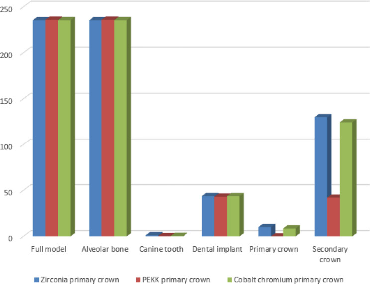 Figure 10
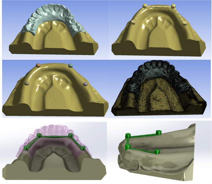 Figure 1
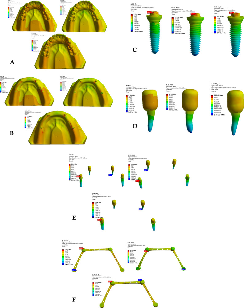 Figure 2
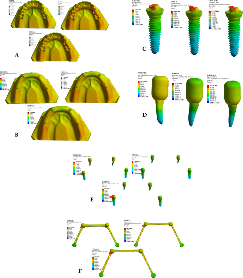 Figure 3
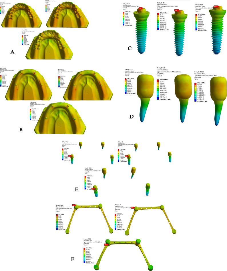 Figure 4
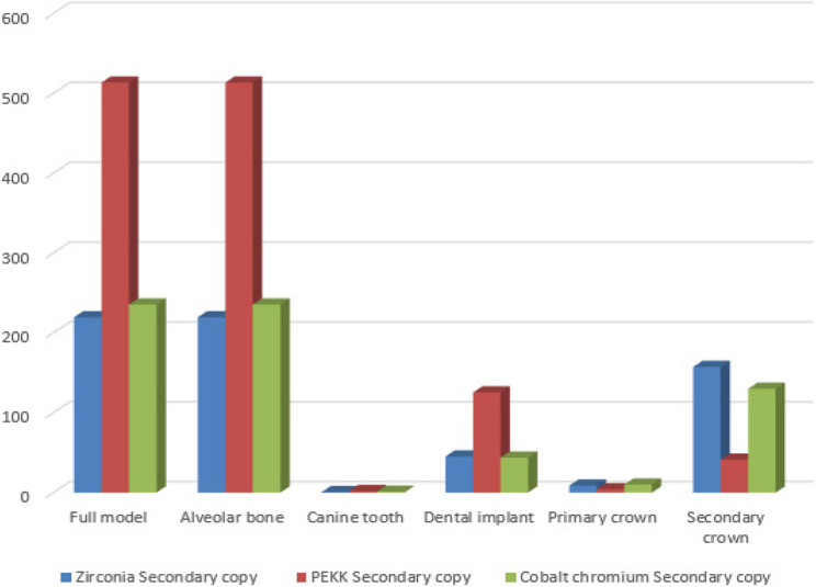 Figure 5
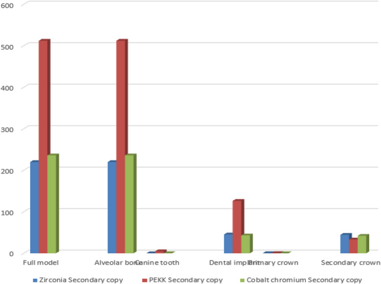 Figure 6
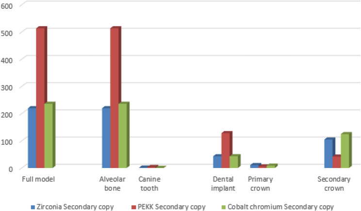 Figure 7
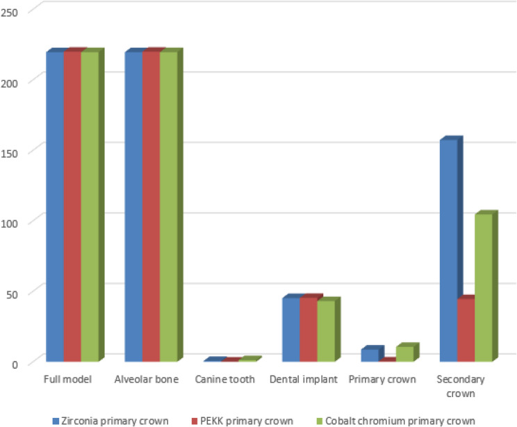 Figure 8
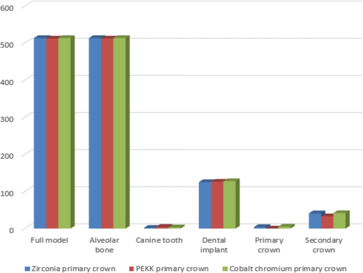 Figure 9
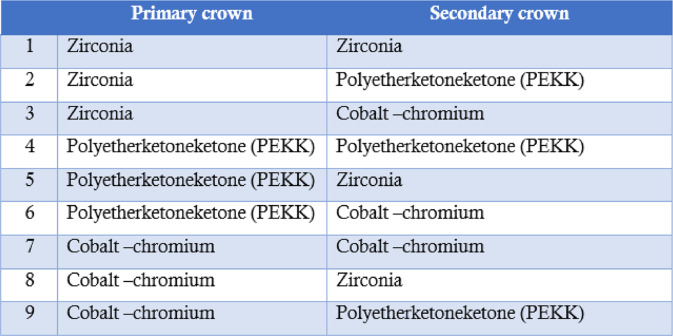 Figure 11
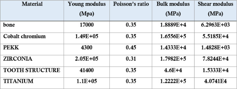 Figure 12
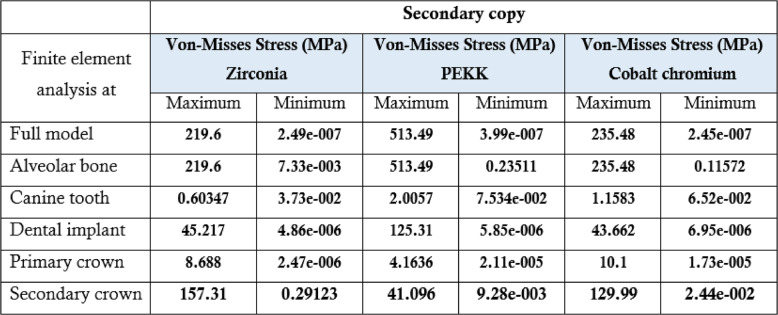 Figure 13
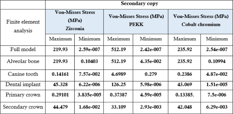 Figure 14
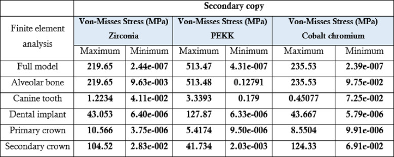 Figure 15
- —Mansoura University
Peer Reviews
No public reviews on file for this paper yet. If you reviewed it on a platform where reviews are public (OpenReview, ICLR, NeurIPS, ICML), you can paste yours below so the community can read it here.
Videos
No videos yet. Explain this paper in a talk, walkthrough, or lecture? Add one.
Taxonomy
TopicsDental Implant Techniques and Outcomes · Dental materials and restorations · Bone Tissue Engineering Materials
Introduction
Prosthetic rehabilitation of partially edentulous patients with a limited number of remaining teeth presents a clinical challenge that requires careful biomechanical planning. Such cases may be effectively managed through the strategic placement of dental implants combined with removable prosthetic solutions, including implant-assisted removable partial dentures and overdentures retained by double-crown (telescopic) systems [1–7]. Telescopic attachments facilitate vertical load transfer and improve prosthesis stability by connecting remaining natural teeth and/or implants, thereby contributing to favorable stress distribution and long-term prosthetic performance [8, 9].
Telescopic attachment systems are widely used in tooth- and implant-supported overdentures due to their versatility and favorable clinical characteristics. These systems offer improved esthetics with reduced implant numbers, flexibility in implant positioning, ease of oral hygiene maintenance, enhanced retention and stability, and improved load distribution with reduced torque on abutments [10, 11]. A telescopic system consists of a primary crown fixed to the abutment and a secondary crown incorporated into the removable prosthesis, with retention primarily achieved through frictional contact between the two components.
Advances in digital dentistry have significantly influenced the fabrication of telescopic prostheses. Computer-aided design and computer-aided manufacturing (CAD/CAM) technologies enable precise and reproducible fabrication of double crowns through subtractive milling and additive manufacturing techniques [12, 13]. These technologies have expanded the range of available materials, allowing the clinical use of advanced ceramics and high-performance polymers with improved mechanical and biological properties [14–16].
Cobalt–chromium (Co-Cr) alloys have traditionally been used for telescopic systems because of their high strength and rigidity; however, their use is associated with esthetic limitations, high thermal conductivity, potential hypersensitivity reactions, and susceptibility to galvanic corrosion [7–18]. Ceramic materials, particularly zirconia, have gained popularity due to their superior esthetics, favorable biocompatibility, and excellent mechanical properties, making them suitable for use in primary crowns, implant abutments, and monolithic restorations [19, 20]. More recently, polyetherketoneketone (PEKK), a high-performance polymer, has emerged as a promising alternative for telescopic prostheses due to its favorable mechanical behavior, hydrolysis resistance, and biocompatibility [9, 21, 22]. PEKK exhibits a semi-crystalline structure and can be processed using digital workflows without altering its chemical properties, enabling its application in CAD/CAM-fabricated prosthetic components.
Finite element analysis (FEA) has been extensively applied in prosthodontics to investigate stress distribution and deformation in dental implants, removable and fixed prostheses, and combined tooth–implant-supported systems [23–29]. By enabling detailed simulation of complex anatomical and prosthetic structures under controlled loading conditions, three-dimensional FEA allows assessment of internal stress patterns that cannot be directly measured in vivo or adequately captured using traditional experimental methods.
Despite the increasing clinical use of telescopic attachments connecting natural teeth and implants, limited data are available regarding the biomechanical influence of different material combinations used for primary and secondary crowns. In particular, the effect of combining zirconia, PEKK, and Co-Cr materials on stress transmission within telescopic systems and supporting structures remains insufficiently investigated [30–36]. Therefore, this study aimed to evaluate the stress distribution and biomechanical behavior of tooth–implant-supported telescopic prostheses using various combinations of zirconia, PEKK, and cobalt–chromium crowns through three-dimensional finite element analysis. The null hypothesis was that the stresses transmitted to prosthetic components and supporting structures would not differ among the evaluated material combinations.
Materials and methods
Study design
This study employed a three-dimensional finite element analysis (3D FEA) approach to evaluate stress distribution in tooth–implant-supported telescopic prostheses using different material combinations for primary and secondary crowns. A mandibular model representing a partially edentulous condition with two canines and two implants in the molar region was selected to reflect a clinically relevant prosthetic scenario. Nine distinct prosthetic configurations were created based on combinations of zirconia, polyetherketoneketone (PEKK), and cobalt–chromium (Co-Cr) alloys used for the primary and secondary crowns (Table 1).Table 1.The nine different groups based on the primary and secondary crown
Three-Dimensional model construction
Three-dimensional geometric models of the mandibular bone, natural teeth, dental implants, and telescopic crown assemblies were generated using SolidWorks 2023 Premium (Dassault Systèmes, France). The anatomical structures were constructed based on segmented computed tomography data and established anatomical landmarks to ensure realistic geometry.
The telescopic system consisted of a primary crown fixed to the abutment and a secondary crown integrated into the removable prosthesis. The primary crown was designed with a total taper angle of 4° (2° per wall), an occlusal thickness of 1.0 mm, and an axial wall thickness of 0.5 mm. The secondary crown was designed with an occlusal thickness of 0.8 mm and a cement space of 0.1 mm. The vertical height of the crown assembly was standardized at 6 mm (Fig. 1). These parameters were selected based on previously validated telescopic prosthesis designs [33, 34].Fig. 1. The 3D models of the prosthetic components including teeth, implants, and double-crown systems
Finite element meshing
The constructed models were imported into ANSYS Workbench 2019 R3 (ANSYS Inc., Canonsburg, PA, USA) for finite element discretization. A three-dimensional tetrahedral mesh using SOLID187 elements was generated. Mesh controls were standardized across all models to ensure consistency, with smoothing set to medium, a transition ratio of 0.272, a maximum of five layers, and a growth rate of 1.2.
The final finite element model comprised 736,006 nodes and 462,183 elements. Mesh convergence was verified by refining the element size from 1.0 mm to 0.8 mm and 0.6 mm, resulting in less than 3% variation in maximum von Mises stress values, confirming numerical stability.
Material properties
Material properties assigned to bone, tooth structure, titanium implants, zirconia, PEKK, and cobalt–chromium were obtained from validated experimental studies and widely cited literature sources (Table 2). All materials were assumed to be homogeneous, isotropic, and linearly elastic to facilitate numerical stability and allow direct comparison among material combinations, consistent with standard practices in prosthodontic finite element studies.Table 2.Material properties used in the study. Table 2 shows Elastic modulus, Poisson’s ratio, shear modulus, and bulk modulus for bone, tooth structure, titanium, zirconia, PEKK, and cobalt-chromium obtained from validated literature sources
Boundary conditions and loading protocol
The inferior border of the mandibular model was fully constrained in all translational and rotational degrees of freedom to prevent rigid-body motion and simulate fixation at temporomandibular and muscular attachment regions. Static axial occlusal loads were applied to represent centric occlusion conditions: a vertical load of 100 N was applied to each canine, and bilateral vertical loads of 250 N were applied to the molar regions.
The magnitude and direction of applied loads were selected based on reported average physiological bite forces and were applied perpendicular to the occlusal plane. All contacts between primary and secondary crowns were defined as frictional to enable realistic load transfer while preventing interpenetration.
Finite element simulation and outcome measures
Finite element simulations were conducted using ANSYS Workbench 2019 R3 for each of the nine prosthetic configurations. Von Mises stress distribution was evaluated within the primary and secondary crowns, dental implants, natural teeth, and surrounding alveolar bone. Stress patterns and peak stress values were recorded and compared across material combinations to assess the biomechanical influence of crown material selection (Figs. 2, 3 and 4).Fig. 2. Von Mises stresses for the first 3 groups of the primary coping zirconia and secondary copy
Fig. 3. Von mises stresses for the first 3 groups of the primary coping PEKK and secondary copy zirconia, PEKK, cobalt
Fig. 4. Von mises stresses for the first 3 groups of the primary coping cobalt chromium and secondary copy
This study was carried out at the Department of Prosthodontics, Faculty of Dentistry, Mansoura University. Ethical approval was obtained prior to commencement (Approval Code: A06012024 RP).
Result
Stress distribution in primary and secondary crowns
Across all material combinations, stress concentrations were predominantly observed at the occlusal surfaces and cervical regions of both primary and secondary crowns. Secondary crowns consistently exhibited higher von Mises stress values than primary crowns under identical loading conditions.
When PEKK was used as a secondary crown, peak stresses within both primary and secondary crowns were reduced compared with zirconia and cobalt–chromium secondary crowns, regardless of the primary crown material. In contrast, zirconia and cobalt–chromium secondary crowns demonstrated comparable stress magnitudes and distribution patterns.
When PEKK was used as a primary crown, stress within the prosthetic components was lower than that observed for zirconia or cobalt–chromium primary crowns; however, this configuration did not substantially alter stress transmission to the supporting structures.
Stress distribution in supporting structures
Stress within the dental implants was consistently higher than that observed in the natural abutment teeth across all prosthetic configurations. Peak stress concentrations were primarily localized at the implant neck region and the crestal bone interface.
Use of PEKK as a secondary crown resulted in increased stress transfer to the supporting structures, including alveolar bone, implants, and natural teeth, compared with zirconia and cobalt–chromium secondary crowns. In contrast, zirconia and cobalt–chromium demonstrated.
similar biomechanical behavior with respect to stress transmission to the supporting tissues.
Effect of material combination on load transfer
Comparative analysis of all nine material combinations demonstrated that crown material selection significantly influenced load distribution within the prosthetic system. While PEKK exhibited favorable stress-reducing behavior within prosthetic components, its lower elastic modulus promoted greater stress transfer to biological structures when used as a secondary crown.
Zirconia and cobalt–chromium, whether used as primary or secondary crowns, produced comparable stress distributions within both prosthetic and supporting components, indicating similar biomechanical performance under static axial loading conditions.
Summary of key findings
A synthesized comparison of peak von Mises stress values across prosthetic and supporting components is presented in Tables 3, 4 and 5. Representative stress contour plots illustrating typical stress patterns for the evaluated material combinations are shown in Figs. 2, 3, 4, 5, 6, 7, 8, 9 and 10.
Fig. 5. The bar chart of the maximum values of von Mises stress (Mpa) for the first 3 groups of the primary coping zirconia using 3D Finite element analysis
Fig. 6. The bar chart of the maximum values of von Mises stress (Mpa) for the second 3 groups of the primary coping PEKK using 3D Finite element analysis
Fig. 7. The bar chart of the maximum values of von Mises stress (Mpa) for the third 3 groups of the primary coping cobalt chromium using 3D Finite element analysis
Fig. 8. The bar chart of the maximum values of von Mises stress (Mpa) for the 3 groups of the secondary crown zirconia using 3D Finite element analysis
Fig. 9. The bar chart of the maximum values of von Mises stress (Mpa) for the 3 groups of the secondary crown PEKK using 3D Finite element analysis
Fig. 10. The bar chart of the maximum values of von Mises stress (Mpa) for the 3 groups of the secondary crown cobalt chromium using 3D Finite element analysis Table 3.The maximum, minimum values of von Mises stress (MPa) for the first 3 groups of the primary coping zirconia using 3 D Finite element analysis at different evaluation sitesTable3 Peak stress generated in each primary crown material under standardized loading conditions for the nine telescopic prosthesis configurations Table 4.The maximum, minimum values of von mises stress (MPa) for the second 3 groups of the primary coping PEKKTable 4 Stress distribution results showing maximum stress concentration in secondary crowns across all zirconia, PEKK, and cobalt-chromium combinations Table 5.The maximum, minimum values of von Mises stress (MPa) for the third 3 groups of the primary coping cobalt chromiumTable5 Comparative analysis of stress transferred to alveolar bone, natural teeth, and implants for all tested double-crown material configurations
Discussion
This three-dimensional finite element analysis investigated the biomechanical behavior of tooth–implant–supported telescopic prostheses fabricated using different combinations of zirconia, PEKK, and cobalt–chromium for the primary and secondary crowns. The findings demonstrate that material selection within telescopic systems significantly influences stress transfer pathways between prosthetic components and supporting biological structures. Accordingly, the null hypothesis was rejected.
Direct in vivo measurement of stress in abutment teeth, implants, and alveolar bone remains impractical due to ethical and technical constraints. Experimental techniques such as strain gauges and Photoelastic analysis provide valuable insights but are limited by localized measurement zones, material requirements, and difficulty in reproducing complex anatomical geometries [27, 37, 38]. Finite element analysis therefore represents a validated and widely accepted approach for investigating biomechanical behavior in prosthodontic systems, enabling controlled evaluation of stress distribution patterns under standardized loading conditions. Numerous experimental–numerical correlation studies have demonstrated that FEA reliably reproduces clinically relevant stress trends when appropriate material properties and boundary conditions are applied [37, 39, 40].
Previous investigations have primarily focused on single-material telescopic systems, whereas limited data exist regarding mixed-material primary–secondary crown combinations. From a clinical perspective, this distinction is important because the primary crown is usually minimally adjusted after cementation, while the secondary crown is modified to achieve optimal frictional retention and path of insertion [41]. Consequently, material pairing rather than isolated material selection may play a decisive role in long-term biomechanical performance.
The present results indicate that PEKK, when used as a secondary crown, generated higher stress levels in the supporting structures—namely alveolar bone, abutment teeth, and implants—compared with zirconia and Co–Cr secondary crowns. This behavior can be attributed to the lower elastic modulus of PEKK, which allows greater deformation at the crown interface, increasing load transfer to the underlying structures. These findings are consistent with previous experimental and numerical studies reporting higher strain levels in PEKK-based telescopic systems compared with Co–Cr alloys [33, 34]. In contrast, the substantially higher stiffness of Co–Cr (≈ 220–230 GPa) limits deformation and thereby reduces stress transmission to supporting tissues.
Frictional behavior between telescopic components further contributes to these observations. PEKK exhibits higher frictional retention compared with zirconia and Co–Cr, which enhances prosthesis stability but simultaneously increases resistance during functional loading [35, 36, 42–45]. Increased friction has been shown to elevate stress transfer within attachment systems, particularly at the implant–bone interface [42]. Therefore, while PEKK may offer favorable retention characteristics, its use as a secondary crown should be carefully considered in cases with compromised supporting structures.
Importantly, despite increasing stress in the supporting tissues, PEKK secondary crowns demonstrated lower stress concentrations within both primary and secondary crowns themselves. This apparent contradiction is resolved by considering the viscoelastic and damping properties of PEKK. The lower elastic modulus and higher energy absorption capacity of PEKK create a cushioning effect that reduces stress accumulation within the prosthetic components, even as greater loads are transferred to the supporting structures [36, 46–48]. This dual behavior explains why PEKK cannot be uniformly classified as biomechanically superior or inferior, but rather as material-dependent on its prosthetic role.
When PEKK was used as a primary crown, stresses within the prosthetic components were reduced without a significant effect on supporting structures. This finding supports previous reports describing favorable stress modulation when PEKK is used as a framework or primary element due to its elastic recovery and toughness [48–50]. In contrast, zirconia and Co–Cr exhibited similar stress distributions regardless of their position as primary or secondary crowns, reflecting their high stiffness and limited energy dissipation capacity.
Across all material combinations, secondary crowns consistently experienced higher stress than primary crowns. This observation aligns with prior FEA and experimental studies demonstrating that secondary crowns act as the primary load-receiving component during mastication and insertion–removal cycles [20, 36, 51]. Additionally, implants were subjected to higher stress levels than natural teeth, which is consistent with the absence of a periodontal ligament and its associated shock-absorbing capacity [52–56].
Localized stress peaks at the crestal cortical bone were observed around implants in all models. Such concentrations are well documented in implant FEA studies and are generally interpreted as numerical singularities resulting from geometric transitions and idealized boundary conditions rather than predictors of immediate clinical failure [57–59]. The reproduction of these stress patterns further supports the validity of the present modeling approach.
The modeling assumptions adopted in this study—homogeneous, isotropic, and linearly elastic material behavior; omission of the periodontal ligament; and application of static axial loads—are consistent with numerous previously validated dental FEA investigations [33, 36, 52–55]. Sensitivity analysis confirmed that moderate variations in elastic modulus and friction coefficients resulted in less than 3% change in peak cortical bone stress, indicating numerical stability. Nevertheless, these assumptions limit direct clinical extrapolation and should be interpreted as representing idealized loading conditions rather than exact physiological behavior.
Future investigations should incorporate dynamic loading, nonlinear material properties, periodontal ligament simulation, and patient-specific geometries to further refine biomechanical predictions. Long-term clinical studies are also necessary to evaluate the influence of wear, fatigue, and cement layer behavior on telescopic prosthesis performance.
Limitations
The present investigation employed a three-dimensional finite element model using established and widely accepted biomechanical assumptions to enable controlled comparison between material combinations. Materials were modeled as homogeneous, isotropic, and linearly elastic, and static axial loading was applied to represent standardized occlusal conditions commonly adopted in comparable finite element studies. Periodontal ligament simulation was not incorporated in order to isolate material-dependent effects and maintain numerical stability of the mixed tooth–implant model. The analysis focused on a single mandibular telescopic prosthesis design to ensure consistency across all simulations. Accordingly, the findings should be interpreted within the context of comparative biomechanical behavior rather than direct clinical outcome prediction. Future studies may expand upon the current model by incorporating dynamic loading conditions, patient-specific geometries, and experimental validation to further refine clinical relevance. [60–64]
Conclusion
This three-dimensional finite element analysis demonstrated that the biomechanical performance of tooth–implant–supported telescopic prostheses is strongly influenced by the material combination used for primary and secondary crowns. The results highlight that material pairing, rather than isolated material selection, plays a decisive role in governing stress distribution within both prosthetic components and supporting biological structures.
PEKK exhibited a material-dependent biomechanical behavior. When used as a secondary crown, PEKK reduced stress concentrations within the prosthetic crowns but increased stress transfer to the supporting bone, teeth, and implants. Conversely, PEKK used as a primary crown contributed to stress modulation within the prosthetic components without significantly affecting the supporting structures. Zirconia and cobalt–chromium demonstrated comparable biomechanical performance regardless of their position within the telescopic system, reflecting their high stiffness and limited damping capacity.
Across all configurations, secondary crowns consistently experienced higher stress than primary crowns, and implants were subjected to greater stress than natural teeth, underscoring the biomechanical influence of periodontal support. These findings provide mechanistic insight into stress transfer pathways in mixed tooth–implant telescopic systems and support informed material selection tailored to individual clinical conditions.
Within the constraints of a controlled numerical model, the present study contributes evidence-based guidance for optimizing telescopic prosthesis design and offers a biomechanical framework for future experimental and clinical investigations.
The reference list from the paper itself. Each links out to its DOI / PubMed record.
- 1de Freitas RF, de Carvalho Dias K, da, Porto Carreiro F, Barbosa A, Ferreira GA. MA. Mandibular implantsupported removable partial denture with distal extension: A systematic review. J Oral Rehabil. 2012;39(10):791–98.10.1111/j.1365-2842.2012.02326.x 22882547 · doi ↗ · pubmed ↗
- 2Kern JS, Hanisch O, Hammächer C, Yildirim M, Wolfart S. Telescopic crowns on implants and teeth: evaluation of A clinical study after 8 to 12 years. Int J Oral Maxillofac Implants. 2019;34(4):977–86. 10.11607/jomi.720431107933 · doi ↗ · pubmed ↗
- 3Alharbi N, Wismeijer D, Osman RB. Additive manufacturing techniques in prosthodontics: where do we currently stand? A critical review. Int J Prosthodont. 2017;30(5). https://pubmed.ncbi.nlm.nih.gov/28750105/10.11607/ijp.507928750105 · doi ↗ · pubmed ↗
- 4Idzior-Haufa M, Pilarska AA, Hędzelek W, Boniecki P, Pilarski K, Dorocka-Bobkowska B. A Comparison of Biomechanical Properties of Implant-Retained Overdenture Based on Precision Attachment Type. Materials (Basel). 2021. Cited 2025. 10.3390/ma 14102598 PMC 815594734067572 · doi ↗ · pubmed ↗
- 5Shahmiri R, Das R, Aarts JM, Bennani V. Finite element analysis of an implant-assisted removable partial denture during bilateral loading: occlusal rests position. J Prosthet Dent. 2014;112(5):1126–33. Cited 16 May 2025. 10.1016/j.prosdent.2014.04.02324951387 · doi ↗ · pubmed ↗
- 6Baccouch Mahboub. Finite element methods and their applications. 2021. Cited 16 May 2025; Available from: https://books.google.com/books/about/Finite_Element_Methods_and_Their_Applica.html?id=o 7Za EAAAQBAJ.
- 7Stock V, Wagner C, Merk S, Roos M, Schmidlin PR, Eichberger M et al. Retention force of differently fabricated telescopic PEEK crowns with different tapers. Dent Mater J. 2016;35(4):594–600. Cited 12 Jun 2025. Available from: https://europepmc.org/article/med/27477224.10.4012/dmj.2015-24927477224 · doi ↗ · pubmed ↗
- 8Mostafa D, Hussein O, Hussein MO. Biomechanical Performance of PEEK and Graphene-Modified PMMA as Telescopic Removable Partial Denture Materials: A Nonlinear 3D Finite Element Analysis. 2022.10.11607/ijp.817736125879 · doi ↗ · pubmed ↗