Correction: ZEB1-mediated biogenesis of circnipbl sustains the metastasis of bladder cancer via Wnt/β-catenin pathway
Yao Kong, Yuanlong Li, Mingjie An, Yuming Luo, Hanhao Zheng, Yan Lin, Jiancheng Chen, Jin Yang, Libo Liu, Baoming Luo, Jian Huang, Tianxin Lin, Changhao Chan

Abstract
Genes, proteins, chemicals, diseases, species, mutations and cell lines named across the full text — each resolved to its canonical identifier and authoritative record.
Click any figure to enlarge with its caption.
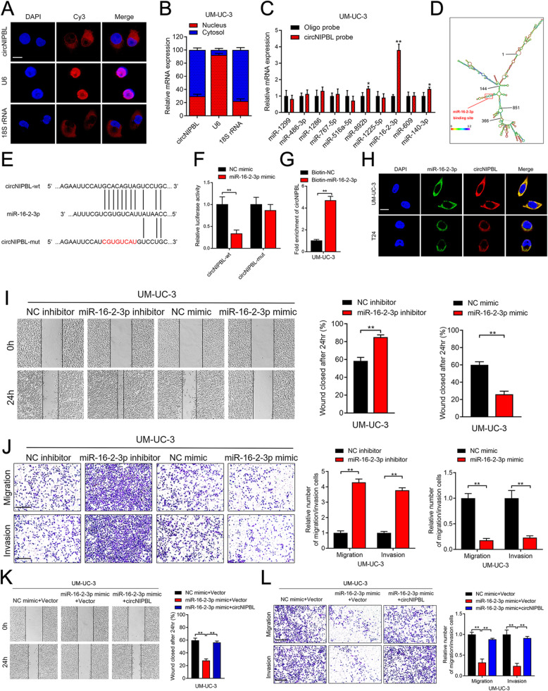 Figure 1
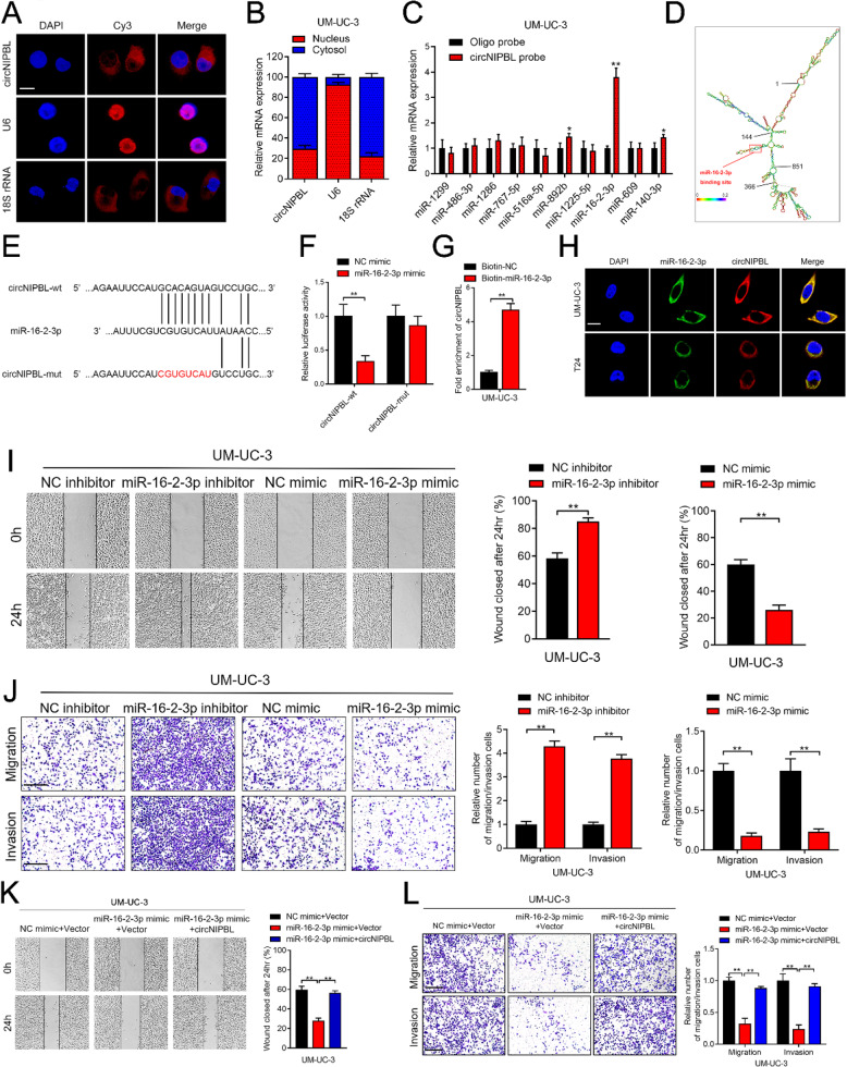 Figure 2
Peer Reviews
No public reviews on file for this paper yet. If you reviewed it on a platform where reviews are public (OpenReview, ICLR, NeurIPS, ICML), you can paste yours below so the community can read it here.
Videos
No videos yet. Explain this paper in a talk, walkthrough, or lecture? Add one.
Taxonomy
TopicsBladder and Urothelial Cancer Treatments · Wnt/β-catenin signaling in development and cancer · Hedgehog Signaling Pathway Studies
Correction: J Exp Clin Cancer Res 42, 191 (2023).
https://doi.org/10.1186/s13046-023-02757-3
Following the publication of the original article [1], the authors identified an unintentionally misused picture in the Fig. 3 caused by incaution of composed type.
The correct figure is presented below:
Incorrect Fig. 3
Fig. 3circNIPBL serves as a sponge for miR-16-2-3p in BCa cells.** A** FISH assay was used to detect the cellular localization of circNIPBL. Scale bar = 5 μm. B Subcellular fractionation assay was used to confirm the cellular localization of circNIPBL in UM-UC-3 cells. U6 was used for the nuclear control and 18S rRNA was used for the cytoplasmic control. C The expression level of ten predicted target miRNAs of circNIPBL were analyzed by qRT-PCR in UM-UC-3 cells. D RNAalifold was used to predict the secondary structure of circNIPBL. E Schematic illustrating the sequence alignment of circNIPBL with miR-16-2-3p. F Dual luciferase reporter assays showed that the luciferase activities of the circNIPBL-wt plasmid or circNIPBL-mut plasmid quantified following co-transfection with either the miR-16-2-3p or control mimics. G qRT-PCR analysis showed the circNIPBL captured by biotinylated miR-16-2-3p. H The co-localization of circNIPBL and miR-16-2-3p was detected by FISH assay. Scale bar = 5 μm. I Representative images and quantification of Wound healing assay showed the migration capability of UM-UC-3 cells transfected with miR-16-2-3p mimics or inhibitors. Scale bar = 100 μm. J Representative images and quantification of Transwell migration and Matrigel invasion assays showed the effect of UM-UC-3 cells transfected with miR-16-2-3p mimics or inhibitors. Scale bar = 100 μm. K,** L** Representative images and quantification of wound healing assay (K), Transwell migration and Matrigel invasion assays (L) by indicated UM-UC-3 cells. Scale bar = 100 μm. The statistical difference was assessed with one-way ANOVA followed by Dunnett tests in K and L; and the two-tailed Student t test in C, F, G, I and J; and the χ^2^ test in B. Error bars show the SD from three independent experiments. *p < 0.05 and **p < 0.01
Correct Fig. 3
Fig. 3circNIPBL serves as a sponge for miR-16-2-3p in BCa cells. A FISH assay was used to detect the cellular localization of circNIPBL. Scale bar = 5 μm. B Subcellular fractionation assay was used to confirm the cellular localization of circNIPBL in UM-UC-3 cells. U6 was used for the nuclear control and 18 S rRNA was used for the cytoplasmic control. C The expression level of ten predicted target miRNAs of circNIPBL were analyzed by qRT-PCR in UM-UC-3 cells. D RNAalifold was used to predict the secondary structure of circNIPBL. E Schematic illustrating the sequence alignment of circNIPBL with miR-16-2-3p. F Dual luciferase reporter assays showed that the luciferase activities of the circNIPBL-wt plasmid or circNIPBL-mut plasmid quantified following co-transfection with either the miR-16-2-3p or control mimics. G qRT-PCR analysis showed the circNIPBL captured by biotinylated miR-16-2-3p. H The co-localization of circNIPBL and miR-16-2-3p was detected by FISH assay. Scale bar = 5 μm. I Representative images and quantification of Wound healing assay showed the migration capability of UM-UC-3 cells transfected with miR-16-2-3p mimics or inhibitors. Scale bar = 100 μm. J Representative images and quantification of Transwell migration and Matrigel invasion assays showed the effect of UM-UC-3 cells transfected with miR-16-2-3p mimics or inhibitors. Scale bar = 100 μm. K, L Representative images and quantification of wound healing assay (K), Transwell migration and Matrigel invasion assays (L) by indicated UM-UC-3 cells. Scale bar = 100 μm. The statistical difference was assessed with one-way ANOVA followed by Dunnett tests in K and L; and the two-tailed Student t test in C, F, G, I and J; and the χ2 test in B. Error bars show the SD from three independent experiments. *p < 0.05 and **p < 0.01
After carefully checking the original data, we confirmed that we mistakenly selected the images during the process of image label and storage. Since the error does not originate from original experiments, this correction does not affect the overall conclusion of this article. The original article has been corrected.