Carvedilol decreases hepatic vascular resistance by reducing fibrogenesis and reversing endothelial dysfunction in cirrhotic rats
Yeldos Nulan, Eric Felli, Sonia-Emilia Selicean, Manuel Prampolini, Annalisa Berzigotti, Jordi Gracia-Sancho, Jaume Bosch

TL;DR
Carvedilol reduces liver vascular resistance and fibrosis in cirrhotic rats more effectively than propranolol, offering potential benefits for treating portal hypertension.
Contribution
This study reveals carvedilol's novel ability to reverse hepatic endothelial dysfunction and reduce fibrosis in cirrhosis, beyond its β-blocking effects.
Findings
Carvedilol significantly reduced portal pressure and liver fibrosis in both early and advanced cirrhosis in rats.
Carvedilol improved endothelial dysfunction and reduced oxidative stress and inflammation in cirrhotic liver cells.
Propranolol had minimal effect on portal pressure and lacked the fibrosis-reducing benefits of carvedilol.
Abstract
Cirrhosis increases hepatic vascular resistance (IHVR) by disrupting liver architecture due to fibrosis, and by elevating hepatic vascular tone due to hepatic endothelial dysfunction. IHVR increases portal pressure (PP), later aggravated by increased portal blood inflow. Carvedilol, a third-generation non-selective β-blocker with anti-α1-adrenergic activity, reduces PP more than propranolol, likely decreasing IHVR. However, its intrahepatic effects remain largely unexplored. This study aimed to address these issues. Human cell lines (LX2 and HUVECs) and primary liver sinusoidal endothelial cells (LSECs) and hepatic stellate cells (HSCs) isolated from cirrhotic rats (12-week thioacetamide [TAA] model) were treated with vehicle, carvedilol (10 μM), or propranolol (10 μM). Nitric oxide release, oxidative stress, and cell contraction were assessed. Cirrhotic rats were treated with vehicle,…
Genes, proteins, chemicals, diseases, species, mutations and cell lines named across the full text — each resolved to its canonical identifier and authoritative record.
Click any figure to enlarge with its caption.
 Figure 1
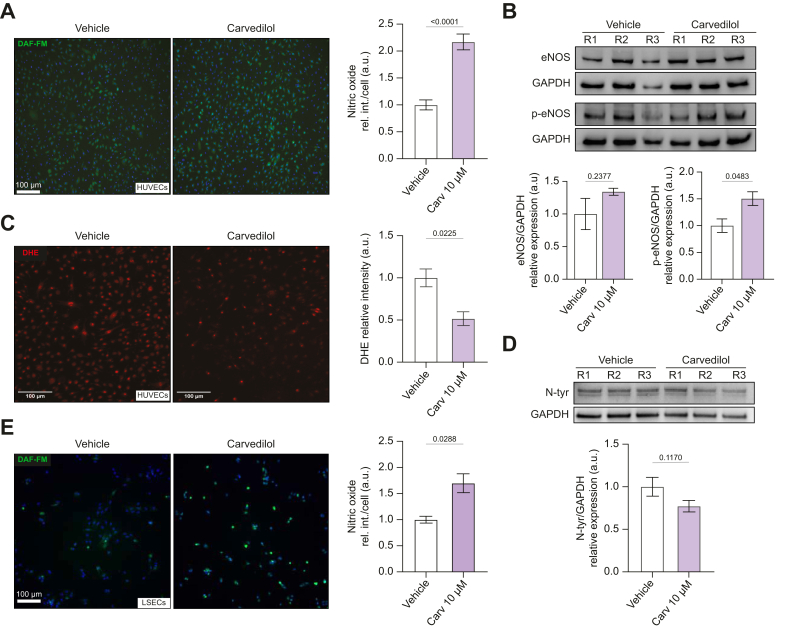 Figure 2
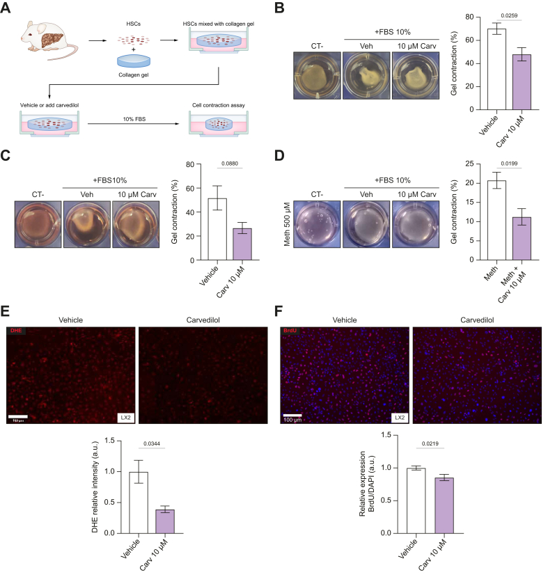 Figure 3
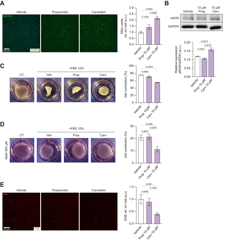 Figure 4
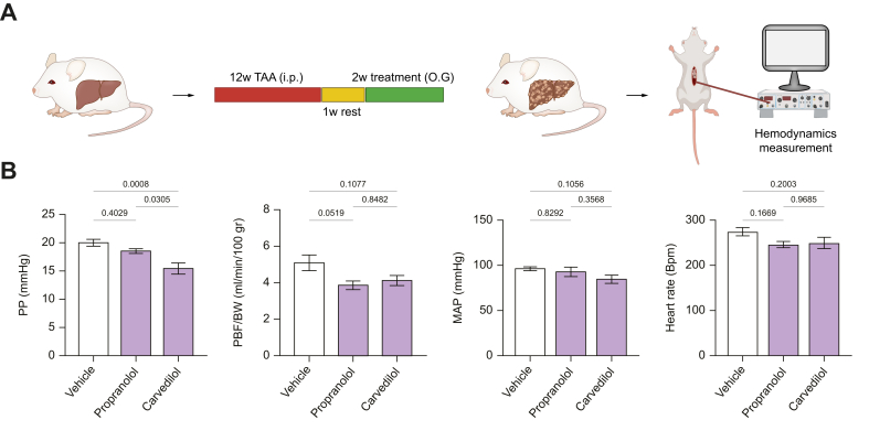 Figure 5
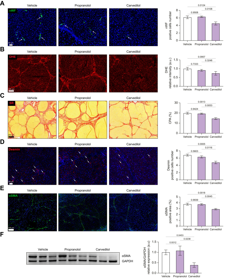 Figure 6
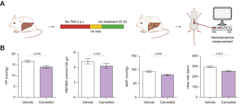 Figure 7
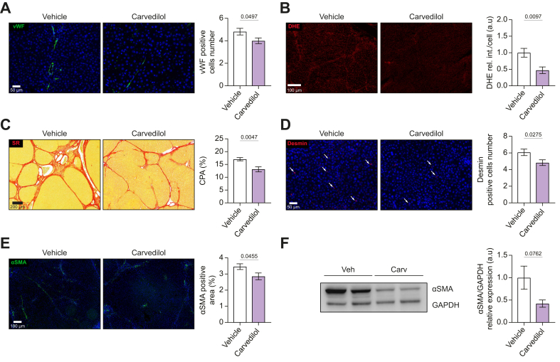 Figure 8
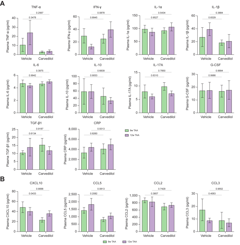 Figure 9
Peer Reviews
No public reviews on file for this paper yet. If you reviewed it on a platform where reviews are public (OpenReview, ICLR, NeurIPS, ICML), you can paste yours below so the community can read it here.
Videos
No videos yet. Explain this paper in a talk, walkthrough, or lecture? Add one.
Taxonomy
TopicsLiver Disease and Transplantation · Organ Transplantation Techniques and Outcomes · Liver physiology and pathology
Introduction
Cirrhosis is a leading cause of mortality worldwide, ranking 5^th^ among adults.1^,^2 The high mortality associated with cirrhosis is due to complications arising from portal hypertension, ascites, variceal bleeding, hepatic encephalopathy, and hepatocellular carcinoma.3 In cirrhosis, portal hypertension occurs because of increased resistance to blood flow within the portal venous system, and is later aggravated by splanchnic vasodilation and increased portal blood inflow. The increased resistance is caused by structural changes in the liver (fibrosis, tissue remodeling, formation of regenerative nodules, and angiogenesis),4^,^5 together with an increased hepatic vascular tone, due to liver sinusoidal endothelial cell (LSEC) dysfunction, characterized by reduced production of vasodilators like nitric oxide (NO)6 and hypersensitivity to vasoconstrictor stimuli.7
Suppression of the cause of cirrhosis is the only therapy that may improve cirrhosis,8 while anti-fibrotic therapies are under development.9 Non-selective β-blockers (NSBBs), such as propranolol or nadolol,10 are the mainstay of therapy for portal hypertension. NSBBs reduce portal pressure by decreasing portal blood inflow by means of lowering cardiac output through β1-adrenoceptor (β1-AR) blockade and by inducing vasoconstriction in the splanchnic circulation via β2-adrenoceptor (β2-AR) blockade. Carvedilol, a third-generation NSBB with intrinsic anti α-1 adrenoceptor activity (α1-AR), is at present the NSBB of choice for portal hypertension because it achieves a greater portal pressure reduction compared to propranolol,11 which is thought to be due to a decrease in the hepatic vascular tone.12 Studies in cardiovascular biology have shown that carvedilol has pleiotropic effects that go beyond adrenergic blockade, including protection against endothelial dysfunction by increasing NO release, anti-oxidant activity, and decreased vascular remodeling.13 These effects suggest that, in cirrhosis, carvedilol might deactivate hepatic stellate cells (HSCs), which are key drivers of disease progression, in part by blocking α1-adrenergic signaling.14 Surprisingly, besides these important connotations, there is scarce knowledge about the intrahepatic effects of carvedilol in cirrhosis, which prompted us to perform the current study.
Materials and methods
Animal models of cirrhosis
Female and male Sprague-Dawley rats were maintained under standard environmental conditions at the University of Bern animal facility. The conditions included a 12-hour light/dark cycle and 10-15 air exchanges per hour. Upon arrival, the rats were acclimatized for 2 weeks before starting any experiments or treatments. Throughout the study, rats had free access to chow diet and water. All experimental procedures were approved by the Bern Cantonal Ethics Committee and adhered to the Laboratory Animal Care guidelines (approval number BE382022). Cirrhosis and portal hypertension was induced by the twice weekly administration of thioacetamide (TAA) (Sigma-Aldrich; Ref. No.172502) (200 mg/kg of body weight; intraperitoneally) for a total of 12 weeks. In addition, other groups of animals received TAA for only 9 weeks to model early cirrhosis. Before each TAA administration, the rats' body weights were recorded, and the TAA dosage was adjusted accordingly.
Isolation of liver endothelial sinusoidal cells and hepatic stellate cells
The isolation of primary liver cells was conducted following methods previously described.15 Further details are given in supplementary methods.
Cell culture
Freshly isolated LSECs were cultured in RPMI 1640 Medium with 10% FBS, 1% antibiotic-antimycotic, 50 μg/ml endothelial cell growth supplement, and 100 μg/ml heparin. HSCs were maintained in Iscove's modified Dulbecco's medium with 10% FBS and 1% antibiotic-antimycotic. HUVECs (P2–P4, C2519A, Lonza) were cultured in Medium 199 with 20% FBS, 1% antibiotic-antimycotic, 50 μg/ml endothelial cell growth supplement, and 100 μg/ml heparin. LX-2 cells (P15–P20, Liver Vascular Biology Research Group, IDIBAPS Biomedical Research Institute) were cultured in Dulbecco's modified eagle medium/nutrient mixture F-12 with 10% FBS and 1% antibiotic-antimycotic. All cells were incubated at 37 °C in a humid atmosphere (95%) with 5% CO_2_. Please refer to the supplementary methods.
Measurement of intracellular nitric oxide levels
Intracellular nitric oxide levels in LSECs and HUVECs were assessed with 4-amino-5-methylamino-2', 7'-dichlorofluorescein diacetate (DAF-FM-DA, ThermoFisher: D23844). LSECs and HUVECs were treated with carvedilol or vehicle for 24 h, and HUVECs with propranolol or vehicle for 24 h. After, cells were loaded with DAF-FM-DA (10 μM) and nucleus biomarker Hoechst (ThermoFisher: 33342) in the incubator for 30 mins. Cells were washed to remove excess probe, replaced with fresh medium, and then incubated for an additional 15 mins period to allow complete de-esterification of the intracellular diacetate. Then, cells were rinsed three times with PBS (ThermoFisher: 10010023), protected from light, and placed on an inverted fluorescence microscope (LEICA-DMI4000). Fluorescence images were obtained by setting the 488 nm (excitation) and 505 to 530 nm (emission) filters for DAF-FM-DA with a 10x or 20x objective.
Cell contraction
Cell contraction assays were conducted using HSCs and LX2 cells in collagen lattices. Freshly isolated HSCs were cultured for 5 days to reach full activation. Collagen gels (1.3 mg/ml) containing HSCs or LX2 cells (1.5 × 10^5^ cells/ml) were prepared in 24-well plates pre-treated with 1% BSA. After gel solidification, serum-free media with 10 μM carvedilol, propranolol or vehicle was added for 24 h. Contraction was induced with 10% FBS for 48 h, with photographs taken at 1, 24, and 48 h. In a separate experiment, 500 μM methoxamine was used to foster α1-adrenergic contraction, and the effects of carvedilol and propranolol were assessed at 1 h. Please see details in supplementary methods.
Cell proliferation assay
The proliferation of LX2 cells was assessed using a bromodeoxyuridine (BrdU) incorporation assay. Cells were cultured in 6-well plates for 24 h, treated with or without carvedilol (10 μM) for 24 h, and labeled with BrdU for 2 h. Following fixation with 4% PFA, cells were permeabilized with 0.1% Triton X-100, treated with hydrochloric acid, neutralized with phosphate/citric acid buffer, and washed. BrdU was detected using a primary antibody (Thermo: MA3-071, 1:100) overnight at 4 °C, followed by a fluorescent secondary antibody (Thermo: A21422, 2 μg/ml) for 1 h. Imaging was performed on EVOS 5000 using the RFP channel. Please see details in supplementary methods.
Hepatic and systemic hemodynamics
Cirrhotic rats were administered either carvedilol, 10 mg/kg/day (Hexal AG, Germany); propranolol, 30 mg/kg/day (Ratiopharm, Germany), or vehicle by oral gavage for 2 weeks. One hour after the final dose, the rats were anesthetized with inhaled isoflurane (Girovet: 469860). Mean arterial pressure was measured by cannulating the femoral artery, and portal pressure was measured by cannulating the mesenteric vein, both using a heparinized P50 catheter (Portex) connected to a pressure transducer. Portal blood flow was determined using a non-constrictive perivascular ultrasonic transit-time flow probe (Transonic Systems Inc.) placed close to the liver hilus. Pressure and flow probes were connected to a PowerLab (4SP) data recording system, and data was displayed using LabChart v5.5.6 software. Hemodynamic parameters were recorded following a 30-minute stabilization period, collecting stable data for 2 minutes. After the hemodynamic study, blood samples were collected. Finally, the animals were euthanized with a pentobarbital overdose (200 mg/kg, Nembutal), and tissue samples were harvested for molecular and histological experiments.
Systemic inflammation multiplex immunoassay
Plasma from cirrhotic rats was collected after hemodynamic measurements, processed via centrifugation, and analyzed using the ProcartaPlex Immunoassay on the Bio-Plex 3D Suspension Array system. The assay involved incubating 25 μl plasma on a customized ProcartaPlex plate, followed by sequential addition and washing of Detection Antibody Mix, Streptavidin-PE, and Reading Buffer, with shaking at room temperature. Data was acquired and analyzed using Bio-Plex Manager Software 6.0. Details are shown in the supplementary methods.
Superoxide detection
Superoxide (O_2_^−^) levels were measured using the fluorescent dye dihydroethidium (DHE) (10 μM) in liver-frozen sections, HUVECs, and LX2 cells. Liver tissue: Frozen sections (10 μm) were prepared using a Leica Cryostat, loaded with DHE, and incubated at room temperature for 30 minutes. Slides were imaged with fluorescent mounting medium. Cell lines: Cells were rinsed with PBS, incubated with DHE for 30 minutes, washed with PBS, and imaged immediately. Fluorescent images were captured using an EVOS 5000 microscope (10x objective), and the signal was quantified with ImageJ software (v1.53). Please see details in supplementary methods.
Liver histology
Liver tissue was fixed in 4% formaldehyde (Sigma), embedded in paraffin, and sectioned. To evaluate fibrosis, the sections were stained with 0.1% Sirius red in an aqueous picric acid solution (Sigma). Then sections were captured using a 3DHistech Slide scanner (Panoramic 250 Flash II). The Sirius red-stained areas were quantified using Qupath-0.4.3, and the results were expressed as collagen proportionate area (CPA), the percentage of the total tissue section area positively stained for fibrosis (red). Septa were classified as thin vs. thick, and incomplete vs. complete.
Immunostaining
Liver tissue was fixed in 4% formaldehyde, paraffin-embedded, sectioned, and dewaxed. Antigen retrieval was performed using Heated EDTA-Tris buffer (pH = 9), followed by blocking with 3% goat serum. Primary antibodies were applied overnight at 4 °C to target alpha smooth muscle actin (α-SMA) (1:400, Thermo) for activated HSCs, desmin (10 μg/ml, Thermo) for proliferating HSCs, and von Willebrand factor (vWF) (2 μg/ml, Thermo) for endothelial cells. Secondary antibodies conjugated to Alexa Fluor dyes were incubated with DAPI for 1 h at room temperature. Sections were mounted with fluorescent medium and scanned using a 3DHistech Slide scanner. Quantification of α-SMA–positive stained area and of desmin- and vWF-positive cells was performed using QuPath v0.4.3. Please see supplementary methods for further details.
Western blot
Cells were lysed in RIPA buffer, and protein concentrations measured using the Pierce™ BCA Kit. Samples (10 μg protein/lane) were separated by SDS-PAGE using Bolt™ Bis-Tris Gels and transferred to PVDF membranes. Membranes were blocked, probed with primary antibodies (endothelial nitric oxide synthase [eNOS], phospho-eNOS, nitrotyrosine, α-SMA, GAPDH), and incubated with HRP-conjugated secondary antibodies. Detection was performed with chemiluminescence and visualized using the Fusion FX system. Liver tissue proteins were similarly processed using the iBind™ Flex Western System. GAPDH served as the loading control. Please see supplementary methods for additional details.
Statistical analysis
Statistical analyses were performed using GraphPad Prism 10 (GraphPad Software). Data represent biological replicates (n), including TAA 12 weeks group (vehicle: n = 8; propranolol: n = 6; carvedilol: n = 8) and TAA 9 weeks group (vehicle: n = 9; carvedilol: n = 10), and are depicted in figures as mean values ± standard error. The frequency distribution of data was assessed using a normality test (Kolmogorov−Smirnov). For samples with a normal distribution, means were compared by Student's t test (two samples) or ANOVA (more than two samples) followed by Tukey's post hoc analysis. Otherwise, non-parametric tests (Kruskal−Wallis) followed by Dunn’s multiple comparisons were used. Differences were considered statistically significant at p <0.05.
Results
Carvedilol increases the release of nitric oxide in LSECs
The effect of carvedilol on NO release was studied in HUVECs using the DAF-FM assay (Fig. 1A). In addition to enhancing NO release, HUVECs treated with carvedilol exhibited increased expression of endothelial nitric oxide synthase (eNOS) and its phosphorylated form (phospho-eNOS) (Fig. 1B). Moreover, decreased NO scavenging by O_2_^−^ further increased the availability of NO. Measuring O_2_^−^ levels by DHE showed that carvedilol had antioxidant activity (Fig. 1C). The reduction in superoxide was associated with decreased peroxynitrite (ONOO^−^) formation, indirectly tested by diminished nitrotyrosinated protein (Fig. 1D). The upregulation of NO availability by carvedilol was further confirmed in freshly isolated LSECs from cirrhotic rats (Fig. 1E).Fig. 1. Carvedilol increases NO release in HUVECs and liver sinusoidal endothelial cells.(A) NO detection using DAF-FM. n = 6. (B) Western blot and the relative semi-quantification of eNOS and p-eNOS from HUVECs treated with and without carvedilol for 24 hours. n = 3. (C) Superoxide levels in HUVECs treated with carvedilol or vehicle. Representative pictures made with EVOS 5000 of in situ detection of O_2_^−^ levels with DHE, n = 3. (D) Representative picture of western blot with relative semiquantitative analysis of nitrotyrosinated protein levels measured in HUVECs treated with carvedilol or vehicle. n = 5. (E) NO levels in primary liver sinusoidal endothelial cells isolated from cirrhotic rats incubated for 24 hours with carvedilol or vehicle. n = 4. DAF-FM live-cell imaging was performed with EVOS 5000 and Leica DMI4000B multipurpose fluorescence system. For each experiment, sample distributions were assessed for normality (Kolmogorov−Smirnov test). Normally distributed data were compared using an unpaired Student’s t test, while non-normally distributed data were compared using the Mann–Whitney test. All data are represented as mean ± SEM. p values <0.05 were considered significant and are depicted in the figure. DAF-FM-DA, 4-amino-5-methylamino-2',7'-dichlorofluorescein diacetate; DHE, dihydroethidium; eNOS, endothelial nitric oxide synthase; HUVECs, human umbilical vein endothelial cells; NO, nitric oxide; O_2_^−^, superoxide.Fig. 1
Carvedilol inhibits HSC contraction via alpha1-blocking activity
A functional cell contraction assay was used to evaluate the response of activated cirrhotic HSCs and LX2 cells to carvedilol (Fig. 2A). Both cell types exhibited a significant reduction in contractility when treated with 10 μM carvedilol (Fig. 2B,C and Fig. S1A). The α1-adrenoceptor expression was verified in LX2 cells (Fig. S1B). The α1-receptor blocking activity of carvedilol was assessed by stimulating LX2 cells with 500 μM of the α1-adrenergic receptor agonist methoxamine (Fig. 2D), where carvedilol significantly inhibited LX2 contraction. In addition, carvedilol showed antioxidant activity in LX2 cells (Fig. 2E). Cell proliferation, assessed by BrdU incorporation in LX2 cells, was significantly decreased by carvedilol (Fig. 2F).Fig. 2. Carvedilol inhibition of hepatic stellate cell contraction via alpha1 blocking activity.(A) Cell contraction assay illustration, cell number is consistent in each gel (1.5x10^5^ cells/ml). (B) Cell contraction assay in LX2. Picture sampled after 48 hours. n = 4. (C) Cell contraction assay in primary HSCs isolated from TAA-cirrhotic rat liver and cultured for 24 hours with carvedilol or vehicle. n = 3. (D) The specific role of α1-receptor blocking activity of carvedilol was assessed by pre-stimulating LX2 cells with α1-adrenoceptor α1-adrenergic receptor agonist methoxamine for 1 h. n = 4. (E) Superoxide levels in LX2 cells treated with carvedilol (cell number = 1,496) or vehicle (cell number = 1,538). Representative pictures made with EVOS 5000 of in situ detection of O_2_^−^ with DHE. n = 3. (F) Effects of carvedilol on cell proliferation (BrdU incorporation assay) in LX2 cells. Representative pictures made with EVOS 5000. n = 9. For each experiment, sample distributions were assessed for normality (Kolmogorov−Smirnov test). Normally distributed data were compared using an unpaired Student’s t test, while non-normally distributed data were compared using the Mann–Whitney test. All data are represented as mean ± SEM. p values <0.05 were considered significant and are depicted in the figure. DHE, dihydroethidium; HSCs, hepatic stellate cells; O_2_^−^, superoxide; TAA, thioacetamide.Fig. 2
Effects of carvedilol vs. propranolol in endothelial cells and HSCs in vitro
A comparative in vitro study was conducted to evaluate the effects of carvedilol and propranolol on HUVECs and LX2 cells. Treatment with 10 μM carvedilol increased NO production in HUVECs, while 10 μM propranolol showed only a small non-significant increase (Fig. 3A). To confirm these findings, eNOS expression was assessed; carvedilol upregulated eNOS expression in HUVECs, whereas propranolol did not (Fig. 3B). In LX2 cells, carvedilol, but not propranolol, inhibited cell contraction (Fig. 3C). To confirm that inhibition of cell contraction involves α1-receptor blocking activity, LX2 cells were stimulated with 500 μM methoxamine for 1 h. Carvedilol inhibited contraction in response to methoxamine, but propranolol did not (Fig. 3D). Finally, unlike propranolol, carvedilol decreased superoxide levels in LX2 cells (Fig. 3E).Fig. 3. Comparison of the effects of carvedilol and propranolol in vitro.(A) NO production in HUVECs by DAF-FM assay with carvedilol or propranolol. Representative images were captured using the EVOS 5000 system. n = 6. (B) Western blot and relative semi-quantification of eNOS from HUVECs treated with carvedilol or propranolol for 24 h. n = 3. (C) Cell contraction assay with LX2. Picture sampled after 48 h. n = 3. (D) LX2 cells treated for 1 h with and without FBS10% plus α1 adrenergic stimulator methoxamine and vehicle, carvedilol, or propranolol. n = 4. (E) Effect of carvedilol on O_2_^−^ levels in LX2 cells treated with and without carvedilol or propranolol. Representative pictures made with EVOS 5000 of in situ detection of O_2_^−^ with DHE. n = 3. For each experiment, sample distributions were assessed for normality (Kolmogorov−Smirnov test). Normally distributed data were compared to vehicle with one-way ANOVA (multiple comparison Tukey), while non-normally distributed data were compared using the Kruskal−Wallis test. All data are represented as mean ± SEM. p values <0.05 were considered significant and are depicted in the figure. DAF-FM-DA, 4-amino-5-methylamino-2',7'-dichlorofluorescein diacetate; DHE, dihydroethidium; eNOS, endothelial nitric oxide synthase; HUVECs, human umbilical vein endothelial cells; NO, nitric oxide; O_2_^−^, superoxide.Fig. 3
Carvedilol reduces portal hypertension more effectively than propranolol
The in vivo effects of 2-week carvedilol vs. propranolol treatment were analyzed in rats with advanced cirrhosis, induced by 12-week TAA (Fig. 4A). We used only the advanced cirrhosis model in this experiment since the effects of propranolol are less marked in early cirrhosis.16 Hemodynamic assessments revealed that carvedilol reduced portal pressure significantly more than vehicle and propranolol (Fig. 4B). Carvedilol and propranolol similarly decreased portal blood flow, implying that the greater fall in portal pressure achieved with carvedilol was due to decreased hepatic vascular resistance. Mean arterial pressure was not significantly modified by carvedilol or propranolol, and both similarly reduced heart rate. Neither drug altered kidney function (Table S1). Carvedilol administration favorably influenced liver function, with decreased alanine and aspartate aminotransferase, and a reduction in bilirubin (Table S1).Fig. 4. Hemodynamic effects of carvedilol and propranolol in experimental cirrhosis.(A) Schematic illustration of the cirrhosis induction program and protocol of the experiments. The animals were treated for 12 weeks with TAA, followed by 1-week rest and 2-week oral gavage with drug or vehicle. (B) Effects of the administration of vehicle (n = 8), propranolol (30 mg/kg/day, n = 6), and carvedilol (10 mg/kg/day, n = 8) for 2 weeks on hemodynamic parameters in cirrhotic rats. Sample distributions were assessed for normality (Kolmogorov−Smirnov test). Normally distributed data were compared to the vehicle with one-way ANOVA (multiple comparison Tukey), while non-normally distributed data were compared using the Kruskal−Wallis test. All data are represented as mean ± SEM. p values <0.05 were considered significant and are depicted in the figure. BW, body weight; MAP, mean arterial pressure; PBF, portal blood flow; PP, portal pressure; TAA, thioacetamide.Fig. 4
Carvedilol, but not propranolol, ameliorates hepatic endothelial dysfunction, reduces liver fibrosis, and promotes intrahepatic remodeling in rats with advanced cirrhosis
After hemodynamics, the liver was collected for further analysis. Carvedilol improved endothelial dysfunction, as indicated by a decreased expression of vWF (Fig. 5A) and reduced oxidative stress (DHE staining) in liver tissue sections (Fig. 5B). Carvedilol reduced collagen deposition, as shown in Sirius red-stained sections. Histologically, thinning and occasional disruption of fibrous septa were noted, which suggests architectural improvement, and partial cirrhosis regression17(Fig. 5C). The percentage of liver tissue occupied by collagen (CPA) decreased by 26% (Table S2). This was likely related to HSC deactivation and decreased proliferation, evidenced by decreased expression of α-SMA and desmin, respectively (Fig. 5D-F). In contrast, propranolol lacks these intrahepatic effects.Fig. 5. Effects of carvedilol and propranolol on intrahepatic remodeling in rats with advanced cirrhosis.(A) Endothelial activation assessed as vWF-positive cells. (B) Oxidative stress (O_2_^−^) was determined in fresh cirrhotic rat liver with dihydroethidium staining. (C–F) Histological and immunostaining evaluation of the liver structural abnormalities was done using Sirius red-stained sections for collagen, αSMA and desmin expression by immunohistochemistry, confirmed by western blot analysis of αSMA and its relative semi-quantification from bulk tissue. Vehicle n = 8; propranolol n = 6; carvedilol n = 8. All images for immunostaining were sampled with a 3DHistech scanner Panoramic 250 Flash II. For each experiment, sample distributions were assessed for normality (Kolmogorov−Smirnov test). Normally distributed data were compared to the vehicle with one-way ANOVA (multiple comparison Tukey), while non-normally distributed data were compared using the Kruskal−Wallis test. All data are represented as mean ± SEM. p values <0.05 were considered significant and are depicted in the figure. O_2_^−^, superoxide; vWF, von Willebrand factor.Fig. 5
Carvedilol consistently reduces portal hypertension and ameliorates liver structure and function in rats with early cirrhosis
To investigate whether the portal pressure reducing effect of carvedilol is consistent across various stages of cirrhosis we tested the effects of a 2-week carvedilol treatment in rats with 9-week TAA intoxication (Fig. 6A). These animals had less structural liver abnormalities, lower CPA, and lower portal pressure than rats receiving TAA for 12-weeks (Table S2). Carvedilol significantly reduced portal pressure, heart rate, and mean arterial pressure compared to vehicle (Fig. 6B). A comparison of carvedilol’s effects in 9-week and 12-week cirrhotic rats is summarized in Table S2. Notably, the mean decrease in portal pressure was similar in both cirrhosis stages (advanced cirrhosis -22% vs. early cirrhosis -17%, p = 0.3621).Fig. 6. Hemodynamic effects of carvedilol in rats with early cirrhosis.A) Schematic illustration of cirrhosis induction program and protocol for hemodynamic procedures. (B) Hemodynamic parameters in rats with early cirrhosis treated with vehicle (n = 9) or carvedilol (10 mg/kg/day, n = 10). Sample distributions were assessed for normality (Kolmogorov−Smirnov test). Normally distributed data were compared using an unpaired Student’s t test, while non-normally distributed data were compared using the Mann–Whitney test. All data are represented as mean ± SEM. p values <0.05 were considered significant and are depicted in the figure.Fig. 6
Analysis in liver tissue in 9-week cirrhotic rats showed that carvedilol reduced the number of vWF-positive cells (Fig. 7A) and oxidative stress (Fig. 7B). Histologically, carvedilol improved the altered liver architecture and liver fibrosis by decreasing collagen deposition, as evidenced in Sirius red-stained sections, and by thinning of fibrous septa, which were frequently incomplete (Fig. 7C). This was accompanied by a significant reduction in the CPA (-23%), and by the deactivation and reduced proliferation of HSCs, as evidenced by reduced expression of α-SMA and desmin (Fig. 7D-F).Fig. 7. Effects of carvedilol on intrahepatic remodeling in early cirrhotic rats.(A) Endothelial activation assessed as vWF positive cells. (B) Oxidative stress (O_2_^−^) determined in fresh liver slices with dihydroethidium staining. (C–F) Histological and immunostaining analyses to assess structural components of cirrhosis in rat liver tissue using Sirius red staining for collagen, and immunostaining for desmin and αSMA, accompanied by a representative Western blot of αSMA with its relative semi-quantification from bulk tissue (vehicle n = 9; carvedilol n = 10). All immunostaining images were captured using a 3DHistech Panoramic 250 Flash II scanner. Sample distributions were assessed for normality (Kolmogorov−Smirnov test). Normally distributed data were compared to the vehicle with one-way ANOVA (multiple comparison Tukey), while non-normally distributed data were compared using the Kruskal−Wallis test. All data are represented as mean ± SEM. P values <0.05 were considered significant and are depicted in the figure. O_2_^−^, superoxide; vWF, von Willebrand factor.Fig. 7
Carvedilol shows anti-inflammatory effects in rats with advanced and early cirrhosis
Chronic inflammation is thought to be a key driver of the progression of cirrhosis. Carvedilol has shown anti-inflammatory effects in various organs.[18], [19], [20] The potential anti-inflammatory effects of carvedilol in cirrhosis were evaluated by measuring plasma cytokines (Fig. 8A) and chemokines (Fig. 8B) in rats with advanced cirrhosis (12-week TAA) and early cirrhosis (9-week TAA). Carvedilol non-significantly decreased the pro-inflammatory cytokine tumor necrosis factor (TNF-α) in both stages and increased the anti-fibrotic cytokine interferon-gamma (IFN-γ) in the advanced stage. With regards to chemokines, carvedilol significantly reduced C-X-C motif chemokine ligand 10 (CXCL10) in early cirrhosis, but not in the advanced stage. Moreover, chemokine ligand 5 (CCL5) was decreased by carvedilol in both stages.Fig. 8. Anti-inflammatory effects of carvedilol in rats with advanced and early cirrhosis.Expression of cytokines (A) and chemokines (B) in plasma from cirrhotic rats receiving vehicle or carvedilol (n = 6-9 per group). Mixed-effects analysis was performed. All data are represented as mean ± SEM. p values <0.05 were considered significant and are depicted in the figure. CCL2, chemokine ligand 2; CCL3, chemokine ligand 3; CCL5, chemokine ligand 5; CRP, C-reactive protein; CXCL10, C-X-C motif chemokine ligand 10; G-CSF, granulocyte colony-stimulating factor; IFN-γ, interferon-gamma; IL-1α, interleukin 1-alpha; IL-1β, interleukin 1-beta; IL-6, interleukin 6; IL-10, interleukin 10; IL-17A, interleukin 17A; TGF-β1, transforming growth factor beta 1; TNF-α, tumor necrosis factor.Fig. 8
Discussion
NSBBs have been the mainstream drug therapy in the management of portal hypertension for decades.21 However, the third-generation NSBB, carvedilol, has become the preferred treatment for portal hypertension22^,^23 after studies demonstrated that it reduces portal pressure to a larger extent and has greater clinical efficacy compared to traditional NSBBs like propranolol and nadolol.24 Moreover, carvedilol has been shown to prevent decompensation and increase survival of patients with compensated cirrhosis in an individual patient data meta-analysis of different studies.25 This improvement in prognosis suggests that besides its effects of lowering portal pressure, carvedilol may favorably influence the underlying liver disease. However, there are few studies on its biological effects on cirrhosis, even if some early investigations reported decreased liver fibrosis in carbon tetrachloride-treated rats.26 Also, although the additional reduction in portal pressure achieved with carvedilol has been attributed to decreased liver vascular resistance,27 no study so far adequately assessed its mechanism(s). The current study aimed to clarify these issues.
Our study demonstrated that carvedilol exerts beneficial effects on LSEC and HSC pathobiology within the cirrhotic liver. Indeed, carvedilol markedly increased NO bioavailability, increased eNOS expression and preserved eNOS phosphorylation, indicating improved LSEC functionality. Moreover, NO availability was further increased due to reduced scavenging of NO by free oxygen radicals to form peroxynitrite (ONOO^−^), which has deleterious effects by causing protein nitrotyrosination.28 Indeed, carvedilol had marked antioxidant activity, as evidenced by reduced O_2_^−^ in HUVEC and LX2 cells, and in fresh-frozen cirrhotic liver slides.5 Notably, carvedilol markedly inhibited HSC contractility, which was not seen after equimolar doses of propranolol. These differential effects are partially due to the presence of a carbazole moiety in the carvedilol molecule, which leads to strong antioxidant activity.29 The mechanism by which carvedilol enhances NOS expression and phosphorylation is incompletely understood, but it has been proposed to involve an enhanced expression of β-arrestin,30 an adaptor protein important for the regulation of G-protein-coupled receptors, a property absent in propranolol.31 However, recent studies showed that the effects of carvedilol can be observed independently of β-arrestin modulation.32^,^33 Finally, we showed that inhibition of HSC contraction by carvedilol is partly due to anti-α1-adrenergic blockade.
Together with an increased portal inflow and hepatic vascular tone, portal hypertension is caused by the architectural distortion of the intrahepatic circulation resulting from collagen deposition, fibrosis, vascular remodeling, and formation of regenerative nodules.6 A major finding of the present study is that carvedilol, but not propranolol, significantly decreases liver fibrosis, as evidenced by reduced CPA and thinning of fibrous septa that frequently become incomplete – hallmarks of cirrhosis regression.34 Reduced liver fibrosis was associated with, and very likely due to, HSC deactivation and decreased proliferation, as suggested by a marked decrease in α-SMA expression, and diminished desmin-positive HSCs. These effects are unique to carvedilol, and were not observed after propranolol administration, either in vitro or in vivo. The remarkable antifibrotic effects of carvedilol, and ensuing tissue remodeling and amelioration of liver structural abnormalities, undoubtedly play a major role in decreasing portal pressure in vivo. Deactivation of HSCs in cirrhosis has been observed previously with other drugs that antagonize LSEC dysfunction, such as simvastatin35 and strong antioxidants such as recombinant human superoxide dismutase.28^,^36^,^37^,^38 HSCs have been reported to be modulated by sympathetic neurotransmitters;14 thus, we hypothesized and verified that the α1-adrenoceptor blocking activity of carvedilol contributes to blunting the contraction of activated HSCs after stimulation of α-1 adrenoceptors by methoxamine.
Our in vivo hemodynamic studies in rats with TAA-induced cirrhosis confirmed that carvedilol significantly reduced portal pressure, and more effectively than propranolol. This reduction was not accompanied by significant differences in the changes in mean arterial pressure, portal blood flow, or heart rate between these two drugs, thus pointing out that the additional portal pressure reduction achieved by carvedilol is not attributable to systemic hemodynamic effects but likely results from its beneficial effects on the intrahepatic circulation, primarily through two mechanisms: ameliorating sinusoidal endothelial dysfunction, thus blunting the increased liver vascular tone of cirrhosis, and through the deactivation of HSCs, thus decreasing liver fibrosis and improving liver structural abnormalities.
Systemic inflammation plays a key role in the progression of chronic liver disease.39 Importantly, carvedilol is believed to have anti-inflammatory effects.40 We explored this by measuring plasma levels of cytokines and inflammatory mediators in cirrhotic rats. The pro-inflammatory factor TNF-α41 was non-significantly reduced by carvedilol both in advanced and early cirrhotic rats, and data showed considerable variability. Importantly, there was an increase in IFN-γ, which has been reported to deactivate HSCs in a fibrotic rat model42 and to abrogate pro-fibrogenic signaling in vitro.43 A recent study identified IFN-γ as a protective factor in humans with cirrhosis, particularly in the decompensated stage.44 Our findings are consistent with this observation since carvedilol increased IFN-γ levels in advanced cirrhotic rats, but not in early cirrhosis. Among chemokines, carvedilol significantly reduced CXCL10 levels in early cirrhotic rats, but not in advanced cirrhosis, consistent with a previous study showing a link between increased CXCL10 and oxidative stress upregulation.45 The stronger antioxidant effect of carvedilol in early cirrhotic rats compared to advanced cirrhotic rats may explain why carvedilol significantly reduced CXCL10 levels in early but not in advanced cirrhotic rats. Notably, CCL5 was the only chemokine that decreased in advanced and early cirrhosis, suggesting its potential as a therapeutic target for modulating inflammation across different stages of chronic liver disease.46
In conclusion, the present study indicates that the beneficial effects of carvedilol over propranolol are not limited to a greater reduction in portal pressure due to hemodynamic effects, but also involve marked improvements in the main intrahepatic determinants of portal hypertension in cirrhosis, including both structural and dynamic components. Carvedilol led to a marked reduction in liver fibrosis, associated with features of partial cirrhosis regression. These changes were accompanied by improvements in hepatic endothelial dysfunction, moderate anti-inflammatory effects, and improved liver function tests. These findings were further substantiated in human cells, underlining the translational potential of our results. Future long-term, longitudinal studies in human cirrhosis are warranted to verify the extent of these benefits, which may not only contribute to the superior efficacy of carvedilol over other drugs in preventing decompensation and improving survival, but also to promoting cirrhosis regression when combined with etiological therapy.
Abbreviations
α1-AR, alpha-1 adrenoceptor; α-SMA, alpha smooth muscle actin; CCL5, chemokine (C–C motif) ligand 5; CPA, collagen proportionate area; CXCL10, C-X-C motif chemokine ligand 10; DAF-FM-DA, 4-amino-5-methylamino-2',7'-dichlorofluorescein diacetate; DHE, dihydroethidium; eNOS, endothelial nitric oxide synthase; HSC, hepatic stellate cell; HUVEC, human umbilical vein endothelial cell; IFN-γ, interferon-gamma; LSEC, liver sinusoidal endothelial cell; NO, nitric oxide; NSBB, non-selective β-blocker; O_2_^−^, superoxide; ONOO^−^, peroxynitrite; TAA, thioacetamide; TNF-α, tumor necrosis factor-alpha; vWF, von Willebrand factor.
Authors’ contributions
Concept and design: JB, EF. Animal procedures: EF, YN, SES, MP. Experimental work: YN, EF, SES, MP. Validation: AB, JGS. Writing of article: YN, EF, JB.
Data availability
The data are available from the authors upon reasonable request.
Financial support
This study was carried out at the Department for Biomedical Research at the 10.13039/100009068University of Bern. This project was supported by own research funds of the Hepatology group, Department of Visceral Surgery and Medicine, Inselspital, Bern. Yeldos Nulan and Prof. Jaime Bosch received support from the Swiss Liver Foundation.
Conflict of interest
The authors declare no conflicts of interest pertaining to this manuscript.
Please refer to the accompanying ICMJE disclosure forms for further details.
The reference list from the paper itself. Each links out to its DOI / PubMed record.
- 1Tsochatzis E.A.Bosch J.Burroughs A.K.Liver cirrhosis Lancet 38399302014174917612448051810.1016/S 0140-6736(14)60121-5 · doi ↗ · pubmed ↗
- 2Jacobs-Wingo J.L.Espey D.K.Groom A.V.Causes and disparities in death rates among Urban American Indian and Alaska Native Populations, 1999-2009 Am J Public Health 106520169069142689016810.2105/AJPH.2015.303033 PMC 4985112 · doi ↗ · pubmed ↗
- 3Engelmann C.Claria J.Szabo G.Pathophysiology of decompensated cirrhosis: portal hypertension, circulatory dysfunction, inflammation, metabolism and mitochondrial dysfunction J Hepatol 75Suppl 12021 S 49S 66Suppl 13403949210.1016/j.jhep.2021.01.002PMC 9272511 · doi ↗ · pubmed ↗
- 4Felli E.Selicean S.Guixe-Muntet S.Mechanobiology of portal hypertension JHEP Rep 511202310086910.1016/j.jhepr.2023.100869 PMC 1056842837841641 · doi ↗ · pubmed ↗
- 5Guixe-Muntet S.Quesada-Vazquez S.Gracia-Sancho J.Pathophysiology and therapeutic options for cirrhotic portal hypertension Lancet Gastroenterol Hepatol 9720246466633864256410.1016/S 2468-1253(23)00438-7 · doi ↗ · pubmed ↗
- 6Gracia-Sancho J.Marrone G.Fernandez-Iglesias A.Hepatic microcirculation and mechanisms of portal hypertension Nat Rev Gastroenterol Hepatol 16420192212343056827810.1038/s 41575-018-0097-3 · doi ↗ · pubmed ↗
- 7Iwakiri Y.Endothelial dysfunction in the regulation of cirrhosis and portal hypertension Liver Int 32220121992132174531810.1111/j.1478-3231.2011.02579.x PMC 3676636 · doi ↗ · pubmed ↗
- 8Reiberger T.Hofer B.S.The Baveno VII concept of cirrhosis recompensation Dig Liver Dis 55420234314413664652710.1016/j.dld.2022.12.014 · doi ↗ · pubmed ↗