Moving Scabies Under Video-Dermoscopy
Meirong Li, Huaiqiu Huang, Peiying Feng

Abstract
Click any figure to enlarge with its caption.
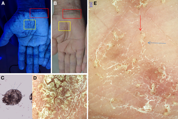 Figure 1
Peer Reviews
No public reviews on file for this paper yet. If you reviewed it on a platform where reviews are public (OpenReview, ICLR, NeurIPS, ICML), you can paste yours below so the community can read it here.
Videos
No videos yet. Explain this paper in a talk, walkthrough, or lecture? Add one.
Taxonomy
TopicsDermatological diseases and infestations · Nail Diseases and Treatments · Acne and Rosacea Treatments and Effects
Norwegian (crusted) scabies is a highly contagious form of scabies in which hyperkeratotic, scaly, highly pruritic lesions teem with mites. This syndrome usually occurs in immune-compromised individuals or in elderly, cognitively impaired, or bedridden individuals.1 Because the condition typically progresses from ordinary scabies, most patients still exhibit classic scabies rashes, such as scattered papulo-vesicles and burrows. The diagnosis is typically established by direct examination of mites or ova from scapings and/or biopsy visualized under light microscopy. With technological advancements, diagnostic methods such as Wood’s lamp examination and dermoscopy have been successively introduced, significantly improving the accuracy of clinical diagnoses.2^,^3 Dermoscopy and video-dermoscopy have the potential to enhance efficient and accurate diagnosis of scabies.
A 76-year-old wheelchair-bound man was admitted to the hospital with a 6-month history of generalized eczematous and pruritus dermatitis. Rash was observed on the neck, trunk, scrotum, buttocks, and extremities. Both wrists exhibited hyperkeratotic, fissured, oyster-shell–like plaques devoid of vesicles.
Two modalities were used to observe the rashes. Wood’s lamp (UV light) examination was used to observe the fluorescent reaction of the mites and their burrows (Figure 1A). Dermoscopy was performed under polarized light (20×, Dermat, Beijing, China); and the images were captured with a smartphone. Polarized dermoscopy showed the sinuous burrow and the brown jet-shaped triangular structure of a translucent mite at the ends of the burrow (Figure 1E). Mites were observed moving rapidly and irregularly (recorded under video-dermoscopy) (Supplemental Video 1). With the guidance of dermoscopic and video-dermoscopic findings, direct microscopic examination confirmed the diagnosis of crusted scabies by the direct observation of mites and their burrows (Figure 1C). The morphology of the mites, their sinuous burrows, and their dynamic movements was notable.4^,^5
The movements of sarcoptic mites on the surface of the skin observed by dermoscopy are different than the static state observed by microscopy. This visualization of actively moving mites rapidly confirms its highly contagious nature and enables efficient initiation of infection control practices.
This patient was successfully treated with a continuous three-day application of 10% sulfur ointment without bathing. An alternative treatment could have been topical permethrin or oral ivermectin. No other cause of immunocompromise was identified. To eliminate the risk of interpersonal transmission, close contacts were required to wear disposable gloves during any physical interaction, and where appropriate, received prophylactic therapy.
Supplemental Materials
10.4269/ajtmh.25-0451Supplemental Materials
The reference list from the paper itself. Each links out to its DOI / PubMed record.
- 1Engelman D, , 2020. The 2020 international alliance for the control of scabies consensus criteria for the diagnosis of scabies. Br J Dermatol 183: 808–820.32034956 10.1111/bjd.18943 PMC 7687112 · doi ↗ · pubmed ↗
- 2Tang J You Z Ran Y, 2017. Simple methods to enhance the diagnosis of scabies. J Am Acad Dermatol 80: e 99–e 100.10.1016/j.jaad.2017.07.03830981314 · doi ↗ · pubmed ↗
- 3Chen XB Wang TT Man XY, 2024. An 80-year-old woman with severe pruritic erythematous and hyperkeratotic plaques. Lancet Infect Dis 24: e 415.38789157 10.1016/S 1473-3099(24)00061-6 · doi ↗ · pubmed ↗
- 4Meduri AR Ciccarese G Viola R Sbarra G Cazzato G Romita P Foti C, 2024. Role of ultraviolet dermoscopy in detecting scabies signs. Skin Res Technol 30: e 70080.39366902 10.1111/srt.70080 PMC 11452253 · doi ↗ · pubmed ↗
- 5Betti S Bassi A Prignano F Lotti T, 2009. Scabies: Should we always perform dermatoscopy? G Ital Dermatol Venereol 144: 313–315.19528911 · pubmed ↗