Clinical resolution of oral lichenoid lesions after amalgam replacement: A systematic review and meta-analysis of observational studies
Harshita Kothari, Ajinkya M. Pawar, Pankaj Gupta, Alexander Maniangat Luke, Mohamed Saleh Hamad Ingafou, Parmeet Banga, Mohmed Isaqali Karobari, Dian Agustin Wahjuningrum

TL;DR
Replacing dental amalgam with biocompatible materials significantly increases the chances of resolving oral lichenoid lesions, though results are affected by study variability and publication bias.
Contribution
This study provides the first quantitative synthesis showing a strong association between amalgam replacement and OLL resolution.
Findings
Amalgam replacement increased lesion resolution odds by over 120 times compared to no replacement.
Subgroup analyses showed greater benefits when lesions had direct amalgam contact and when ceramics or gold were used.
Publication bias and moderate heterogeneity suggest the need for more rigorous, standardized studies.
Abstract
Oral lichenoid lesions (OLLs) can mimic oral lichen planus but are often linked to contact with dental amalgam. Replacement with biocompatible materials has been associated with lesion resolution, yet prior evidence lacked quantitative synthesis. Following PRISMA 2020 (PROSPERO: CRDXXXXXXXXXX2), eight databases (1986–2024) were searched for adult in vivo studies reporting OLL resolution after amalgam replacement. Two reviewers independently screened, extracted data, and assessed bias (ROBINS-I, Newcastle–Ottawa Scale, JBI). Random-effects meta-analysis estimated pooled odds ratios (OR) for clinical resolution; certainty was graded with GRADE. Of 2368 records, 35 studies were qualitatively synthesized and 11 entered meta-analysis (N = 365). Amalgam was replaced with glass ionomer, ceramics, composite resin, or gold alloys. Replacement markedly increased odds of lesion resolution (OR =…
Genes, proteins, chemicals, diseases, species, mutations and cell lines named across the full text — each resolved to its canonical identifier and authoritative record.
Click any figure to enlarge with its caption.
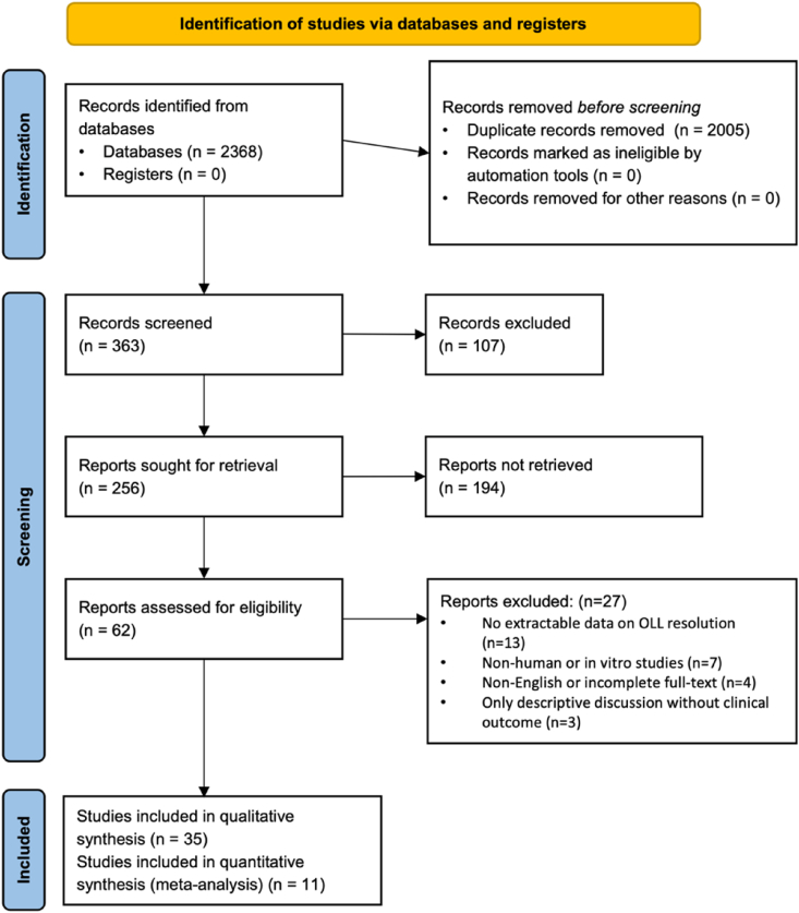 Figure 1
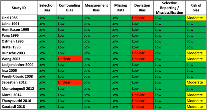 Figure 2
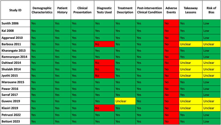 Figure 3
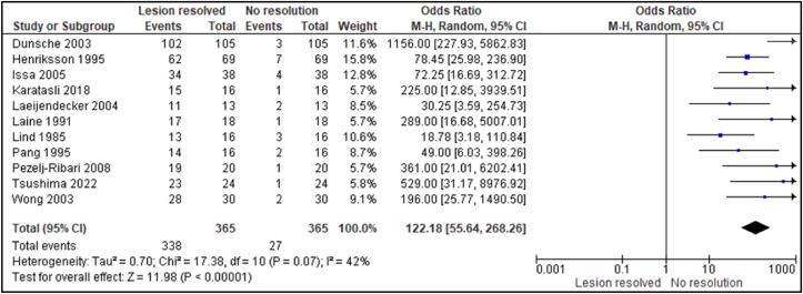 Figure 4
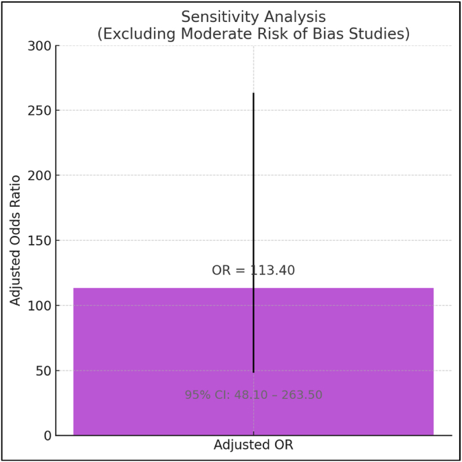 Figure 5
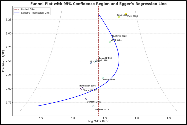 Figure 6
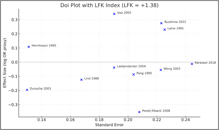 Figure 7
Peer Reviews
No public reviews on file for this paper yet. If you reviewed it on a platform where reviews are public (OpenReview, ICLR, NeurIPS, ICML), you can paste yours below so the community can read it here.
Videos
No videos yet. Explain this paper in a talk, walkthrough, or lecture? Add one.
Taxonomy
TopicsOral Health Pathology and Treatment · Oral health in cancer treatment · Oral and Craniofacial Lesions