Vein of Galen Aneurysmal Malformation: A Case Report With Insights Into Radiological Diagnosis Using Ultrasonography and Magnetic Resonance Imaging
Atul Kumar, Pragya Chaturvedi, Shrea Gulati, Sushma Pandey

TL;DR
This case report describes a rare brain malformation in a newborn diagnosed using ultrasound and MRI, emphasizing the importance of these imaging techniques for diagnosis and treatment planning.
Contribution
The paper provides a detailed case study demonstrating the complementary use of ultrasonography and MRI in diagnosing vein of Galen malformation in neonates.
Findings
Ultrasonography rapidly identified a large midline anechoic structure with intense flow in a neonate.
MRI and MRA precisely mapped the neurovascular anatomy, including arterial supply and absent deep venous drainage.
The case underscores the roles of ultrasonography and MRI in diagnosis and pretreatment planning for vein of Galen malformation.
Abstract
This report details the comprehensive radiological evaluation of a vein of Galen aneurysmal malformation in a neonate, using ultrasonography and magnetic resonance imaging. Vein of Galen malformation is a rare, congenital cerebral arteriovenous fistula and the leading cause of life-threatening high-output cardiac failure in neonates. A late preterm male infant presented with worsening tachypnea and generalized body swelling. Initial cranial ultrasonography revealed a large, midline anechoic structure with intense flow on color Doppler. Subsequent multiplanar magnetic resonance imaging and magnetic resonance angiography precisely delineated the neurovascular anatomy, showing a markedly dilated median prosencephalic vein, arterial supply from the left posterior cerebral artery, and the absence of normal deep venous drainage. This case highlights the critical, complementary roles of…
Genes, proteins, chemicals, diseases, species, mutations and cell lines named across the full text — each resolved to its canonical identifier and authoritative record.
Click any figure to enlarge with its caption.
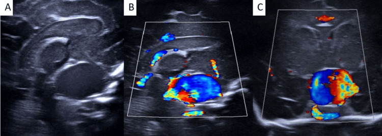 Figure 1
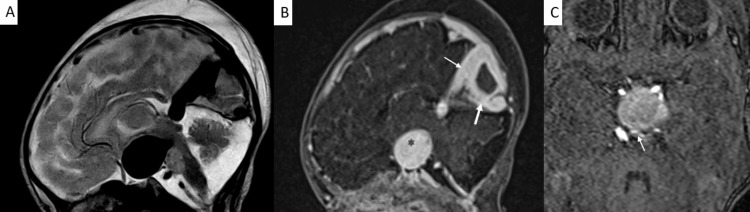 Figure 2
Peer Reviews
No public reviews on file for this paper yet. If you reviewed it on a platform where reviews are public (OpenReview, ICLR, NeurIPS, ICML), you can paste yours below so the community can read it here.
Videos
No videos yet. Explain this paper in a talk, walkthrough, or lecture? Add one.
Taxonomy
TopicsVascular Malformations Diagnosis and Treatment · Intracranial Aneurysms: Treatment and Complications · Meningioma and schwannoma management