Label-free assessment of a microfluidic vessel-on-chip model with visible-light optical tomography reveals structural changes in vascular networks
Devin Veerman, Carlos Cuartas-Vélez, Tarek Gensheimer, Tomas van Dorp, Andries van der Meer, Nienke Bosschaart

TL;DR
This paper shows that optical coherence tomography can monitor vascular network changes in a lab model, offering insights into diseases like retinal disorders.
Contribution
The study introduces optical coherence tomography as a label-free, real-time imaging method for vessel-on-chip models.
Findings
Optical coherence tomography can detect and quantify vascular network changes over multiple days.
The technique provides non-invasive, label-free imaging of vascular structures in organ-on-chip systems.
It has potential to correlate clinical metrics with disease mechanisms in models like retinal diseases.
Abstract
Microvascular dysfunction is characterized by impaired structure and function of small blood vessels, contributing to disease-related tissue and organ damage, such as in the retina. Optical coherence tomography is a widely used clinical technology to detect, monitor and diagnose disorders of the retina and choroid, such as diabetic retinopathy, macular degeneration, and various inherited retinal diseases. Currently, there are limited experimental platforms that correlate observed changes in clinical metrics with underlying mechanisms of disease progression. Organ-on-chips have the potential to offer a platform for correlative studies. Previous studies have demonstrated that the three-dimensional complexity of the microvasculature can be captured in a vessel-on-chip. Yet, current vessel-on-chip imaging analysis is based on end-point read-outs that provide limited dynamic information and…
Genes, proteins, chemicals, diseases, species, mutations and cell lines named across the full text — each resolved to its canonical identifier and authoritative record.
Click any figure to enlarge with its caption.
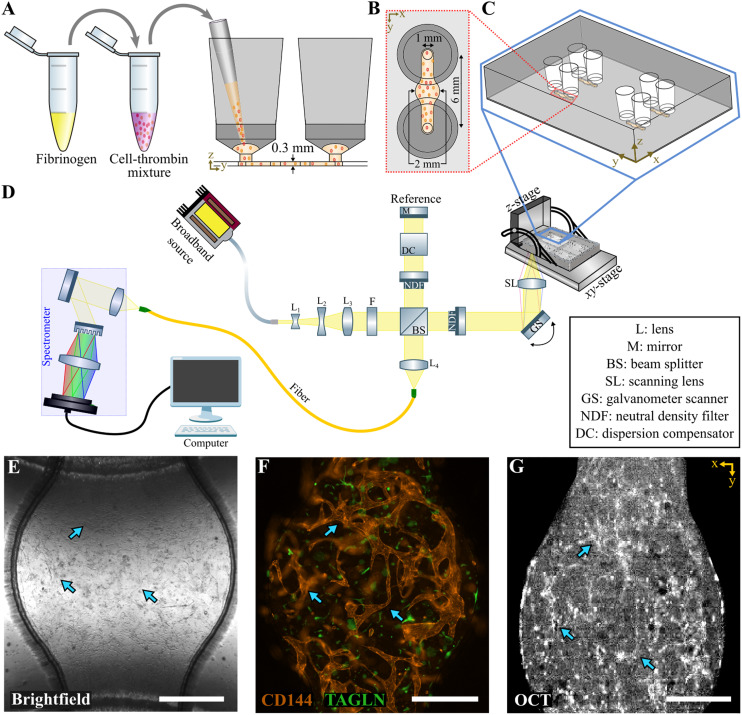 Figure 1
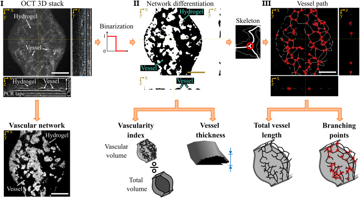 Figure 2
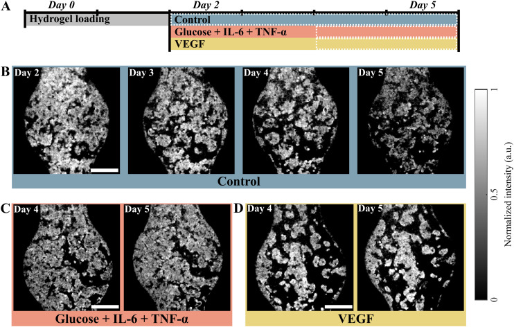 Figure 3
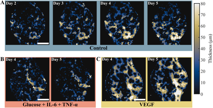 Figure 4
 Figure 5
 Figure 6
 Figure 7
 Figure 8
Peer Reviews
No public reviews on file for this paper yet. If you reviewed it on a platform where reviews are public (OpenReview, ICLR, NeurIPS, ICML), you can paste yours below so the community can read it here.
Videos
No videos yet. Explain this paper in a talk, walkthrough, or lecture? Add one.
Taxonomy
TopicsOptical Coherence Tomography Applications · Digital Holography and Microscopy · Optical Imaging and Spectroscopy Techniques