A corona-like distribution and patchy pattern of cerebellar infarcts identify patients with giant cell arteritis
Carolin Beuker, Jan-Kolja Strecker, Veith Jungmann, Nils Werring, Tobias Brix, Christian Thomas, Maximilian Christian Wankner, Antje Schmidt-Pogoda, Paul Stracke, Bernd Eckert, Thomas Raphael Meinel, Marcel Arnold, Jens Schaumberg, Schulamith Krüger, Milani Deb-Chatterji

TL;DR
This study identifies unique brain infarct patterns in patients with giant cell arteritis, which can help improve diagnosis and treatment timing.
Contribution
The study introduces a novel imaging-based diagnostic signature for intracranial giant cell arteritis using cerebellar infarct patterns.
Findings
A corona-like infarct pattern showed 79% sensitivity and 64% specificity for GCA.
A patchy infarct pattern was present in 53% of GCA cases with 93% specificity.
Combining both patterns increased specificity to 98% but reduced sensitivity to 47%.
Abstract
Cerebrovascular events are a potentially serious complication of giant cell arteritis (GCA) with intracranial involvement. However, diagnosing GCA in this context remains challenging, as classical clinical features may be absent. To identify characteristic cerebellar infarct patterns associated with intracranial GCA and to differentiate them from other common causes of posterior circulation stroke. Multicenter retrospective study. A total of 125 patients with cerebellar infarctions of various etiologies were included. Among these, 19 patients had confirmed intracranial GCA. Infarct patterns were compared to those seen in strokes of cardioembolic origin (n = 42), arterio-arterial embolism from proximal vertebral artery atherosclerosis (n = 13), local atherosclerotic stenosis of the V4 segment (n = 21), and vertebral artery dissection (n = 30). Infarct topography was assessed using…
Genes, proteins, chemicals, diseases, species, mutations and cell lines named across the full text — each resolved to its canonical identifier and authoritative record.
Click any figure to enlarge with its caption.
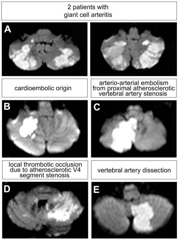 Figure 1
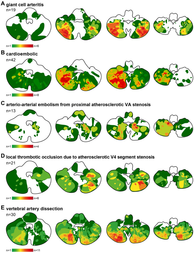 Figure 2
 Figure 3
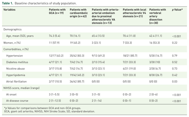 Figure 4
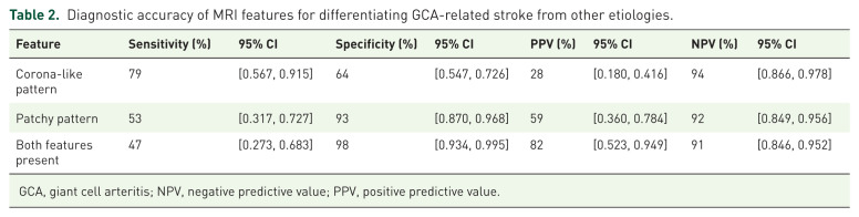 Figure 5
Peer Reviews
No public reviews on file for this paper yet. If you reviewed it on a platform where reviews are public (OpenReview, ICLR, NeurIPS, ICML), you can paste yours below so the community can read it here.
Videos
No videos yet. Explain this paper in a talk, walkthrough, or lecture? Add one.
Taxonomy
TopicsVasculitis and related conditions · Otitis Media and Relapsing Polychondritis · Whipple's Disease and Interleukins