Impact of low-energy virtual monoenergetic imaging in photon-counting CT for pre-TAVI pelvic arteries visualization
Leona S. Alizadeh, Christian Booz, Thomas J. Vogl, Ludovica R. M. Lanzafame, Simon S. Martin, Ibrahim Yel, Leon D. Gruenewald, Vitali Koch, Tommaso D’Angelo, Silvio Mazziotti, Kerstin Smolka, Grit Braunegger, Daniel Dillinger, Leonhard Kaatsch, Daniel Overhoff, Niklas Verloh

TL;DR
This study shows that low-energy virtual monoenergetic imaging in photon-counting CT improves image quality for planning heart valve procedures.
Contribution
The study demonstrates that 55 keV virtual monoenergetic images outperform traditional reconstructions in vascular imaging for TAVI.
Findings
Low-keV VMI reconstructions showed significantly higher SNR and CNR than polychromatic images.
55 keV VMI images scored highest in qualitative assessments for TAVI access site suitability.
VMI techniques may allow future reductions in contrast agent use for TAVI imaging.
Abstract
This study aimed to assess the impact of photon-counting computed tomography (PCCT) virtual monoenergetic images (VMI) on quantitative and qualitative parameters in abdominal and pelvic vascular imaging for transcatheter aortic valve implantation (TAVI) planning. A retrospective analysis of 125 patients undergoing dual-source PCCT scans before TAVI procedures was conducted. Reconstructions included polychromatic (T3D) images, leveraging multiple photon energy levels and VMI series spanning 40–100 keV in 15 keV increments. Quantitative parameters (signal-to-noise ratio [SNR] and contrast-to-noise ratio [CNR]) were evaluated. Qualitative assessments by three radiologists used clinically relevant five-point scales for overall image quality, TAVI access site suitability, and confidence in TAVI measurements. VMI reconstructions, particularly at 40 and 55 keV, demonstrated significantly…
Genes, proteins, chemicals, diseases, species, mutations and cell lines named across the full text — each resolved to its canonical identifier and authoritative record.
Click any figure to enlarge with its caption.
 Figure 1
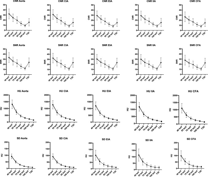 Figure 2
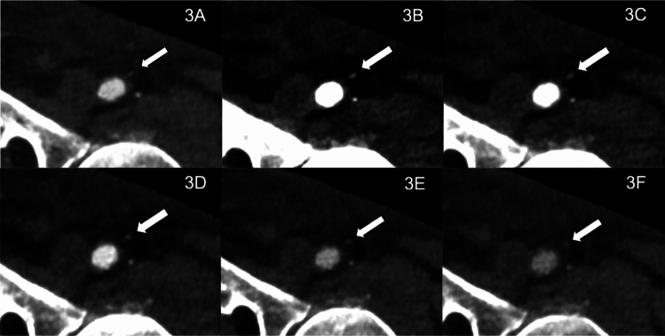 Figure 3
 Figure 4
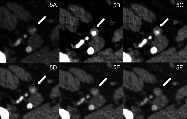 Figure 5
Peer Reviews
No public reviews on file for this paper yet. If you reviewed it on a platform where reviews are public (OpenReview, ICLR, NeurIPS, ICML), you can paste yours below so the community can read it here.
Videos
No videos yet. Explain this paper in a talk, walkthrough, or lecture? Add one.
Taxonomy
TopicsAdvanced X-ray and CT Imaging · Radiation Dose and Imaging · Cardiac Imaging and Diagnostics