Age-dependent progenitor switching shapes adult brown adipose tissue heterogeneity
Hai-Bin Ruan, Chenxin Gu, Zengdi Zhang, Shaolei Xiong, Jiawen Ma, Zuoxiao Shi, Zan Huang, Sunhye Shin, Aneesh Swaminathan, Ifrah Aden, Zahra Moazzami, Xiaoli Wu, Christina Camell, Yuwei Jiang

TL;DR
Brown fat tissue in adults is shaped by different progenitor cells that change with age, affecting metabolism.
Contribution
Discovery of an age-dependent progenitor switch that controls brown adipose tissue development and heterogeneity in adults.
Findings
Pdgfra+ progenitors contribute to BAT in early life but not in adults at room temperature.
Myl1+ progenitors emerge with age and generate brown fat cells with a unique metabolic profile.
Disruption of Myl1+ progenitors leads to reduced brown fat and impaired glucose metabolism in adult mice.
Abstract
The ontogeny of brown adipose tissue (BAT) begins during embryogenesis and continues into the postnatal period and throughout adulthood. Distinct populations of adipose progenitor cells (APCs) have been identified to support BAT development and thermogenesis; however, the division of labor and temporal relationship between different APCs, particularly during adulthood and aging, remain undetermined. Here, we showed that Pdgfra+ APCs establish BAT in early life but have a limited contribution to brown adipogenesis in adult mice housed at room temperature. Using integrative single-cell analysis and lineage tracing, we identified a distinct population of Myl1-expressing cells that emerge in an age-dependent manner and function as committed BAT progenitors in adult and middle-aged mice. Myl1+ APC-derived brown adipocytes possess a unique molecular signature that links to more dependence on…
Genes, proteins, chemicals, diseases, species, mutations and cell lines named across the full text — each resolved to its canonical identifier and authoritative record.
Click any figure to enlarge with its caption.
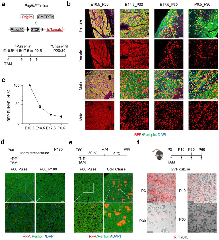 Figure 1
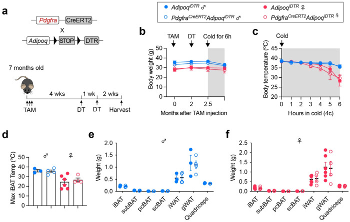 Figure 2
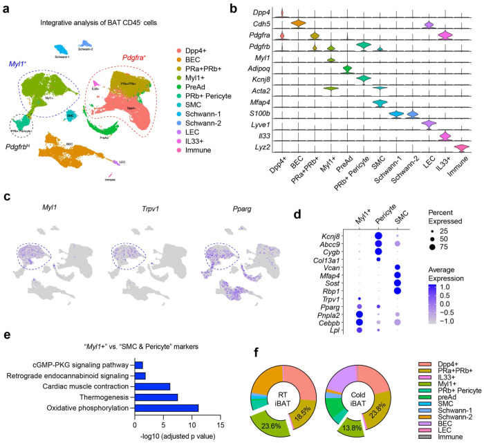 Figure 3
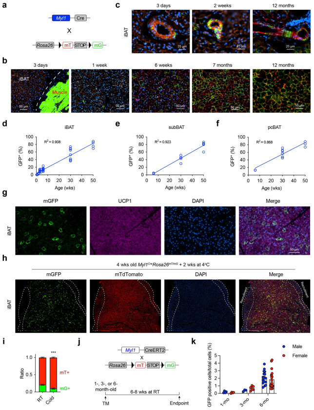 Figure 4
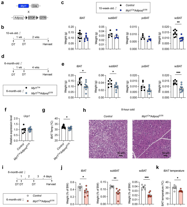 Figure 5
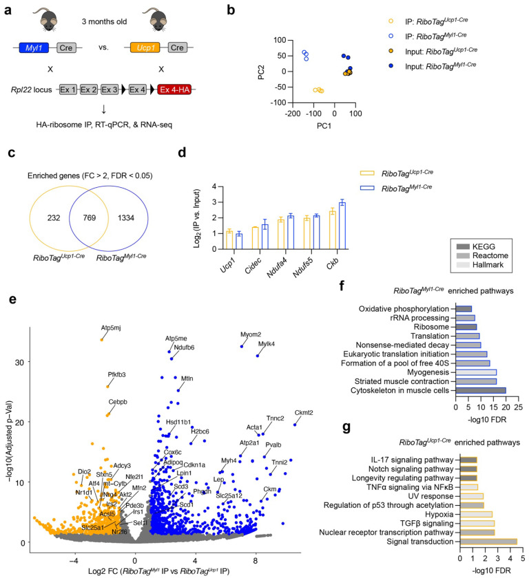 Figure 6
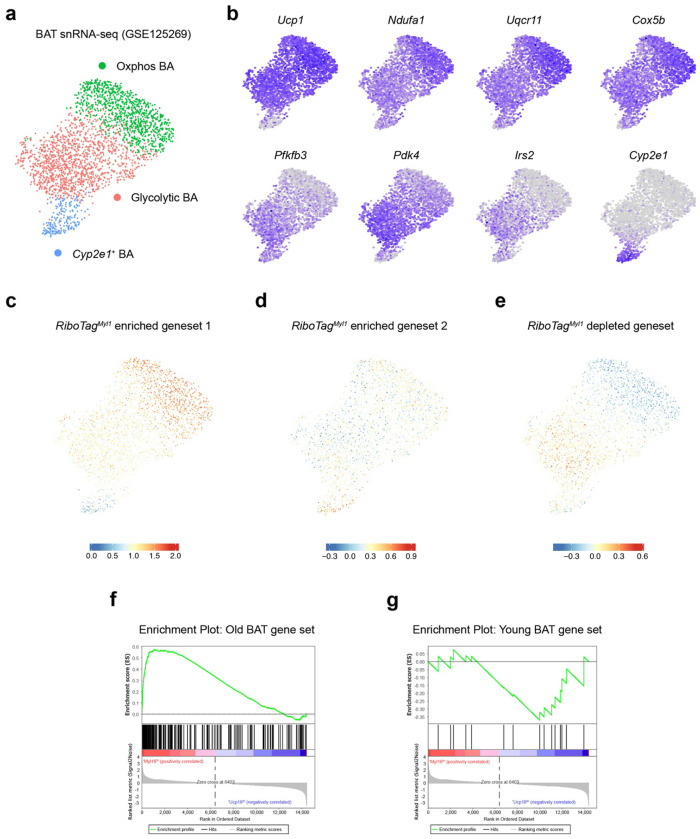 Figure 7
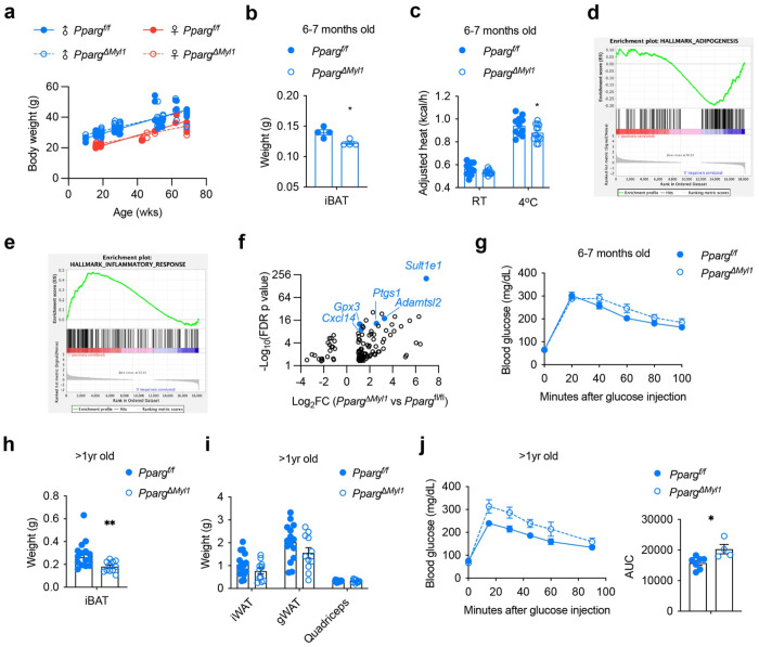 Figure 8
Peer Reviews
No public reviews on file for this paper yet. If you reviewed it on a platform where reviews are public (OpenReview, ICLR, NeurIPS, ICML), you can paste yours below so the community can read it here.
Videos
No videos yet. Explain this paper in a talk, walkthrough, or lecture? Add one.
Taxonomy
TopicsAdipose Tissue and Metabolism · Adipokines, Inflammation, and Metabolic Diseases · Cardiovascular Disease and Adiposity
Introduction
Brown adipose tissue (BAT) is a thermogenic organ conserved in placental mammals, and its prevalence is associated with metabolic health in adult humans^1–3^. Upon activation by cold, brown adipocytes consume large amount of glucose, fatty acids, and other fuels to generate heat via UCP1-dependent mitochondrial uncoupling and ATP-consuming futile cycles^4,5^. As an endocrine organ, BAT also secretes “batokines” and “lipokines” to regulate systemic homeostasis^6,7^. For these reasons, BAT transplantation and cell-based engineering have been shown to improve glucose metabolism and confer physiological benefits in preclinical models^7,8^. However, the prevalence of human BAT progressively decreases with age^9–11^, challenging the thermogenic capacity and therapeutical potential of BAT in middle-aged and elderly individuals. Mechanistic insights into the impact of age on BAT are urgently needed but significantly lacking.
Brown adipocytes share the same embryonic origins as myogenic cells^12^. The dermomyotome marked by Myf5 gives rise to dorsal BAT, including the interscapular depot^13–18^, while Tbx1^+^ progenitors within the pharyngeal mesoderm develop into supraclavicular BAT^19^. Single-cell RNA sequencing (scRNA-seq) and lineage tracing experiments have defined distinct populations of adipose progenitor cells (APCs) that control BAT development and homeostasis. Apparently, though not unequivocally tested, there are temporal waves of brown adipogenesis from different APCs. Pdgfra-expressing APCs, including Dpp4^+^ fascial cells, form interscapular BAT starting around embryonic day 14.5 in mice^20,21^. BAT expands rapidly after birth^22^, and Pdgfrb^+^ and Tbx18^+^ pericytes are the early postnatal APCs for BAT growth^23^. While the adipogenic potential of Pdgfra^+^ and Pdgfrb^+^ APCs can be unlocked by cold in adult mice^24–26^, their contribution to BAT homeostasis in animals housed at room temperature (RT) is rather limited^26–30^. In addition, a group of smooth muscle-like cells expressing Trpv1 and Myh11 is also an adipogenic source of interscapular and perivascular BAT^21,31^. And a subset of UCP1^+^ brown adipocytes can proliferate and contribute to BAT expansion^32,33^. However, the temporal relationship and division of labor between these heterogeneous groups of APCs are not known. Importantly, it remains undetermined if specific APCs exist during aging, in order to replenish dead brown adipocytes and support tissue turnover.
Growing evidence starts to reveal the metabolic heterogeneity of brown adipocytes. Compared to classic Ucp1-highly expressing brown adipocytes, a group of low-thermogenic brown adipocytes co-exist in BAT with larger lipid droplets and lower mitochondrial content^34^. Low-thermogenic brown adipocytes become abundant in thermoneutrality and aging^34,35^. On the other hand, cold exposure actively alters the composition of brown adipocyte subtypes^36^, and a group of lipogenic brown adipocytes may mediate thermogenic memory^37^. Furthermore, a subpopulation of Cyp2e1+ adipocytes, though rare in BAT, can robustly regulate the thermogenic capacity of classic brown adipocytes^38^. Despite these rapid advances in our understanding of the metabolic plasticity of BAT, it is unknown if such heterogeneity within mature brown adipocytes is established and influenced by progenitor diversity.
In this study, taking advantage of scRNA-seq, lineage tracing, and genetic cell ablation in mice, we uncover an age-dependent switch from embryonic Pdgfra^+^ APCs to adult Myl1^+^ APCs in maintaining BAT homeostasis. Brown adipocytes derived from Myl1^+^ APCs display unique molecular signatures, constitute BAT heterogeneity, and are essential for preserving BAT mass and metabolic fitness in midlife. Our study establishes the Myl1^+^ lineage as a preferable target for future strategies to recruit or engineer BAT.
Results
Minimal BAT adipogenesis from Pdgfra+ APCs in adult mice at room temperature.
The temporal contribution of Pdgfra^+^ APCs to BAT early development and adult homeostasis has not been explicitly investigated. Using the Pdgfra^RFP^(Pdgfra^CreERT2^; Rosa26^tdTomato^) fate mapping mice, we recently showed that Pdgfra^+^ APCs are essential to postnatal adipose tissue development but not to its adult formation^28^. Here, we performed temporal “pulse-chase” lineage tracing of BAT using the *Pdgfra^RFP^*mice housed at room temperature (RT, Fig. 1a). Brown adipocytes derived from Pdgfra^+^ APCs, as assessed by RFP/UCP1/Perilipin co-staining, were predominant in interscapular BAT (iBAT) at postnatal day 20 (P20) when pulsed at the embryonic day 10.5 (E10.5) (Fig. 1b). Notably, the contribution of Pdgfra^+^ APCs to BAT progressively declined if they were traced at later stages, including E14.5, E17.5, and postnatal day 0.5 (P0.5), in both females and males (Fig. 1b, c). Strikingly, the P60–P180 chase in adult mice produced minimal RFP^+^ adipocyte labeling in BAT (Fig. 1d), suggesting that Pdgfra^+^ APCs make little to no contribution to brown adipocytes in adulthood.
The low tracing rate of Pdgfra^+^ APCs in adult mice was unlikely due to the lack of adipogenesis, since multiple reports have demonstrated active BAT turnover during adulthood even at RT^23,34^. To confirm the fidelity of the Pdgfra^RFP^ line, we pulsed P60 mice and housed them at thermoneutrality (TN, 30°C) for 2 weeks, followed by another 2 weeks of cold challenge at 4°C (Fig. 1e). As expected, many iBAT adipocytes were labelled RFP (Fig. 1e), indicative of active adipogenesis from Pdgfra^+^ APCs induced by cold.
To directly test the adipogenic capacity of Pdgfra^+^ APCs, we labeled them with tamoxifen in RT -housed Pdgfra^RFP^ mice at P3, P10, P30, and P60, isolated and differentiated stromal vascular fraction (SVF) cells in vitro (Fig. 1f). By assessing RFP overlapped with lipid-containing adipocytes, we confirmed that Pdgfra^+^ cells exhibited minimal brown adipogenic potential in adulthood when compared to the early postnatal period (Fig. 1f). We conclude from these results that Pdgfra^+^ APCs are crucial for early development but do not actively differentiate in adult adipose tissue. Recruitment and activation of Pdgfra^+^ APCs require adipogenic stimuli such as high-fat feeding (in WAT) and increased thermogenic demand (in brown and beige adipose tissue) ^25,39–41^.
Pdgfra+ APCs contribute little to adult BAT homeostasis.
To determine the function of Pdgfra^+^ APCs in adult BAT, we generated an inducible adipocyte “deleter” mice (Adipoq^iDTR^) that express diphtheria toxin receptor (DTR) under the control of the Adipoq promoter in a Cre-dependent manner and crossed them to the Pdgfra^CreERT2^ (Fig. 2a). Tamoxifen (TAM) injection to 7-month-old Pdgfra^CreERT2^ Adipoq^iDTR^ mice induced DTR expression in newly formed adipocytes derived from Pdgfra^+^ APCs over a 4-week chase period, followed by two doses of diphtheria toxin (DT) to ablate these adipocytes. No differences in body weight between Adipoq^iDTR^ (control) and Pdgfra^CreERT2^ Adipoq^iDTR^ mice were observed in either males or females (Fig. 2b). Pdgfra^CreERT2^ Adipoq^iDTR^ mice were able to maintain their body temperature (Fig. 2c) and iBAT temperature (Fig. 2d) during acute cold challenge. Ablation of Pdgfra^+^ APC-derived adipocytes, if any, did not change tissue weight of various BAT and WAT depots, including iBAT, subscapular (sub) BAT, posterior cervical (pc) BAT, supraclavicular (sc) BAT, inguinal (i) WAT, and gonadal (g) WAT (Fig. 2e, f). Together, these data suggest that Pdgfra^+^ APCs play a minimal role in maintaining BAT homeostasis and function at the steady state.
Identification of Myl1-expressing APCs.
To identify alternative sources of brown adipogenesis within the stromal compartment, we performed an integrated analysis of scRNA-seq data from BAT SVF cells from three groups (Granneman^24^, Tseng^31^, and Seale^21^). Clustering of CD45 negative cells identified all major stromal subtypes (Fig. 3a, b, and Fig. S1a, b), including Pdgfra^+^/Dpp4^+^ interstitial progenitors^42^, Pdgfra^+^/Pdgfrb^+^ APCs^23^, Il33^+^/Pdgfra^+^ stromal cells^43^, Pdgfrb^hi^ pericytes^44^, Acta2^+^/Myh11^+^ smooth muscle cells (SMCs), Adipoq^+^/Ucp1^+^ brown (pre)adipocytes, blood endothelial cells (BECs), lymphatic endothelial cells (LECs), and Schwann cells. Notably, within this landscape, we identified a discrete population of Acta2^+^/Myh11^+^/Pdgfra^−^/Pdgfrb^lo^ cells uniquely labelled by Myl1, which encodes a striated muscle myosin light chain (Fig. 3b, c). These Myl1^+^ cells did not express canonical markers for SMCs (e.g., Kcnj8 and Abcc9) or pericytes (e.g., Vcan and Mfap4); instead, they expressed vascular smooth muscle-derived progenitor marker Trpv1^31^, together with adipogenic regulators (Pparg and Cebpb) and lipid metabolism genes (Lpl and Pnpla2) (Fig. 3c, d, and Table S1). Compared to Pdgfrb-expressing SMCs and pericytes, pathway analysis indicated that genes enriched in the Myl1^+^ cluster (fold change > 2; >50% expressing) were associated with oxidative phosphorylation, thermogenesis, and muscle contractile programs (Fig. 3e). Moreover, Myl1^+^ cells were the dominant stromal population that express niche cytokines, including Pdgfa, Notch3, and Jag1 (Fig. S1c), ligands known to restrain brown adipogenesis ^26,28,45^, suggesting potential regulatory crosstalk between Myl1^+^ cells and classical Pdgfra^+^/Pdgfrb^+^ APCs.
To assess depot specificity, we interrogated an integrated scRNA and single-nucleus(sn) RNA-seq WAT atlas containing ~300,000 cells from visceral and subcutaneous WAT^46^. We found no expression or enrichment of Myl1 in any stromal or mature adipocyte populations (Fig. S2), suggesting that Myl1^+^ cells are uniquely enriched in BAT. Finally, we also quantified stromal populations found in iBAT scRNA-seq from mice housed at RT or cold for 2-7 days. As previously reported, cold exposure increased Pdgfra^+^/Pdgfrb^+^ APC proportions; however, a higher percentage of Myl1^+^ cells was observed at RT than cold housing (Fig. 3f), suggesting that Myl1^+^ progenitors might be more active at the steady state when mice are adapted to the mild cold stress at RT.
Age-dependent adipogenesis from Myl1+ APCs
To examine if Myl1^+^ cells could function as brown APCs, we first performed cell marking by crossing a constitutive Myl1^Cre^ to the Rosa26^mT/mG^ reporter mice (Fig. 4a). As a myocyte-specific line, Myl1^Cre^ consistently labeled skeletal muscle cells as expected (Fig. 4b). Notably, a subpopulation of mGFP+ perivascular cells could be observed in iBAT from newborn to middle-aged mice (Fig. 4c). No mGFP+ brown adipocytes were detected at postnatal day 3, but scattered lineage-marked adipocytes started to be seen in 1-2 weeks old pups (Fig. 4b). The percent of Myl1-labeled adipocytes increased to ~15% at 6 weeks, ~50% at 7 months, and ~80% at 12 months in iBAT (Fig. 4b, d). A similar pace and percentage of Myl1^+^ marked brown adipocytes could also be observed in the subscapular (sub) and posterior cervical (pc) BAT depots (Fig. 4e, f). To confirm that mGFP-labeled adipocytes were bona fide brown adipocytes, we co-stained for UCP1 and found that mGFP^+^ and unlabeled adipocytes expressed comparable levels of UCP1 protein (Fig. 4g), validating their brown identity. We next asked whether age-related changes in Myl1 expression might account for the increased lineage labeling. Compared with the SVF fraction, mature brown adipocytes expressed little MYL1 protein, and this low level did not increase with aging (Fig. S3a). Myl1 transcription in SVF cells was likewise comparable between young and old mice (Fig. S3b). Thus, the age-dependent rise in labeling reflects cumulative differentiation from Myl1^+^ progenitors rather than increased Myl1 expression in mature adipocytes. Together, these data indicate that Myl1^+^ APCs contribute minimally to neonatal BAT but become the dominant progenitor pool in adulthood.
To assess whether Myl1^+^ APCs contribute to cold-induced adipogenesis, we challenged 4-week-old Myl1^Cre^; Rosa26^mT/mG^ mice with 2 weeks of cold exposure to induce de novo brown adipogenesis. As previously reported^25^, newly formed brown adipocytes were on the dorsal edge of the iBAT depot and stained strongly with DAPI (Fig. 4h). Notably, these cold-recruited regions were largely devoid of mGFP^+^ adipocytes (Fig. 4h), and cold exposure modestly reduced the overall proportion of Myl1-derived adipocytes (Fig. 4i). This aligns with scRNA-seq data showing a relative depletion of Myl1^+^ APCs in cold-stimulated BAT (Fig. 3f) and supports the existing dogma that Pdgfra^+^ and Pdgfrb^+^ progenitors are the primary source of cold-induced brown adipogenesis.
To perform temporal “pulse-chase” lineage tracing, we bred the Rosa26^mT/mG^ reporter to an inducible Myl1^CreERT^ line (Fig. 4j). Myl1^CreERT2^; Rosa26^mT/mG^ mice were pulsed with tamoxifen at 1, 3, and 6 months of age and chased for 6-8 weeks at RT. In both males and females, we observed an age-dependent increase in the differentiation of Myl1^+^ APCs into mature adipocytes (Fig. 4k and Fig. S3c). Together, these findings demonstrate that Myl1^+^ cells are bona fide APCs that contribute minimally during early life but increase their contribution to brown adipogenesis under homeostatic conditions in adulthood.
Myl1+ APCs are essential sources of adult brown adipocytes
To evaluate the functional relevance of Myl1^+^ APCs to BAT, we utilized the inducible adipocyte “deleter” mice (Adipoq^iDTR^) and crossed them to Myl1^Cre^ (Fig. 5a). We first ablated Myl1^+^ APC-derived adipocytes in young 10-week-old males with two doses of DT (Fig. 5b). No differences in body weight, WAT, or muscle mass (Fig. S4a) were noticed, suggesting no overall adversity or leakage of the model. Consistently with the low Myl1^+^ labeling at this age, BAT depots in interscapular, subscapular and posterior cervical regions were not evidently perturbed by DT/DTR-mediated ablation (Fig. 5c). Interestingly, a modest reduction of supraclavicular BAT (scBAT) in young Myl1^Cre^ Adipoq^iDTR^ mice was observed, suggesting that Myl1^+^ APCs may participate in the early establishment of this anatomically distinct and clinically relevant depot, a possibility warranting further investigation.
We next performed adipocyte deletion in 6-month-old Myl1^Cre^ Adipoq^iDTR^ male mice (Fig. 5d). Administration of DT did not alter the body weight, WAT, or skeletal muscle (Fig. S4b). Instead, a 30-50% reduction of BAT mass was evident in all depots examined (Fig. 5e), matching the lineage marking percentage at this age (Fig. 4). Despite the BAT paucity in these animals, relative Ucp1 gene expression in the remaining tissue was compared to controls (Fig. 5f). Reduced iBAT mass in DT-injected Myl1^Cre^ Adipoq^iDTR^ mice resulted in a reduction of iBAT temperature during cold challenge (Fig. 5g). Histological examination of iBAT from Myl1^Cre^ Adipoq^iDTR^ mice revealed a profound depletion of intracellular lipids after cold exposure (Fig. 5h), suggesting increased fat utilization to compensate for the BAT mass loss. It also indicates a different scenario in which Myl1^+^ APCs give rise to a distinct subset of brown adipocytes that disproportionately rely on lipid metabolism to support thermogenesis in adulthood.
In addition, we also deleted Myl1-lineage adipocytes in 6-month-old females (Fig. 5i), using an acute DT-mediated ablation protocol^47^. We observed a significant reduction of iBAT, subBAT, and scBAT weight (Fig. 5j), without negative impact on body weight change, WAT, or muscle weight (Fig. S4c). As a result, iBAT from female Myl1^Cre^ Adipoq^iDTR^ mice displayed lower temperature after 6 hours of cold challenge (Fig. 5k). These results demonstrate that Myl1^+^ APCs are important sources for adult BAT maintenance.
Myl1+ APC-derived brown adipocytespossess a unique molecular signature
To determine if brown adipocytes derived from Myl1^+^ APCs exhibit distinct molecular features, we performed ribosomal profiling to analyze cell-type-specific mRNA translation^48^. The “RiboTag” mice carrying a Cre-dependent HA-tagged ribosomal protein L22 (Rpl22) locus were crossed to Myl1^Cre^, allowing HA immunoprecipitation (IP) to isolate actively translating polyribosomes from Myl1^+^ APCs and their descendants (Fig. 6a). For comparison, Ucp1^Cre^ mediated RiboTag mice were generated to profile active mRNA translation in all brown adipocytes. Total iBAT samples from 3-month-old adult animals that had an adequate number of (~30%) Myl1^+^ APC-derived adipocytes were collected for analysis. RT-qPCR showed a similar enrichment of Ucp1, Dio2, and Fabp4 genes in *RiboTag^Myl1-Cre^*and *RiboTag^Ucp1-Cre^*ribosomes when normalized to input mRNA (Fig. S5a), while stromal genes (e.g., Fstl1^49^) were depleted, confirming experimental validity. We then performed RNA-seq, and principal component analysis (PCA) showed a distant separation between ribosomal IP samples from *RiboTag^Myl1-Cre^*and *RiboTag^Ucp1-Cre^*BAT (Fig. 6b). As expected, 4-way analysis revealed a significant overlap between *RiboTag^Myl1-Cre^*and *RiboTag^Ucp1-Cre^*ribosomal genes (Fig. 6c and Table S2), many of which were adipocyte, mitochondrial, and thermogenic markers, such as Ucp1, Adipoq, Cidec, Fabp4, Ndufa4, Ndufa6, and Ckb (Fig. 6d and Fig. S5b). On the other hand, markers for the SVF compartment, including APCs, endothelial cells, and immune cells, were similarly depleted between RiboTag^Myl1-Cre^ and RiboTag^Ucp1-Cre^ ribosomes (Fig. S5b-d).
We then analyzed differential genes between RiboTag^Myl1-Cre^ and *RiboTag^Ucp1-Cre^*IP samples (Fig. 6e, Fig. S5e, and Table S3). Pathway analysis revealed that RiboTag^Myl1-Cre^ ribosomes enriched genes involved in myogenesis and muscle contraction (likely due to the targeting of intra-BAT myocytes by Myl1^cre^), RNA/ribosome processing and translation, and oxidative phosphorylation (Oxphos) (Fig. 6f). Significantly upregulated genes from RiboTag^Myl1-Cre^ ribosomes encodes many Oxphos complex genes, such as Ndufb6, Ndufa12, Ndufv3, Uqcr11, Cox5b, Cox6c, and Cox7b (Fig. 6e and Table S3). Conversely, signaling transduction pathways relevant to thermogenesis, such as TGFβ, nuclear receptor, Notch, and TNFα signaling, were overrepresented in RiboTag^Ucp1-Cre^ ribosomal genes (Fig. 6g). Notably, genes related to insulin signaling (e.g., Irs1, Irs2, and Akt2) and glycolysis (e.g., Pfkfb3 and Pdk1) were less actively translated by RiboTag^Myl1-Cre^ ribosomes (Fig. 6e and Table S3). Together, these data reveal that Myl1^+^ APC-derived brown adipocytes adopt a distinct translational and metabolic identity characterized by enhanced oxidative phosphorylation and reduced glycolytic and insulin signaling programs.
Myl1+ APCs may give rise to Oxphos-high brown adipocytes
To determine how Myl1^+^ APC-derived adipocytes relate to known brown adipocyte subtypes, we reanalyzed a previously published snRNA-seq dataset of BAT^34^. After excluding non-adipocytes (Fig. S6a, b), three major subsets of brown adipocytes emerged at the UMAP space (Fig. 7a and Table S4): The first comprised Ucp1-high “Oxphos” adipocytes, which were enriched for mitochondrial respiratory chain genes (e.g., Ndufa1, Uqcr11, Cox5b). The second consisted of “glycolytic” adipocytes expressing glycolytic enzyme genes (Pfkfb3, Pdk4, Gk) and insulin signaling components (Irs2, Foxo1) (Fig. 7b). The third group, marked uniquely by Cyp2e1, corresponded to the recently described regulatory adipocyte population^38^.
We next compared these transcriptional subtypes with the *RiboTag^Myl1-Cre^*ribosome-enriched genes and listed the top ones that were expressed by mature adipocytes (Table S5). These genes mapped predominantly to Oxphos or Cyp2e1^+^ adipocytes (Fig. 7c, d). On the other hand, *RiboTag^Myl1-Cre^*ribosome-depleted genes were expressed by glycolytic and Cyp2e1^+^ adipocytes (Fig. 7e and Table S6). These patterns raise the possibility that Oxphos and glycolytic brown adipocytes arise preferentially from Myl1+ and non-Myl1^+^ APCs, respectively, whereas the regulatory Cyp2e1^+^ subtype may have mixed lineage origins.
To assess whether Myl1-derived adipocytes become more prominent with age, we analyzed the transcriptomes of BAT from young (2–4 months) and old (24 months) mice in two independent datasets^50,51^. Differentially expressed gene sets were used for GESA analysis (Table S7). We found that gene sets upregulated with aging were significantly enriched for *RiboTag^Myl1-Cre^*ribosomal genes (Fig. 7f), whereas aging-downregulated gene sets were enriched for *RiboTag^Ucp1-Cre^*ribosomal genes (Fig. 7g). These findings support the conclusion that Myl1^+^ APC-derived brown adipocytes increasingly dominate the adipocyte landscape during aging.
Myl1+ APC adipogenesis is required to maintain BAT integrity and glucose control in aging
To assess the physiological importance of the differentiation from Myl1^+^ APCs into adipocytes, we knocked out the master adipogenic transcription factor PPARγ driven by the Myl1^Cre^. No change in body weight was observed in both female and male Pparg^ΔMyl1^, compared to Pparg^f/f^ controls (Fig. 8a). By 6 to 7 months of age, a modest but significant reduction in iBAT weight was observed in male Pparg^ΔMyl1^ (Fig. 8b). This was associated with reduced heat production when challenging Pparg^ΔMyl1^ males with cold (Fig. 8c). RNA-sequencing followed with GESA analysis revealed that PPARγ deficiency was negatively correlated with adipogenesis, while positively linked to inflammation (Fig. 8d, e, and Table S8). Several upregulated transcripts were markers of Pdgfra^+^ APCs, including Gpx3, Cxcl12, Ptgs1, Sult1e1, and Adamtsl2 (Fig. 8f), suggesting that compensatory recruitment of Pdgfra^+^ APCs may occur when Myl1^+^ APCs differentiation is blocked. When subjected to a glucose tolerance test, Pparg^ΔMyl1^ animals showed a subtle increase in blood glucose levels (Fig. 8g).
We next examined the impact of long-term loss of Myl1-lineage adipogenesis. After more than 1 year of aging, a consistent reduction in iBAT mass was found in both male (Fig. 8h) and female Pparg^ΔMyl1^ mice (Fig. S7a). As expected, WAT and skeletal muscle weight remained unaffected in old males (Fig. 8i) and females (Fig. S7b), suggesting that Myl1^+^ APC was not an essential source for white adipogenesis and PPARγ was dispensable for myocytes. Notably, aged Pparg^ΔMyl1^ males displayed significantly impaired glucose tolerance (Fig. 8j). Collectively, these findings demonstrate that adipogenesis from Myl1^+^ APCs is essential for maintaining BAT mass and supporting metabolic health during aging, highlighting their importance as a protective progenitor pool in later life.
Discussion
BAT undergoes continuous remodeling across the lifespan, yet the progenitor sources sustaining this turnover have remained largely undefined. Here we identify an age-dependent progenitor-switching mechanism in which embryonic and early postnatal Pdgfra^+^ APCs establish BAT, whereas a distinct population of Myl1^+^ APCs progressively becomes the dominant source of brown adipocytes in adulthood. This transition provides a mechanistic framework that explains why early APCs contribute minimally to homeostatic brown adipogenesis in adulthood but are rather primarily recruited in response to strong thermogenic stimuli.
Waves of cellular differentiation from distinct types of stem and progenitor cells across the lifespan are common phenomena. In many systems, fetal programs and progenitors can be reactivated following injury and stress^52^. For instance, Lgr5^+^ intestinal stem cells are replaced by fetal-like revival stem cells upon helminth infection, which is crucial for type 2 immune regulation^53,54^. Similar principles likely apply to adipose tissues. We propose that acute cold-induced adipogenic differentiation from early Pdgfra^+^ and/or Pdgfrb^+^ APCs represents a stress-activated fetal programming in BAT. While in mice housed at thermoneutrality or adapted to the mild cold at RT, adipogenesis from Myl1^+^ APCs supports BAT homeostasis and turnover. Such temporal transitions (developmental vs. adult) and context-dependent utilization (homeostasis vs. obesity and aging) of APCs are also increasingly recognized in WAT^55,56^.
Myl1^+^ APCs form a transcriptionally distinct, BAT-restricted stromal population enriched for Oxphos genes, niche ligands, and adipogenic regulators, while lacking canonical markers of SMCs. Consistent with the findings from scRNA-seq integration, lineage tracing experiments have repeatedly shown that SMCs labelled by Myh11 or Acat2 do not give rise to iBAT adipocytes^21,57,58^. Instead, a distinct population of SMC-like cells marked by Trpv1^31^ and Myl1 (this study) are adipogenic. Our current study demonstrates for the first time the age-dependent recruitment of Myl1^+^ APCs. More importantly, progenitor origin underlies metabolic heterogeneity within BAT: Myl1^+^ APCs generate OxPhos-high adipocytes, whereas non-Myl1 progenitors contribute more glycolytic populations, with regulatory Cyp2e1^+^ adipocytes likely reflecting a mixed lineage. Future experiments incorporating fate mapping with single-cell metabolic and functional profiling are required to determine the route of BAT heterogeneity.
Myl1^+^ APCs are essential for maintaining BAT mass, preserving thermal homeostasis, and sustaining glucose control during aging. Conditional deletion of Pparg in Myl1^+^ APCs abolished their differentiation into brown adipocytes, resulting in marked BAT loss, cold intolerance, and impaired glucose metabolism. These findings indicate that Myl1^+^ APCs are not only recruited in adulthood but also function as obligately PPARγ-dependent progenitors whose adipogenic output is particularly critical for BAT integrity during aging.
Despite these insights, several mechanistic questions remain. The developmental origin of Myl1^+^ APCs is still unresolved. They may represent a fate-restricted derivative of embryonic Pdgfra^+^ stromal progenitors that undergo lineage maturation, or an independent mesenchymal lineage emerging from muscle-associated or vascular smooth muscle–derived progenitors. The stability of Myl1 expression across age and its absence in neonatal APCs argue against simple activation of the Pdgfra^+^ lineage. Resolving whether this lineage transition is developmentally programmed or environmentally induced will require a temporally resolved dual-recombinase that provides resolution beyond what classical Cre-based lineage tracing can achieve.
Our data also raises the question of what signals activate Myl1^+^ APCs. Their enrichment at room temperature and depletion during acute cold exposure suggest that mild, chronic thermogenic demand, not β-adrenergic stimulation, governs their recruitment. Myl1^+^ APCs express PDGF, Notch, and TGFβ ligands, which are capable of modulating progenitor competence, raising the possibility of autocrine or paracrine regulation. The mitochondrial metabolic state may further influence APC identity; a high-Oxphos, low-sympathetic niche may favor the activation of Myl1^+^ APCs over classical progenitors.
Our data support a model of reciprocal crosstalk among APC pools. The expression of inhibitory ligands, such as Pdgfa, Notch3, and Jag1, in Myl1^+^ APCs suggests that they may restrain adipogenesis in Pdgfra^+^/Pdgfrb^+^ APCs, coordinating a temporal handoff from neonatal to adult progenitor dominance. The compensatory activation of Pdgfra^+^ signatures when Myl1-lineage adipogenesis is blocked further supports a flexible, competitive hierarchy. Aging adds another layer of complexity. Our transcriptomic analyses reveal that aging-upregulated BAT gene sets are enriched for Myl1-lineage translational signatures, indicating that Myl1^+^ APCs may compensate for age-associated declines in other APC pools. Myl1^+^ APCs may therefore represent a stress-resistant progenitor population uniquely suited to preserving BAT function in late life. Whether their dominance reflects intrinsic increases in adipogenic potential or preferential survival of Myl1-derived adipocytes remains unresolved.
Collectively, our findings uncover a previously unrecognized progenitor-switching mechanism that governs adult BAT renewal and shapes brown adipocyte heterogeneity. We establish Myl1^+^ APCs as a specialized adult progenitor pool with distinct metabolic, molecular, and functional properties, whose PPARγ-dependent differentiation is indispensable for maintaining BAT homeostasis during aging. These insights highlight new opportunities to target progenitor identity and stromal crosstalk to preserve BAT function in metabolic disease.
Methods
Animals
All animal experiments were approved by the institutional animal care and use committee (IACUC) of the University of Minnesota and of University of Illinois Chicago and adhered to the NIH Guide for the Care and Use of Laboratory Animals. All the mice were group-housed in a light/dark cycle (12/12 h), temperature (21.5 ± 1.5 °C), and humidity-controlled (30-70%) room, and had free access to water and regular chow. Myl1^Cre^ (Jax #024713), Myl1^CreERT2^ (Jax #025670), Pdgfra^CreERT2^ (Jax #032770). Pparg^f/f^ (Jax #004584), Rosa2^LtdTomato^(Jax #007914), Rosa26^LSL-mT/mG^(Jax #007676), RiboTag (Jax #029977) mice were from Jackson Lab. The Adipoq-LSL-DTR (T058436) line was purchased from GemPharmatech.
For cold treatment, mice were housed in a temperature-controlled room (4°C) with free access to water. Core body temperature was measured using an electronic thermometer with an anal probe (Physitemp). Interscapular skin temperature was measured by anesthetizing mice with isoflurane and quickly capturing images using a FLIR C2 thermal camera. The average skin temperature within the interscapular region was analyzed using FLIR Thermal Studio.
For the glucose tolerance test, mice were fasted overnight for 16 hours and then intraperitoneally injected with 1.5 g/kg body weight of glucose. Blood glucose levels were measured using a glucometer at the indicated time points after injection.
Tamoxifen preparation
Tamoxifen (Sigma, T5648) was dissolved at a concentration of 20 mg/mL in corn oil containing 10% ethanol. Briefly, tamoxifen powder was weighed into an amber tube, wetted with ethanol, and brought to volume with sterile corn oil. The suspension was vortexed thoroughly and then sonicated or incubated at 37 °C with intermittent mixing until it was fully dissolved. The stock solution was aliquoted into light-protected tubes and stored at −20 °C for up to several weeks. On the day of use, aliquots were equilibrated to room temperature and vortexed immediately before injection to ensure homogeneity.
Timed breeding
For embryonic induction, timed matings were established by pairing one Pdgfra^RFP^ male with two sexually mature Pdgfra^RFP^ females in the late afternoon. The following morning, females were examined for the presence of a copulation plug by gently lifting the tail and visually inspecting the vaginal opening under bright light. Females with a visible vaginal plug were designated as embryonic day 0.5 (E0.5), separated into individual cages, and monitored daily for general health. Tamoxifen administration was scheduled based on this staging, with injections performed at the desired gestational days (e.g., E10.5, E14.5, or E17.5).
Histology
Adipose tissues were fixed in a formalin or PFA solution at 4°C for 24 hours. Tissue embedding, sectioning, and hematoxylin and eosin (H&E) staining were performed at the Comparative Pathology Shared Resource of the University of Minnesota. For immunostaining, antigen retrieval was performed in Citric buffer using a 2100 Retriever (Aptum Biologics). After incubation with blocking buffer (3% BSA in PBS) for 1 hour, sections were incubated overnight at 4°C with the primary antibody in blocking buffer. For immunofluorescence, PBS-washed slides were incubated with a fluorescent secondary antibody at room temperature for 1 hour and then mounted with VECTASHIELD Antifade Mounting Medium with DAPI after three washes in PBS. A Keyence all-in-one fluorescence microscope was used for imaging.
RT-qPCR
After weight measurement, BAT tissues were homogenized in TRIzol (Thermo Scientific) for RNA isolation, following the manufacturer’s protocol. RNA concentrations were measured with a NanoDrop spectrophotometer. Reverse transcription was performed with the iScript^™^ cDNA Synthesis Kit. Real-time RT-PCR was conducted using iTaq^™^ Universal SYBR^®^ Green Supermix and gene-specific primers on a Bio-Rad C1000 Thermal Cycler. Relative expression was normalized to the housekeeping Rplp0 gene.
SVF isolation, culture, and differentiation
BAT depots were collected and minced in 10 ml of digestion buffer (DMEM/F12 with 1mg/ml Collagenase I, 1% FBS, 1% HEPES, and 1% Pen-Strep). After shaking at 37°C and 100 rpm for 45 min, the digested tissues were filtered through 70-μm strainers and centrifuged at 1,500 rpm for 3 min. The pellets were resuspended in ACK buffer and put on ice for 5 min to remove red blood cells. The ACK buffer was neutralized with 5 ml of DMEM/F12 plus 10% FBS and removed after a 1,500-rpm centrifugation for 3 min. SVF cells were cultured with DMEM/F12 containing 20% FBS, 1% Pen-Strep, and 10 μg/ml Ciprofloxacin.
To test intrinsic adipogenic capacity, SVF cells were isolated from Pdgfra^RFP^ mice pulsed at P3, P10, P30, or P60. SVF cultures were differentiated in vitro under standard adipogenic conditions. Briefly, confluent cells were induced with DMEM/F12 containing 10% FBS, 1x Pen-Strep, 20 nM insulin, 1 nM T3, 0.5 mM IBMX, 5 μM dexamethasone, and 125 μM indomethacin. Two days later, the cells were maintained in DMEM/F12 containing 10% FBS, 1x Pen-Strep, 20 nM insulin, and 1 nM T3. Medium was changed every other day until lipid droplets appeared.
Ribosomal profiling and RNA-seq
RiboTag experiments were carried out following an established protocol^59^. Briefly, iBAT samples from RiboTag^Myl1-Cre^ and RiboTag^Ucp1-Cre^ mice were harvested and lysed in ice-cold homogenization buffer with Dounce tissue grinders. Lysate was cleared by centrifugation for 10 min at 10,000 g, 4°C. A small aliquot was stored at −80°C for subsequent Input analysis. The remaining lysate was incubated with anti-HA antibody and protein A/G beads for overnight at 4°C. Next day, beads were centrifuged and washed with high salt buffer for 3 times. RNA was extracted using the Qiangen RNeasy mini kit as described in the instructions. RNA quality was determined by an Agilent 2100 Bioanalyzer. Total RNA from Input and IP samples were subjected to dual-indexed TruSeq stranded mRNA library preparation and sequenced on a NovaSeq 2x150-bp run at the University of Minnesota Genomics Center. A pipeline developed and maintained by the Research Informatics Solutions (RIS) group at the University of Minnesota Informatics Institute (UMII) was used for RNA-seq analysis. Differentially expressed genes were detected using DEseq2 or EdgeR.
To compare BAT transcriptome of young and old mice, two publicly available bulk RNA-seq datasets (GSE135391 and GSE141252) were used. Raw gene counts from GSE135391 (including 3- and 24-month-old mice at RT) were extracted from ARCHS^4^ (All RNA-seq and ChIP-seq sample and signature search)^60^, and merged with those from GSE141252 (BAT from 4 and 24 month old mice) using R. Differentially expressed genes (fold change > 2 and adjust P value < 0.1) were determined with iDEP (integrated Differential Expression & Pathway analysis, v 2.20.5) with the correction for batch effect^61^. Significantly upregulated and downregulated gene sets were created (Table. S7) for Gene Set Enrichment Analysis (GSEA, v 4.4.0)^62^.
Analysis of scRNA-seq and snRNA-seq
Seurat (v5.0.0) on RStudio (2023.12.0, R version 4.3.2) was used for integrative scRNA-seq analysis of GSE207706 (iBAT from mice housed at RT or cold 4 days)^24^, GSE160585 (iBAT from mice housed TN for 1 week, RT, or cold for 2 or 7 days)^31^, and GSM5068995 (paBAT from adult mice)^21^. Individual samples were filtered (> 100 features, < 4, 000 features and < 10% of genes mapped to mitochondria) and normalized with the SCTransform function. Integration was performed with the FindIntegrationAnchors function with “rpca” reduction and the IntegrateData function with “SCT” as the normalization method. After finding neighbors, clusters, and markers, the subset of lineage-negative cells was created and re-clustered (dims = 1:30, resolution = 0.1). UMAP and tSNE were then used for two-dimensional visualization of the resulting cluster. Marker genes were identified using the FindAllMarkers function (only.pos = TRUE, min.pct = 0.25, logfc.threshold = 0.25). Violin plots, dot plots, heatmaps, and individual tSNE and UMAP plots for the given genes were generated by using the VlnPlot, DotPlot, DoHeatmap, and FeaturePlot functions, respectively.
snRNA-seq data of freshly isolated mouse brown adipocytes (GSM3567479) was ^ltered with > 200 features, < 3, 000 features and < 75% of genes mapped to mitochondria. Seurat was used to NormalizeData, ScaleData, FindVariableFeatures, RunPCA, FindNeighbors, FindClusters (resolution = 0.2), and RunUMAP (dims = 1:30). The subset of brown adipocytes was clustered again to FindAllMarkers. RiboTag^Myl1-Cre^ ribosome-enriched and -depleted genes were visualized in the UMAP space with the AddModuleScore and FeaturePlot functions.
Quantification and statistical analysis
Results are shown as mean ± SEM or ± SD. N values (biological replicates) and statistical analysis methods are described in the figure legends. The statistical comparisons were carried out using two-tailed Student’s t-test and one-way or two-way ANOVA with indicated post hoc tests with Prism (Graphpad). Differences were considered significant when p < 0.05. *, p < 0.05; **, p < 0.01; ***, p < 0.001.
Supplementary Material
This is a list of supplementary files associated with this preprint. Click to download.
The reference list from the paper itself. Each links out to its DOI / PubMed record.
- 1Cannon B. & Nedergaard J. Brown adipose tissue: function and physiological significance. Physiol Rev 84, 277–359 (2004).14715917 10.1152/physrev.00015.2003 · doi ↗ · pubmed ↗
- 2Kajimura S., Seale P. & Spiegelman B. M. Transcriptional control of brown fat development. Cell Metab 11, 257–262 (2010). 10.1016/j.cmet.2010.03.00520374957 PMC 2857670 · doi ↗ · pubmed ↗
- 3Becher T. Brown adipose tissue is associated with cardiometabolic health. Nat Med 27, 58–65 (2021). 10.1038/s 41591-020-1126-733398160 PMC 8461455 · doi ↗ · pubmed ↗
- 4Townsend K. L. & Tseng Y. H. Brown fat fuel utilization and thermogenesis. Trends Endocrinol Metab 25, 168–177 (2014). 10.1016/j.tem.2013.12.00424389130 PMC 3972344 · doi ↗ · pubmed ↗
- 5Roesler A. & Kazak L. UCP 1-independent thermogenesis. Biochemical Journal 477, 709–725 (2020). 10.1042/bcj 2019046332059055 · doi ↗ · pubmed ↗
- 6Ziqubu K. Brown adipose tissue-derived metabolites and their role in regulating metabolism. Metabolism 150, 155709 (2024). 10.1016/j.metabol.2023.15570937866810 · doi ↗ · pubmed ↗
- 7Li V. L., Kim J. T. & Long J. Z. Adipose Tissue Lipokines: Recent Progress and Future Directions. Diabetes 69, 2541–2548 (2020). 10.2337/dbi 20-001233219098 PMC 7679773 · doi ↗ · pubmed ↗
- 8White J. D., Dewal R. S. & Stanford K. I. The beneficial effects of brown adipose tissue transplantation. Mol Aspects Med 68, 74–81 (2019). 10.1016/j.mam.2019.06.00431228478 PMC 6708446 · doi ↗ · pubmed ↗