Longitudinal changes in gut microbiota across reproductive states in wild baboons
Chelsea A. Southworth, Logan Barrios, Mauna R. Dasari, Laurence R. Gesquiere, Jack A. Gilbert, Susan C. Alberts, Jenny Tung, Elizabeth A. Archie

TL;DR
The study shows that gut microbiota in wild baboons changes significantly during different reproductive states, with pregnancy causing the most dramatic shifts.
Contribution
The study provides new insights into how gut microbiota dynamics are linked to reproductive states and steroid hormone levels in wild baboons.
Findings
Pregnancy in baboons is associated with distinct changes in gut microbiota compared to other reproductive states.
Microbial shifts during pregnancy are linked to taxa associated with host immunity, weight gain, and hormone levels.
Host identity is the strongest predictor of gut microbiota composition, especially during pregnancy.
Abstract
In humans and other mammals, female reproduction is linked to extensive changes in physiology, immunity, hormones, and behavior. These changes likely shape, and may be shaped by, the composition of gut microbial communities. Characterizing the dynamics of gut microbial change across reproductive states, including its relationship to female physiology, is important for understanding how the gut microbiota influences female and offspring health. Here we characterize longitudinal changes in gut microbiota across reproduction by combining 16S rRNA gene sequencing data from 4,462 stool samples (spanning 14 years of sample collection) with life history data on multiple reproductive events in 169 female baboons. These baboons were members of a well-studied, natural baboon population in Kenya where reproductive state (ovarian cycling, pregnancy, and postpartum amenorrhea) is tracked daily and…
Genes, proteins, chemicals, diseases, species, mutations and cell lines named across the full text — each resolved to its canonical identifier and authoritative record.
Click any figure to enlarge with its caption.
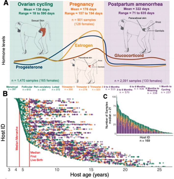 Figure 1
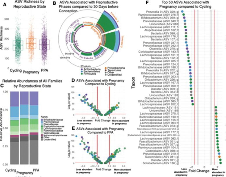 Figure 2
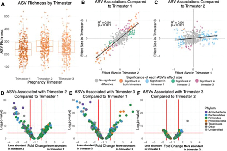 Figure 3
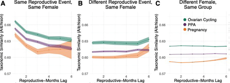 Figure 4
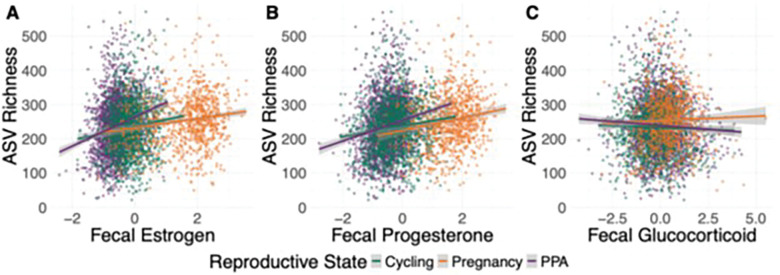 Figure 5
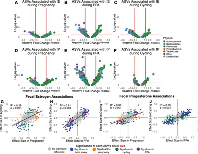 Figure 6
- —National Science Foundation (NSF)
- —National Institutes of Health (NIH)
- —Jack Kent Cooke Foundation Graduate Scholarship, NSF Integrated Data Science Fellowship, Duke University Population Research Institute
- —Notre Dame’s Eck Institute for Global Health, Environmental Change Initiative, Max Planck Institute for Evolutionary Anthropology, Duke University, Princeton University, and the University of Notre Da
Peer Reviews
No public reviews on file for this paper yet. If you reviewed it on a platform where reviews are public (OpenReview, ICLR, NeurIPS, ICML), you can paste yours below so the community can read it here.
Videos
No videos yet. Explain this paper in a talk, walkthrough, or lecture? Add one.
Taxonomy
TopicsGut microbiota and health · Reproductive tract infections research · Epigenetics and DNA Methylation
INTRODUCTION
Mammalian gut microbiota are highly complex, personalized microbial communities whose dynamics both reflect their host’s physical state and can have direct effects on host health [1, 2]. Changes in female reproductive state may be some of the most important drivers of these dynamics, but current evidence on the importance of reproductive state remains cross-sectional and largely limited to lab and domestic animals [3–6]. As female mammals transition from conceiving, to gestating, to caring for offspring, they experience changes in physiology, immunity, and behavior that could alter gut microbial composition, particularly during pregnancy [6–14]. Females’ social relationships also change across reproduction, and changes in the rate of social interaction could also change microbial exposures [8, 13, 15–18]. Gut bacteria may also play a causal role in some of these changes, influencing hormone levels, immunity, metabolism, and behavior [19–23]. For instance, experiments that transplanted microbiota from pregnant women to germ free mice implicate gut bacteria in weight gain during pregnancy[24]. Understanding gut microbial dynamics across female reproduction is therefore relevant to maternal health, as well as to reproductive outcomes in humans and other animals, including preeclampsia, preterm birth, litter size, and the likelihood of entering estrus [25–28].
Despite multiple potentially important roles for the gut microbiota in female reproduction, studies that trace longitudinal changes in female gut microbial communities as females transition between reproductive states are lacking. Such data are necessary to understand how gut microbial dynamics align with female reproduction, which aspects of these dynamics are shared across hosts, and which are unique to individual hosts [29, 30]. However, collecting longitudinal data on gut microbial composition is challenging: in humans, collecting repeated samples from the same individual over time and across reproductive states is difficult, while in most other mammals, one or more stages of female reproduction are cryptic, including ovulation and early pregnancy [31, 32]. This limitation makes it difficult to assign a given microbiota sample to a specific reproductive state.
Given that changes in steroid hormone physiology (e.g., estrogen, progesterone, glucocorticoids) are intimately involved in driving changes in mammalian reproductive state, studies of the reproductive dynamics of the gut microbiota would ideally take hormonal changes into account. However, data on hormone levels and gut microbial composition are rarely paired, especially in longitudinal study designs [33, 34]. Where paired data are available, studies have typically focused on a narrow band of women’s lives (e.g., menopause) or hormonal disorders that disrupt normal reproduction [35–37]. Nevertheless, existing work points to the importance of estrogen, progesterone, and glucocorticoids in altering gut microbiota [38–42]. Indeed, administering estrogen and progesterone to ovariectomized mice maintains the gut microbial diversity found in control mice, while diversity decreases in non-hormone treated ovariectomized individuals [40]. Some gut microbes may also directly contribute to hormone production: administering butyric acid, a short chain fatty acid produced by gut microbes, to porcine granulosa cells stimulates cellular secretion of both estrogen and progesterone in pigs [39]. However, while these studies indicate that hormone levels can be mechanistically linked to the composition and function of gut microbiota, whether steroid hormone levels and the gut microbiota vary across female reproduction in natural populations remains unclear. To our knowledge, only one study has connected hormones and gut microbiota longitudinally during normal reproductive transitions in a natural mammal population [43]. Mallot et al. (2020) tested relationships between the gut microbiota, fecal estrogen, and fecal progesterone using 68 fecal samples across cycling, pregnancy, and lactation from 10 wild female Phayre’s leaf monkeys. This study revealed that progesterone might play a role in linking reproductive state to microbial diversity and composition, but the small sample size made it challenging to detect associations between individual taxa and either reproductive state or hormone levels [43].
Here we address some of the gaps in the current literature using a 14-year longitudinal dataset of 16S rRNA-based gut microbial profiles from 169 wild female baboons in Kenya (n = 4,462 fecal samples with median samples per host = 21 and range = 1–100 samples; a sub-set of a previously published data set [44, 45]). We tracked both the female baboons and their gut microbiota as the females naturally transited between reproductive states [46] (Fig. 1). Baboons are a valuable reproductive model for human reproduction because, like humans, they breed year-round and have a 30–40 day ovarian cycle. However, unlike humans and most other mammals, they exhibit readily observable visual signals that allow accurate, non-invasive identification of the follicular and luteal phases of ovarian cycling, menstruation, pregnancy, and postpartum amenorrhea (PPA) [47–51] (Fig. 1A and Methods). Transitions between these reproductive states occur alongside well-characterized shifts in steroid hormones, including an estrogen spike before ovulation, increased progesterone in the luteal phase, and sharp increases in both hormones during the first trimester of pregnancy [48, 52–54] (Fig. 1A).
Leveraging near-daily reproductive state data from the Amboseli population, our primary objective was to trace gut microbial changes as female baboons transitioned between ovarian cycling, pregnancy, and PPA. Our goal was to describe, in finer detail than available to date, both how and when the microbiota change across phases of female. We first tested for differences in microbiota between these states, controlling for known predictors of gut microbiota in this population, including season, weather, host diet, social group membership, and age [44, 45, 55–59]. Based on prior research [11, 24, 60], we expected that gut microbiota would be most distinct during pregnancy because pregnancy is linked to marked changes in immunity, metabolism, and hormone levels [7, 10, 61]. We also tested how gut microbiota change within each reproductive state, but we did not have strong expectations about the patterns we would observe.
Next, we tested whether female gut microbiota exhibit reproductive personalization. It is well known that gut microbial communities are highly personalized (i.e., individual identity is often a key driver of gut microbiota composition) [45, 58, 62–66]. However, it is not known if this personalization is stronger during some reproductive phases than others or if individual females have particular microbial communities they return to in subsequent reproductive events across their lifetime. Because of the strong and predictable physiological changes during pregnancy, we expected that gut microbial composition would be the least personalized during pregnancy compared to ovarian cycling and PPA.
Finally, we tested how steroid hormone levels were associated with gut microbiota during each reproductive state, leveraging a unique feature of this data set: data on the concentration of fecal metabolites of estrogen, progesterone, and glucocorticoids measured in all 4,462 fecal samples [52, 67–70]. Based on previous research, we expected positive relationships between estrogen/progesterone levels and gut microbial alpha diversity, and a negative relationship between glucocorticoid concentrations and alpha diversity [35, 36, 71, 72]. Together, our results provide insight into gut microbial dynamics as a function of reproductive state, personalization, and hormones, highlighting the role of individualized host responses and host hormones in shaping this relationship. Our results will guide future research into the mechanistic basis and cross-mammalian consistency of these relationships.
METHODS
Study population and subjects
The female baboons we studied were the subjects of individual-based research by the Amboseli Baboon Research Project (ABRP) in Kenya. Baboons in this population are admixed between the yellow baboon (Papio cynocephalus) and the anubis baboon (P. anubis), with majority yellow baboon ancestry [73–75], but prior research has found no link between host genetic ancestry and gut microbial composition in this system [76]. Since 1971, the ABRP has been collecting continuous observations of the baboons’ demography, behavior, and environment [46, 77]. The baboons are identified individually by experienced observers who collect data on each baboon social group 2 to 4 times per week; during the study period represented here, subjects could have lived in one or more of 10 different social groups [77]. Our study subjects were adult female baboons, i.e., females that had attained menarche. In Amboseli, menarche occurs at a median age of 4.50 years of age; the median age at first live birth is 5.97 years, and females continue reproductive activity into old age without evidence for systematic menopause [78, 79]. Data on female reproductive states and events (e.g., changes in ovarian cycle state, miscarriage, conception etc.) are collected from the study subjects on a near-daily basis, allowing for a fine-grained assignment of each subject to a reproductive state on each day during the study.
This research was approved by the IACUCs at Duke University and the University of Notre Dame and the Ethics Council of the Max Planck Society. It adhered to all the laws and guidelines of Kenya.
Fecal sample collection, DNA extraction, and 16S data generation
Sample collection.
The 4,462 gut microbial profiles in this analysis were a subset of 17,277 profiles that were previously described in [44, 45] (Fig. 1B and 1C). These 4,462 samples were collected from 169 individual adult females. The same fecal samples were also used to assay the concentrations of estrogen, progesterone, and glucocorticoid metabolites, providing paired microbiota-hormone data [69, 80, 81] (see below).
Fecal samples were collected, stored, and extracted as described previously [69, 80, 81]. Briefly, feces were collected from known individual baboons within 15 minutes of defecation and preserved in 95% ethanol. Samples were stored in Amboseli at ambient temperature then transported to the University of Nairobi where they were freeze-dried, sifted, and stored at −20°C until transportation to the United States. Once in the US, samples were stored at −20°C until fecal powder was loaded into MoBio and QIAGEN PowerSoil kit for 96-well plates [82] (~ 0.05 g fecal powder per well). After sample loading, plates were sealed and stored at −80°C until DNA extraction.
DNA extraction and sequencing.
DNA was extracted using the MoBio and QIAGEN PowerSoil kit for 96-well plates [82] with modifications described in [44, 45]. To construct 16S rRNA gene sequencing libraries, polymerase chain reaction was used to amplify a ~ 390 bp-long fragment encompassing the V4 region [29, 83]. Amplicons were quantified via the Quant-iT PicoGreen dsDNA Assay Kit (ThermoFisher/Invitrogen cat. no. P11496) and equal amounts of amplicon DNA from each sample (70 ng) were pooled and cleaned using AMPure XP beads (Beckman Coulter). Libraries were sequenced on the Illumina HiSeq 2500 using the Rapid Run mode (2 lanes per run) and sequences were single indexed on the forward primer and 12 bp Golay barcoded to enable a high level of multiplexing [83].
Analysis pipeline and quality filtering.
Sequences were processed using the Illumina demultiplexing protocol and DADA2 pipeline [44, 45, 84]. During demultiplexing, we used the --create-fastq-for-index-reads argument to prevent barcode misassignment, retained sequences with a maximum error estimate (maxEE) < 0.1, and applied a minimum length = 150 bases for Illumina library adapter removal and to remove too-short reads [84–86]. Read pairs were merged using DADA2 and we removed potential chimeric reads. Taxonomic identification was performed using the DECIPHER package [87] against the Silva reference database SILVA_SSU_r132_March2018.RData [88]. Additional quality control steps in the original, 17,277 profile data set revealed no strong relationship between sample storage length and DNA concentration (b = 2.0×10^−4^, p = 0.064) [44]. Using sequencing technical replicates (182 replicates from 30 samples) across the original 40 plates, we also confirmed that technical replicates clustered together in a Bray-Curtis dissimilarity matrix rather than with their sequencing plate, indicating that true biological differences between samples were stronger than plate-based batch effects [44]. Finally, we removed samples that had DNA extraction concentrations < 4X the blank on that sample’s plate [44]. Prior to analysis, we excluded any amplicon sequence variants (ASVs) found in fewer than 5% of the 4,462 samples used in this analysis.
Quantifying gut microbial features.
To measure within-sample (alpha) diversity, we calculated the ASV richness for each sample by summing the total number of unique ASVs using the alpha function from the microbiome package in R [89, 90]. To measure differences in microbial community composition between samples, we centered log-ratio (CLR) transformed ASV relative abundances with the microbiome package’s transform function[89]. We then constructed an Aitchison distance [91] matrix using the microbiome package’s dist function [89]. This distance metric is the Euclidean distance between CLR-transformed samples and is an appropriate metric for handling the compositionality of microbial data; it is also robust to sub-setting samples [45, 92, 93]. We also used the CLR-transformed relative abundances of individual microbial taxa to measure their differential abundance between samples. We focused on taxa that were commonly found across samples, specifically 401 ASVs, 50 families, and 14 phyla present in 20% or more of the 4,462 samples.
Measuring predictors of the gut microbiota across reproductive states.
Our main aims in this analysis were to understand how the microbiota changes as a function of reproductive state, and whether and to what degree individual identity and hormone profiles were associated with these changes. Hence, our primary variables of interest were: (i) host reproductive state and phase; (ii) the identity of the reproductive event (i.e., all the reproductive states and phases leading to the birth of a given offspring were treated as distinct from other states/phases that led to a different offspring); and (iii) the concentrations of estrogen, progesterone, and glucocorticoids in a given sample. Below we describe how these variables were measured, followed by a description of the other variables we included in our analyses.
Reproductive variables. (i) Reproductive state and phase. Following menarche, female baboons move through three reproductive states: ovarian cycling, pregnancy, and postpartum amenorrhea (PPA), all of which are easily distinguishable by external signals [48, 50, 94] (Fig. 1A). Each fecal sample for microbial analysis was assigned to one of these states, with further sub-categories within each state described below.
Ovarian cycle data set.
An ovarian cycle consists of four phases: (1) the follicular phase, during which estrogen concentrations rise and the sexual skin begins to swell; (2) the peri-ovulatory phase, i.e., the five-day ovulation window when estrogen peaks and the sexual swelling reaches its maximum size and turgescence; (3) the luteal phase, which is marked by the deturgescence of the sexual skin and a concurrent decrease in estrogen and increase in progesterone; and (4) the anestrus/menstrual phase, where the sexual skin becomes flat and menstrual blood may be observed if a female does not conceive [48, 67, 68, 94]. The mean duration of ovarian cycling from cycle resumption to conception for females in the Amboseli population is 138 days (SD = 82, range = 18–590 days), which equates to 6–8 cycles to conception [67, 68].
For samples in this ovarian cycle data set (hereafter cycling samples), we only included normal ovarian cycles where the swelling length (i.e., the combined follicular phase and 5-day periovulatory phase) was between 10 and 39 days long. This criterion excluded 4% of the total samples in the pre-filtered data set, including unusually long and short cycles that occur in especially young or old females. Our final data set for cycling females therefore included a total of 1,470 fecal samples from 165 female baboons collected during 407 distinct periods of ovarian cycling.
Pregnancy data set.
Pregnancy is identified by the cessation of sexual cycling (i.e. the absence of sexual skin swellings and menstruation) and a transition of the paracallosal skin (PCS) from gray to pink during the first several months following conception (the baboon “pregnancy sign”) [52, 94]. While this visual assessment correctly identifies 97% of endocrinologically-confirmed pregnancies, we are likely to miss pregnancies where fetal loss occurs early in pregnancy [52]. In Amboseli, the average gestation for a live birth is 178 days (SD = 6, range = 157–194), which we divided into three trimesters of 60 days each [52, 68]. We only included samples from pregnancies that resulted in a live birth, resulting in a total of 901 fecal samples collected during 359 distinct pregnancies from 128 females.
Post-partum amenorrhea data set.
Once a female gives birth, her PCS typically turns from pink back to dark gray (although females in our population and in other baboon species can retain some permanent pink after a pregnancy, and the amount of ‘permanent pink’ skin can increase with each parturition) [52, 94–96]. Females then enter a period of post-partum amenorrhea (PPA) during which their PCS remains flat. After giving birth to an infant, females whose infants survived their first year of life spent an average of 322 days (SD = 87, range = 71–635 days) in PPA before cycling resumption [68].
We divided PPA into five periods based on the energetic demands of the infant and physiology of the mother. Each fecal sample for microbiota and hormone analysis was assigned to one of these five periods: (1) 0–3 months postpartum, when infants are completely reliant on the mother for nutrition and transportation; we excluded the first seven days postpartum due to the high hormone levels that persist immediately after a recent pregnancy; (2) 3–6 months postpartum, when infants begin to ingest food other than milk but still heavily rely on maternal carrying; (3) 6–9 months postpartum, when infants became more independent but still ingest milk and are still carried during periods of rapid movement despite their larger size; (4) 9 months postpartum to infant weaning (~ 70 weeks postpartum), when infants are largely eating and traveling independently; and (5) one month prior to cycling resumption, when female energetic condition begins to improve after the metabolic demands of lactation [68, 69, 97–99]. Note that this fifth category overrode the others: e.g., if a female resumed cycling in month 8, a fecal sample in month 8 was assigned to the 30 days prior to resumption rather than the 6–9 months postpartum period.
We excluded samples from PPAs than lasted more than 496 days (> 2 SD from the mean of 337.5 days in our pre-filtered data set). Likewise, because we excluded pregnancy samples if a miscarriage occurred, we also excluded samples from the (typically brief) PPA periods that followed miscarriages. If a female gave birth to a live infant that died while the female was in PPA, samples from that PPA were included if the sample was collected prior to the infant death or if the sample was collected after the infant death but within 30 days of the female’s cycling resumption. Samples collected after the infant death but > 30 days before cycling resumption were excluded, as they did not fit into any of the PPA phase categories as we have defined them above. The final PPA samples therefore included a total of 2,091 fecal samples from 133 females collected during 394 periods of PPA.
(ii) Reproductive event. We used the term “unique reproductive event” (n = 551) to refer to the complete set of reproductive states a female experienced starting with cycle resumption (after the birth or fetal loss of a previous infant), continuing through the conception and birth of an offspring and the subsequent lactation period, and ending when the female resumed cycling again in preparation for the next conception [68] (Fig. 1. A full reproductive event for a female whose infant survived to one year was on average 638 days long (SD = 116, range = 333–1084 days[68]). When a female resumed cycling after a conception, whether because the fetus or infant died or because the mother resumed cycling once the infant survived to near-independence, the first day of cycling marked the beginning of the next reproductive event. Because the baboons were observed on a near-daily basis, the length of each unique reproductive event in our data set was accurate to within a few days’ error.(iii) Steroid hormone concentrations. Steroid hormone concentrations of fecal estrogen (fE), progesterone (fP), and glucocorticoid (fGC) metabolites were measured in all fecal samples via I-125 radioimmunoassay, using a well-established protocol described in [69, 80, 81]. Hormone metabolites were expressed as nanograms of hormone per gram of lyophilized, sifted fecal sample.
Non-reproductive variables known to predict microbiota composition. In addition to the three reproductive variables discussed above (reproductive state/phase, reproductive event, and steroid hormone concentrations), we also included as predictors in our models several other variables known to predict gut microbial composition in our study population [44, 45, 55–59]. The results for these variables were largely consistent with prior studies [44, 45, 55–59]; hence, in the results, we focused solely on outcomes for reproductive state and hormone concentrations. These non-reproductive variables included: (iv) host traits such as host identity, age, and social group membership, (v) measures of host diet in the 30 days before sample collection, (vi) rainfall in the 30 days before sample collection, and (vii) technical aspects related to 16S rRNA profile data generation. Although gut microbiota phenotypes are heritable in our study population[44], we did not include genotype because these heritability effects are quite small and we sought to avoid overparameterizing our models, which are already complex.
(iv) Host traits (individual identity, age, social group membership). All baboons (n = 169) were individually recognizable on sight by ABRP observers and born into continuously observed study groups. As a result, age for most baboons (n = 158) was known to within a few days’ error. For eleven females, age was estimated to within six months’ error, i.e., the birth date was estimated with an error of ± three months. Group membership was recorded from near-daily censuses of all group members (n = 10 social groups) [77]. Group membership and age are known to predict microbiota composition in the Amboseli baboons and were included in our analyses as controls [44, 45, 55–57, 59].
(v) Diet. Host diet composition was measured as described in [44, 45] and included in our analyses to control for known diet-dependent variation in microbiota composition. Briefly, diet composition data were collected during 10-minute focal animal samples of adult females and juveniles [77, 100]. When the focal animal was observed eating, the food type and part was recorded; these food types were grouped into 14 categories (e.g., grass blades, grass seed heads, blossoms, etc.). Because feeding data collected within each focal animal sample are autocorrelated, we randomly generated 1,000 subsets of 1 feeding value per focal sample. An individual’s diet was estimated using feeding observations from all focal samples in the individual’s social group during the 30 days prior to the collection of each fecal sample. Each fecal sample was thus assigned a specific set of values representing the proportion of time spent feeding on each of the 14 food categories. Several studies support the use of social group-level diet data to represent individual-level diet in the Amboseli baboons [101–106]. To ensure that diet composition data were reliable, we only included fecal samples for microbial analysis when at least 15 focal samples were collected in an individual’s social group in the 30 days prior to when the fecal sample was collected [44]. We calculated the dietary Shannon’s diversity for each fecal sample using the diversity function in the R vegan package to quantify diversity and evenness of the diet composition associated with each fecal sample [90, 107]. To measure compositional variation in diet between fecal samples, we conducted a principal components analysis (PCA) on the diet matrix of the 14 food categories using the base R prcomp function.
(vi) Rainfall. Rainfall is highly seasonal in the Amboseli ecosystem and has profound effects on food availability for the baboons [77, 101, 103, 108]. Hence, rainfall was included as a predictor variable in all our models to control for known rainfall-dependent variation in microbial composition [44, 45, 55, 56, 59]. Rainfall data were collected using a rain gauge at the ABRP camp and represented as the sum of total rainfall during the 30 days prior to sample collection [77].(vii) Technical variables (sequencing depth, plate ID). Sample sequencing depth (read count) was included to control for apparent differences in microbial diversity and abundance due to technical variation and was quantified using the DADA2 pipeline [84] (range = 1,028–477,241). Samples were loaded onto 166 different extraction plates and plate identity was included as a predictor variable to control for batch effects.
Statistical analyses
All statistical analyses were performed in the R statistical environment (version 4.2.0 [90]).
Aim 1: Testing gut microbiota changes between and within reproductive states.
To examine the relationship between microbial alpha diversity, reproductive state (cycling, pregnant, or in PPA), and phases within states (pregnancy trimesters, phases of cycling or stages of PPA), we ran a series of linear mixed effects models using the lme4 and lmerTest packages [109, 110]. The response variable in each model was ASV richness. The key fixed effect was (1) female reproductive state or phase (e.g., cycling, pregnant, or in PPA, or pregnancy trimester for models sub-set to pregnant females). To control for other known sources of microbial composition we also included fixed effects of: (2) host age in years; (3) total rainfall in the 30 days prior to sample collection in mm; (4) the sequencing depth of the sample; (5) the collection date of the sample as a running count from the start of the sample collection period; (6) dietary Shannon’s diversity calculated using the vegan package [107]; and (7–11) the first five principal components of the dietary PCA from the base R prcomp function, which explained a cumulative 55.36% proportion of the variance in diet between samples (we selected the minimum number of PCs that explained > 50% of the variance in diet composition to avoid overparameterization). To facilitate comparison of effect sizes, all fixed effects were standardized to mean zero and variance of 1 across the entire data set. We also modeled random effects of host baboon identity, the host’s social group at time of collection, and the sample extraction plate as a technical control.
To test for differences in microbial community composition between hosts in different reproductive states and phases, we ran ANOVAs using sequential marginal tests (9999 permutations) on a redundancy analysis (RDA) models predicting variance in the CLR-transformed microbial abundance matrix using the vegan package [107]. Individual identity was included as a stratum in the ANOVA, while the same variables used in the taxonomic richness model were included as fixed effects in the RDA (host reproductive phase, host age, rainfall, sample read count, sample collection date, measures of host diet, host social group, and sample extraction plate). We chose this method because the variance in the distribution of the distance matrix was significantly different between phases of PPA (F = 4.23, p = 0.002, from betadisper in vegan), violating a key PERMANOVA assumption.
To test the effect of reproductive state and phase on the abundances of individual microbial taxa, we ran linear mixed effects models on the CLR-transformed relative abundances of the 401 ASVs, 50 families, and 14 phyla that were present in 20% or more of samples using MaAsLin2 [111]. The model structure was the same as the linear models of ASV richness. We used the Benjamini-Hochberg correction at a q-value < 0.05 to control for the false discovery rate. For interpretability, we focus our presentation of the results on taxa that had relatively strong effect sizes (> 0.4 or <−0.4, equivalent to a fold change ~ 1.5 or −1.5) and a q-value < 0.05. However, it is possible that changes below this threshold could be biologically important, and we therefore present results for all taxa in our supplementary tables.
Aim 2: Testing for gut microbiota personalization between and within reproductive states.
To test the effect of individual identity and reproductive state on gut microbial beta diversity between reproductive states, we subset our data to the ten best-sampled hosts (range = 67–100 samples per female) and ran ANOVAs (sequential marginal tests, 9999 permutations) on RDAs for each female’s samples separately. Predictors included host reproductive state as well as most of the other host, environmental, and technical variables described in Aim 1. These analyses differ from those in Aim 1 in that we simplified the model structure for these smaller data sets to avoid overfitting. To do so, we removed the host social group and age variables as there was insufficient variation in either variable at the within-host level; host age also explained only a small portion of variation between samples.
To measure personalization within reproductive states, we then subset the full data set to adult females who had at least 10 total samples with at least two samples from each reproductive state, and at least two samples that were from the same reproductive event. These criteria resulted in 4,137 samples from 116 females and 452 reproductive events. We then used temporal autocorrelation analysis to reveal the extent to which longitudinal patterns of microbial change were specific to individual reproductive events, to individual females, or to the reproductive state in general (i.e., across females and reproductive events). To accomplish this goal, we first transformed the Aitchison distances between samples into Aitchison similarities bounded from 0 to 1, where a similarity of 1 indicates that two samples are identical and a similarity of 0 indicates that two samples are completely different [45]. We then tested similarities between three different categories of sample pairs in each reproductive state: (1) pairs of samples from the same reproductive event in the same female, (2) pairs of samples from different reproductive events in the same female, and (3) pairs of samples from different females in the same social group during the same reproductive state.
We tested similarities between these categories of pairs based on lag times of reproductive-months. For example, two samples collected on day 40 and day 60 of pregnancy would be within one reproductive-month lag of each other, regardless of what calendar dates the samples were collected on. For instance, for category 1, these samples would have been collected within on the 40th and 60th day of the same pregnancy. For category 2, these samples would have been collected 40th and 60th day of pregnancy, but likely in different calendar years (e.g. one sample in March 2005 on the female’s 40th day of one pregnancy and one sample in November 2010 on the female’s 60th day of a different pregnancy). Category 1 comparisons were necessarily collected chronologically close in time to each other (a one reproductive-month lag is also one chronological-month lag) and category 2 comparisons were necessarily always further in chronological time to each other (a one reproductive-month lag is always longer than a one chronological-month lag, due to the time between different reproductive events in the same female). Category 3 comparisons include samples of varying chronological-month lags relative to the reproductive-month lags.
Within each of the three comparison categories (within female, same pregnancy; within female, different pregnancy; between females, same social group), we considered pairwise comparisons of samples collected with reproductive-month time lags ranging from 1 month to six months. Reproductive-month time lags of one month correspond to samples collected within 0–30 days of each other, based on the timeline of reproduction (not chronological time). Time lags of two months correspond to a 30–60 day difference on the reproductive timeline, and so on, up to a maximum difference of 150–180 days (six reproductive-months). Because we consider a maximum of six months, the length of a typical successful pregnancy, this approach means that we track more pairwise comparisons for time lags of one month than for six months.
For all comparisons, we calculated the mean and upper and lower confidence intervals across samples in each reproductive state (considering pregnancy, cycling, and PPA) [112]. For example, we calculated the mean Aitchison similarity and confidence intervals across pairs of pregnancy samples from the same pregnancy (category 1) at one, two, three, four, five, and six reproductive-month time lags, then for pairs of pregnancy samples from different pregnancies in the same female (category 2) across each of the six time lags, and finally for pairs of pregnancy samples from pregnancies in different females (category 3) across each of the six reproductive-month time lags. We repeated these calculations for cycling and PPA samples. To test the statistical significance of differences between similarity means within each reproductive state at each reproductive-month time lag, we conducted pairwise t-tests with Benjamini-Hochberg corrected p-values [113].
Aim 3: Testing gut microbiota associations with steroid hormone estrogen, progesterone, and glucocorticoid metabolite concentrations.
To examine the relationship between fE, fP, and fGC and microbial alpha diversity, we ran linear mixed effects models with the general structure described in Aim 1 (including host reproductive state or phase and host, environmental, and technical covariates). Here, we modeled ASV richness as the response variable, with the addition of fE, fP, and fGC metabolite concentrations as fixed effect predictors. For each hormone, its fecal concentration (ng/g dry fecal matter) was first natural log transformed and modeled as a function of days from collection to extraction and days from extraction to assay (log fecal hormone concentration ~ days to extraction + days to assay) to control for known effects of time in storage [114]. We extracted the residuals from these models and mean-centered each set of residuals to zero for each hormone separately. While hormones are correlated both with each other and with reproductive states and phases (Fig. 1A), Variance Inflation Factor scores [115] were all < 5, a conservative estimate for mathematically problematic multicollinearity. We modeled all samples from all reproductive states together, including interaction effects between each hormone and reproductive state to test if the effects of hormones on alpha diversity varied between reproductive states. We further investigated the effects of all three hormones within each reproductive state, controlling for reproductive phase, by modeling ASV richness within only pregnancy, only PPA, and only cycling samples.
To test for a relationship between fE, fP, fGC, and beta diversity, we ran ANOVAs (sequential marginal tests, 9999 permutations) on RDAs of all samples and within each reproductive state separately. As described in Aim 1, the RDA models included reproductive state or phase, fixed effects of host, environmental, and technical covariates, in addition to standardized fE, fP, and fGC concentrations corrected for technical effects of collection and extraction time in the models. As for our analyses of alpha diversity and steroid hormone metabolites, when modeling all samples together, we included interaction effects between each hormone and reproductive state to statistically test if the relationships between hormones on beta diversity varied between reproductive states. Individual identity was included as a stratum in the ANOVAs.
To test for a relationship between fE, fP, fGC, and the abundances of individual microbial taxa, we ran linear mixed effects models on CLR-transformed taxonomic abundances at the phylum, family, and ASV levels for taxa present in 20% or more of samples. These models included standardized fE, fP, and fGC concentrations corrected for technical effects, in addition to the variables described in the methods for Aim 1 including host reproductive state or phase and measures of host, environmental, and technical covariates. We ran these models across all samples to test for generalized, consistent relationships between hormones and microbial taxa, and additionally ran these models within each reproductive state separately to test differences between states in hormone-taxon relationships.
RESULTS
Gut microbial composition changes between reproductive states, and the pregnancy microbiota is distinct.
After quality control and filtering, we detected a total of 898 microbial ASVs with a prevalence > 5% across all 4,462 samples. On average, each sample had 240 ASVs (SD ± 79) with representatives from a range of microbial families, including members of Bifidobacteriaceae, Prevotellaceae, Lachnospiraceae, and several other families typically observed in primate gut microbiota.
Gut microbiota change predictably between reproductive states. Baboon gut microbiota exhibited predictable changes in composition as females transitioned between reproductive states (Fig. 2). Microbial diversity was highest in pregnant females, who harbored 13 more ASVs, on average, than cycling females, and 10 more ASVs than females in PPA, or about a 5% increase from the average sample’s alpha diversity; while small, this effect exceeds that of host age, measures of host diet, and cumulative rainfall (Fig. 2A; Table S1). While changes in overall between-sample beta diversity were small (R^2^ = 0.0146, p = 0.001; Fig. S1; Table S2), the number of microbial taxa that changed in abundance between reproductive states was relatively large—especially during pregnancy (Fig. 2B–2F). For instance, when females transitioned from cycling to pregnancy they exhibited changes in the abundances of 92 ASVs (22.9% of tested ASVs), as well as higher abundances of 4 families (vadinBE97, Succinivibrionaceae, Burkholderiaceae, and an unidentified family in the order Betaproteobacteriales) and the phylum Lentisphaerae (Fig. 2D, 2F; Table S4). Furthermore, when females transitioned from pregnancy to PPA, they exhibited changes in the abundances of 97 ASVs (24.2% of tested ASVs) and 2 families (Succinivibrionaceae and Burkholderiaceae, both in lower abundance), but no phyla (Fig. 2E; Table S4). By contrast, as females transitioned from PPA to ovarian cycling, they exhibited changes in the abundances of just 8 taxa, including lower abundances of the Lentisphaerae phylum and its family vadinBE97 as well as changes in the abundances of 6 ASVs (1.5% of tested ASVs; Fig. S2; Table S4).
Gut microbiota shifts during pregnancy emerge in the second and third trimesters. Baboon gut microbiota also changed within pregnancy. The first trimester largely resembled the cycling microbiota, while the larger microbial differences that characterized pregnancy as a whole emerged during trimesters 2 and 3 (Fig. 2B, Fig. 3; Table S5–7). ASV richness rose slightly over the course of pregnancy, with trimesters 2 and 3 showing higher ASV richness than trimester 1 (Fig. 3A; Table S5; trimesters 2 and 3 did not differ in richness). We observed small changes in microbial beta diversity between trimesters (R^2^ = 0.0064, p = 0.001; Table S6). In terms of individual taxa, 69 ASVs (17.2% of tested ASVs) and 5 families differed in relative abundance both in the pairwise comparisons of trimester 1 and 2, and when comparing trimester 1 and 3 (Fig. 3B, 3D-E; Table S7). An additional 30 ASVs, 4 families, and 2 phyla varied in abundance between trimesters 1 and 2 (but not 1 and 3), while an additional 27 ASVs and 2 families differed in abundance between trimesters 1 and 3 (but not 1 and 2) (Fig. 3B, 3D-E; Table S7). In contrast, only 17 ASVs (4.2% of tested ASVs) and two families differed in abundance between trimesters 2 and 3 (Fig. 3C, 3F; Table S7). These patterns indicate that major pregnancy microbial shifts begin early in the second trimester and stabilize by the third. See Table S7 for details on specific taxa.
Early postpartum amenorrhea (PPA) differs microbially from later PPA. Baboon gut microbiota in early PPA were similar to those at the end of pregnancy, with the microbial features that characterized PPA emerging after 3 months postpartum. For example, in an extension of the elevated alpha diversity in trimesters 2 and 3 (Fig. 3A), ASV richness was significantly higher in the first 3 months of PPA compared to females in all later phases of PPA (β=−12.72 to −6.63, p≤0.026; Fig S3A; Table S5). Small changes in beta diversity also occurred as females transitioned between these periods of PPA (R^2^ = 0.0030, p = 0.001; Table S6), and in terms of individual taxa, 96 ASVs (23.9% of tested ASVs), 16 families, and 3 phyla changed in relative abundance between the first three months of PPA and at least one other period of PPA (Fig. S3B-E; Table S8). See Table S8 for details on specific taxa.
Gut microbiota do not show strong changes across ovarian cycle phases. Despite the fact that baboon female physiology and behavior change across the 4 phases of the ovarian cycle [18, 67, 116], we found very few changes in baboon gut microbiota. ASV richness did not change as a function of ovarian cycle phase (Fig. S4A; Table S5), nor did beta diversity (R^2^ = 0.0023, p = 0.178; Table S6). Few taxa varied between the ovarian cycle phases (Fig. S4B-D; see Table S9 for details).
Changes in the microbiota with reproductive state are individualized, especially during pregnancy.
Many studies have shown that gut microbial communities are highly personalized to individual hosts, including in the Amboseli baboons [45, 58, 66]. Thus, we wondered if the effects of reproductive state on gut microbial composition were stronger when inferred based on samples collected from the same female host. In support, when we subset our data to the 10 best-sampled female hosts (those with ≥65 samples; range = 67–100 samples), the percent variance in community composition attributable to reproductive state was 2 to 3.6 times higher for samples from a single host than for all hosts combined (Fig. S5; Table S10). The percent variance explained by reproductive state was statistically significant within all hosts and ranged from 2.8% to 5.4% (Fig. S5; Table S10). These values exceeded the variance explained when considering samples across all females (R^2^ = 0.0146 in Table S2). However, these differences are still quite small and may be due to the fact that removing the variance attributed to individual identity inherently increases the variance that may be attributable to other predictor variables.
Given this personalization, we tested if gut microbiota similarity changed between the same and different female baboon hosts and different reproductive states over time using temporal autocorrelation analyses. Within categories of comparison, samples from pregnant females had the lowest Aitchison similarities of all reproductive states, whether those samples were from the same female and same pregnancy (Fig. 4A), the same female but a different pregnancy (except at the 6 reproductive-months lag; Fig. 4B), or a different pregnant female baboon (Fig. 4C; Table S11). The strongest gut microbial similarity occurred between samples collected from the same female host, in the same reproductive state, within 30 days of each other (left-hand side of Fig. 4A), though this similarity declined with increasing time between samples (Fig. 4A). In comparison, we observed lower similarity within each reproductive state between samples from different reproductive events from the same female host (i.e., different pregnancies, different ovarian cycles in the same female etc.; Fig. 4B). The slight increase at the 6 reproductive-months lag may be an artifact of the reduced number of comparisons possible at this time lag. While many samples can be collected within thirty reproductive days, relatively fewer samples can be collected between 150 and 180 reproductive days of each other, especially considering pregnancy lasts ~ 180 days in total. Sample similarity within each reproductive state was the lowest between samples from different female hosts in the same reproductive state, highlighting host-specific personalization (Fig. 4C).
Fecal steroid hormone concentrations are associated with gut microbial composition, but the effects vary between reproductive states.
Lastly, we tested how variation in steroid hormones—mean-centered fecal estrogen (fE), fecal progesterone (fP), and fecal glucocorticoid (fGC) concentrations—were associated with variation in gut microbiota, controlling for reproductive state and other covariates. We found that high fE and fP concentrations were significantly associated with high ASV richness (fE β = 13.74–18.75, p < 0.001 and fP β = 14.01–23.53, p < 0.001 in Table S12; Fig. 5A-B). However, these effects varied depending on reproductive state. For instance, the relationships of fE and fP to ASV richness were weaker during pregnancy than during PPA (fE and pregnancy interaction β=−13.58, p < 0.001 and fP and pregnancy interaction β=−11.90, p = 0.002). In addition, the relationship between fE and ASV richness was weaker during pregnancy than during ovarian cycling for (fE and pregnancy interaction β=−8.57, p = 0.025; Fig. 5A-B; Table S12). This difference is perhaps because fE and fP are already high during pregnancy (Fig. 1A, Fig. 5A-B). In contrast, high fGC concentrations were significantly associated with lower ASV richness (β=−4.47, p = 0.001 with cycling reference state and β=−6.91, p < 0.001 with PPA reference state). These results for fGC were largely consistent between reproductive states, with a small significant interaction effect only found in one pairwise comparison (pregnancy versus PPA: β = 4.73, p = 0.021; Fig. 5C; Table S12). All three hormones were associated with statistically significant variation in beta diversity (i.e., between sample variation in microbiota composition), though these effects were very small (R^2^ range = 0.0022–0.0049, p = 0.001 in Table S13). As with alpha diversity, we found small but significant interaction effects for beta diversity between reproductive state and fE (R^2^ = 0.0016, p = 0.001), fP (R^2^ = 0.0006, p = 0.016), and fGC (R^2^ = 0.0007, p = 0.002; Table S13).
In terms of individual microbial taxa, fE concentrations were associated with differential abundances of 135 ASVs (33.7% of tested ASVs), 13 families, and two phyla, controlling for reproductive state and other covariates (Fig. 6A-6C, 6G-H; Table S14–17). Likewise, fP concentrations were associated with differential abundances of 163 ASVs (41.6% of tested ASVs), 25 families, and two phyla (Fig. 6D-6F, 6I-J; Table S14–17). The relationships of fE and fP to the abundances of individual taxa varied across reproductive states (Fig. 6). For instance, fE was associated with 93 ASVs during PPA, but only 42 ASVs during pregnancy and 54 during cycling, and fP was associated with 123 ASVs in PPA samples but only 79 ASVs in pregnancy and 24 ASVs in cycling samples (Fig. 6). Four taxonomic families – Christensenellaceae, Spirochaetaceae, an unidentified Mollicutes RF39 family, and an unidentified Mollicutes family – were positively associated with both fE and fP; the last two of these families belong to phylum Tenericutes (Table S14–17). Six families – Lactobacillaceae, Pasteurellaceae, Brachyspiraceae, Atopobiaceae, Bifidobacteriaceae, and Coriobacteriaceae – were negatively associated with both fE and fP; the last three of these families belong to phylum Actinobacteria (Table S14–17). At the phylum level, Tenericutes was positively associated with fE within PPA and with fP across all samples and within each state (Table S14–17). Actinobacteria was negatively associated with fE during PPA and with fP during PPA and pregnancy (Table S14–17).
In contrast to fE and fP, fGC concentrations predicted the relative abundances of relatively few taxa. Across all samples and within each reproductive state, fGC was strongly associated with only five ASVs (1.2% of tested ASVs), one family in the order Gastranaerophilales, and no phyla (Table S14–17).
DISCUSSION
The gut microbiota is connected to several aspects of female physiology that change during reproduction (e.g., immunity, metabolism)— yet scientific understanding of how and why the gut microbiota shapes and is shaped by these changes is still developing [3, 34, 117]. Leveraging a large, longitudinal dataset of samples from 169 female baboons across 14 years, we found strong, predictable changes in females’ gut microbiota as they move through cycling to conception, pregnancy, and lactation. These changes were evident both between reproductive states (cycling, pregnant, and postpartum amenorrhea) and between trimesters within pregnancies. While the gut microbiota changed relatively little during ovarian cycling, once females conceived, their microbiota shifted in microbial diversity and composition, especially during the 2nd and 3rd trimesters. This “pregnancy microbiota” was personalized to individual females. The signature of pregnancy on the microbiota then faded in the first months of postpartum amenorrhea (PPA), so that as females in PPA approached cycling resumption, their microbiota returned to the composition they exhibited during ovarian cycling. Reproductive hormones were also important predictors of gut microbial composition, perhaps because these hormones affect microbial environments or because their production is influenced by gut microbes directly [118–120]. Below we discuss the implications of these results, focusing on the pregnancy microbiota, its personalization, and the connections between hormones and gut microbial composition. Together, our research provides a critical springboard to investigate the role of the gut microbiota in mammalian reproduction and its evolutionary consequences.
A pregnancy microbiota in baboons.
Across mammals, female gut microbiota are reshaped during pregnancy, with distinct effects on microbial taxonomic diversity and abundances [4, 5, 34, 121–123]. Such “pregnancy microbiota” have been identified and studied, mostly cross-sectionally, in humans, captive animals, and a handful of wild mammal populations [43, 60, 122, 124–134]. Here we document a pregnancy microbiota in wild baboons, largely driven by community shifts in mid- and late-pregnancy. We also confirm that the gut microbial signature of pregnancy is consistent both within and across individuals, and we identify specific windows during which the characteristic pregnancy microbiota develops.
Across mammals, many taxa associated with pregnancy, such as those in the phyla Proteobacteria and Firmicutes, have known functions related to pregnancy physiology [127, 130–132, 135]. For instance, tight regulation of the immune system is an important aspect of pregnancy, with early and late pregnancy characterized as pro-inflammatory compared to the anti-inflammatory second trimester [21, 136, 137]. Consistent with this pattern, we found that the Proteobacteria family Burkholderiaceae was more abundant during late pregnancy in baboons. A recent study found that a Burkholderia strain stimulates the immune system in an insect model [138]; our results motivate testing whether related microbial taxa interact with the immune system in pregnant mammals. Proteobacteria as a whole are broadly implicated in host inflammation [139, 140] and are more abundant during pregnancy in baboons. Pregnancy-associated variation in the phylum Firmicutes in our sample also may be related to immune function and/or host metabolism. Some Firmicutes taxa promote weight gain through increased capacity for energy harvest and production of the short-chain fatty acid butyrate [141–143], which in turn has anti-inflammatory effects on the host [34, 144]. Other Firmicutes taxa, including members of the family Lachnospiraceae that we identified as pregnancy-associated, such as members of the genera Ruminococcus and Roseburia, can induce a pro-inflammatory state via the upregulation of Treg and Th2 cells [3, 127, 141].
Understanding the relationship between pregnancy and alpha diversity is important because higher gut microbial diversity is often assumed to be healthier for both hosts and their microbiota [145, 146]. This idea is largely based on theoretical work predicting that microbial diversity produces greater functional redundancy and hence resilience in the face of perturbations [147–149]. More diverse microbiota might also be more challenging for pathogens to invade [150, 151]. However, the relationships between alpha diversity and pregnancy thus far reported in the literature vary between species, suggesting that biological variation in species’ physiology, phylogenetic histories, and/or environments at the time of sampling may be important in determining this relationship [152–154]. For instance, our observation that pregnant baboons have higher microbial alpha diversity during pregnancy contrasts with the growing consensus that humans have lower diversity during pregnancy compared to non-pregnancy and in later pregnancy compared to earlier pregnancy [3, 11, 60, 122, 141] (but see [155–157]). Our result, in combination with other studies of non-human animals that show considerable variation in patterns of pregnancy-associated gut microbial diversity [43, 125–130, 132–135, 158–167], emphasizes the complexity of the relationship between pregnancy and alpha diversity across mammals.
One likely source of this variability is the well-documented variation across mammals in patterns of maternal energetic investment during pregnancy, which is affected by the level of placental invasiveness, fetal brain and body size, the number of fetuses in a single pregnancy, and the length of gestation [168–172]. Primates exhibit a unique suite of pregnancy characteristics compared to other mammals of similar body size, including longer gestations, slower rates of fetal growth, and larger neonate bodies and brains [169, 173]. These traits allow pregnant primates, including humans, great flexibility in adjusting to the physiological demands of pregnancy, because the daily energetic requirements for fetal maintenance and development are relatively low [169, 173]. Studying other species with shorter gestations, faster rates of fetal growth, and/or smaller neonates will be essential for comparisons in a literature largely dominated by work on humans. A second source heterogeneity may be methodological: the timing of sample collection in cross-sectional studies of pregnancy is highly variable and often only includes a single time point. This sampling regime may contribute to the conflicting results in the literature. As more research investigates the gut microbiota and female reproduction in other mammals, comparative longitudinal studies will be a productive path to understanding what shapes the relationship between reproductive state and gut microbial alpha diversity across species.
Gut microbial personalization during reproduction.
We expected that the well-documented personalization of the gut microbiota [45, 62] would be least marked during pregnancy, based on the reasoning that the relatively consistent suite of physiological changes required to carry a fetus to term would lead to consistent microbial changes across females. Contrary to this expectation, personalization was strongest during pregnancy, with gut microbiota being less similar between pregnancy samples—whether from the same or from different females—than during other states. Typical explanations for between-host microbiota differences, including host genotype, priority effects, and functional redundancy [13, 45, 148], likely contribute to some of this variation. However, three additional processes may contribute to personalization during pregnancy in particular.
First, pregnant females exhibit much greater variance in estrogen and progesterone levels than females in other reproductive states. Specifically, variance in fE is significantly higher during pregnancy than PPA (variance in pregnancy = 0.838, SD = 0.916; variance in PPA = 0.182, SD = 0.427; Levene’s test F = 577.72, Bonferroni-corrected q < 0.001); the same pattern occurs when we compare pregnancy to cycling (variance in cycling = 0.205, SD = 0.453; F = 377.12, q < 0.001). Variance in fP is also significantly higher during pregnancy than PPA (variance in pregnancy = 0.502, SD = 0.708; variance in PPA = 0.232, SD = 0.482; F = 171.92, q < 0.001), although not compared to cycling (variance in cycling = 0.442, SD = 0.665; F = 3.62, q = 0.17). Because pregnant females differ from each other in their steroid hormone levels more than females in other reproductive states, and these hormones are connected to gut microbial composition, pregnant females’ gut microbiota might likewise be more personalized. The pattern of greater variance during pregnancy could also hold true for other traits that influence reproductive physiology and vary across individuals and over the lifetime, including psychosocial stress, gene expression, and metabolic status [174–176], though we cannot explicitly test this prediction in our data set.
Second, the unique genotype of each fetus might also contribute to greater personalization during pregnancy. This possibility follows from the observation that fetal genotype can have direct impacts on maternal physiology, including stimulating maternal appetite [177, 178]. Third, interactions between fetal and maternal characteristics could further contribute to the diversity of physiological responses to pregnancy [178]. For instance, in humans hyperemesis gravidarum (severe nausea and vomiting during pregnancy) is caused by an increase in serum levels of hormone GDF15, which is largely produced by the feto-placental unit, not the mother herself [179]. However, pre-pregnancy maternal GDF15 levels, which have a genetic basis, modulate the risk of developing hyperemesis gravidarum even as pregnancy GDF15 levels vary [179]. Given the gut microbiota’s ties to immune, metabolic, and psychosocial health [21, 142, 180], we suspect that both fetal and maternal variation play a role in shaping personalized pregnancy-related microbial dynamics.
Estrogen and progesterone predict gut microbiota.
Fluctuations in steroid hormones may be one factor driving reproduction-associated changes in gut microbial communities. Our results support this idea for the two sex steroid hormones we evaluated—estrogen and progesterone, which predict increases in gut microbial alpha diversity—but not for glucocorticoids. Empirical studies thus far have disagreed about the nature of the correlations between sex steroid hormone levels and gut microbial diversity: some studies have reported positive relationships, others have reported negative relationships, and still others have found no relationship [37, 119, 120, 181–187]. Conflicts in the literature may arise in part from the special nature of the populations under study (i.e., women with hormonal and metabolic disorders, menopausal women, or ovariectomized lab animals [119, 181, 182], instead of population-typic or natural levels), or from species-level differences. For example, in the one other study that focused on hormone levels and gut microbiota across reproduction in a natural primate population, progesterone was negatively correlated with gut microbial Shannon diversity, estrogen showed no relationship with gut microbial diversity, and alpha diversity was also lowest during pregnancy, when progesterone levels are highest [43]. All three patterns differ from our observations, highlighting the complexity of the reproductive hormone-microbiota interface. More longitudinal research across a larger array of species will be important to elucidate the extent and drivers (e.g. host phylogeny, physiology, behavior [34, 62, 152, 153]) of species differences in the relationships between reproductive hormones and the gut microbiota.
Nevertheless, our study does reveal internally consistent effects for several hormone-associated taxa with the potential to affect host health. For instance, we found an inverse relationship between fP and Proteobacteria, which is consistent with the idea that progesterone suppresses host immunity [136, 188] (some Proteobacteria are pathogenic and pro-inflammatory [139, 140]). Matching patterns found in pregnant animals, Firmicutes taxa are often positively associated with fE and fP. Clostridiaceae ASVs tend to have positive associations with both hormones, which is expected given that taxa in this family have β-glucuronidase genes, which enable bacteria to deconjugate biologically-inactive conjugated estrogen into its biologically active form, which is then reabsorbed by the host and returned to circulation [119, 182, 189]. Likewise, some species in this family can upregulate progesterone [38]. The Firmicutes family Christensenellaceae and ASVs in this family are also positively associated with fE and fP. Butyrate, the SCFA mainly produced by Firmicutes taxa, has direct effects on sex hormones, increasing their secretion from ovarian granulosa cells [39, 41, 144].
We found that the relationship between reproductive hormones and gut microbial community composition—especially alpha diversity—differed across reproductive states. Specifically, estrogen and progesterone were less robust predictors of alpha diversity during pregnancy than other reproductive states, a novel finding of this study. One explanation for this pattern is that both of these hormones are already high during pregnancy (Fig. 1A), and the relationship between hormone levels and bacterial diversity may plateau at high hormone levels. Alternatively, other physiological changes that are unique to pregnancy, such as tight regulation of immunity and metabolism, may exert effects on the microbiota that outweigh those of hormones [34].
Conclusions and future directions.
Here we leveraged a unique, longitudinal data set from a wild mammal population to demonstrate that the gut microbiota is distinct during pregnancy and tied to host physiological shifts in pregnant baboons. During pregnancy, the gut microbiota is especially personalized, which may be due to the effects of variation in, and interactions between, maternal and fetal traits that impact microbial composition. Hormonal fluctuations may be a mechanism by which the host directly influences the gut microbiota and the gut microbiota in turn influences host physiology. However, the relationship between hormones and the microbial community is not consistent across reproductive states and is likely modulated by other aspects of host physiology like immune and metabolic function.
Moving forward, metagenomic sequencing techniques will be useful in elucidating what genes and microbial functions are present in gut microbes associated with reproductive states and hormones, rather than potentially present based on taxonomy. Longitudinal data sets with paired hormone and microbial measurements will be critical to advancing our understanding of these relationships, particularly in light of the personalization found in the gut microbiota during reproduction. In particular, despite a growing understanding of the importance of the gut microbiota during pregnancy, there is considerable disagreement across studies and species about how community diversity and taxonomic abundances shift during gestation. Longitudinal data collected from different mammalian species during pregnancy will help reveal if these differences between studies represent true biological differences due to host phylogeny, physiology, or ecology, or are artifacts of cross-sectional sampling. Further, future research connecting gut microbiota to fitness will help propel the field past descriptions of patterns and towards an improved evolutionary understanding of host-microbe associations.
Supplementary Material
Supplementary Files
This Ishida a list of supplementary files associated with this preprint. Click to download.
• SouthworthetalGutmicrobiotareproductionsupplementalfigures.docx
• SouthworthetalGutmicrobiotareproductionsupplementaltables.xlsx
The reference list from the paper itself. Each links out to its DOI / PubMed record.
- 1Mc Fall-Ngai M, Hadfield MG, Bosch TCG, Carey HV, Domazet-Lošo T, Douglas AE, Animals in a bacterial world, a new imperative for the life sciences. Proc Natl Acad Sci. 2013;110:3229–36. 10.1073/pnas.1218525110.23391737 PMC 3587249 · doi ↗ · pubmed ↗
- 2Thomas S, Izard J, Walsh E, Batich K, Chongsathidkiet P, Clarke G, The Host Microbiome Regulates and Maintains Human Health: A Primer and Perspective for Non-Microbiologists. Cancer Res. 2017;77:1783–812. 10.1158/0008-5472.CAN-16-2929.28292977 PMC 5392374 · doi ↗ · pubmed ↗
- 3Christopoulos P, Tsarna E, Domali E. The Controversial Interplay of Gut Microbiome and Reproductive Function in Humans. In: Gazouli M, Theodoropoulos G, editors. Gut Microbiome-Related Diseases and Therapies. Cham: Springer International Publishing; 2021. p. 265–97. 10.1007/978-3-030-59642-2_9. · doi ↗
- 4Comizzoli P, Power ML, Bornbusch SL, Muletz-Wolz CR. Interactions between reproductive biology and microbiomes in wild animal species. Anim Microbiome. 2021;3:87. 10.1186/s 42523-021-00156-7.34949226 PMC 8697499 · doi ↗ · pubmed ↗
- 5Garcia-Garcia RM, Arias-Álvarez M, Jordán-Rodríguez D, Rebollar PG, Lorenzo PL, Herranz C, Female reproduction and the microbiota in mammals: Where are we? Theriogenology. 2022;194:144–53. 10.1016/j.theriogenology.2022.10.007.36252450 · doi ↗ · pubmed ↗
- 6Williams CL, Garcia-Reyero N, Martyniuk CJ, Tubbs CW, Bisesi JH. Regulation of endocrine systems by the microbiome: Perspectives from comparative animal models. Gen Comp Endocrinol. 2020;292:113437. 10.1016/j.ygcen.2020.113437.32061639 · doi ↗ · pubmed ↗
- 7Wade GN, Schneider JE. Metabolic fuels and reproduction in female mammals. Neurosci Biobehav Rev. 1992;16:235–72. 10.1016/S 0149-7634(05)80183-6.1630733 · doi ↗ · pubmed ↗
- 8Wallen K.Sex and Context: Hormones and Primate Sexual Motivation. Horm Behav. 2001;40:339–57. 10.1006/hbeh.2001.1696.11534996 · doi ↗ · pubmed ↗