CDKN2AIPNL: a potential pan-cancer biomarker
Yulin Yuan, Sheng-Xiao Ma, Heshi Liu, Yang Gong, Xuan Sun, Quan Wang, Weifu Zhang

TL;DR
CDKN2AIPNL is a protein that can both suppress and promote cancer, and this study shows it could be a valuable biomarker for predicting cancer outcomes across many types.
Contribution
This study is the first to systematically analyze CDKN2AIPNL's expression, prognosis, and molecular mechanisms across multiple cancer types.
Findings
CDKN2AIPNL is upregulated in some cancers and downregulated in others, showing high tumor-specific expression patterns.
High CDKN2AIPNL expression correlates with poor survival in several cancers like liver and breast cancer.
CDKN2AIPNL interacts with proteins like MYC and is linked to cancer metabolism and immune suppression.
Abstract
Cancer progression involves dynamic crosstalk between tumor-intrinsic pathways and microenvironmental remodeling, and identifying pan-cancer biomarkers is critical for precision oncology. CDKN2AIPNL exhibits a paradoxical role in cancer, acting as a tumor suppressor in myeloid malignancies but promoting solid tumor progression, yet its systematic pan-cancer characteristics remain unelucidated. This study aimed to comprehensively analyze CDKN2AIPNL’s expression patterns, prognostic value, genetic alterations, and molecular mechanisms across multiple tumor types using public datasets including TCGA, GTEx, HPA, and tools such as GEPIA2, cBioPortal, TIMER2, STRING, and BioGRID. We performed expression difference analysis, survival analysis (overall survival, disease-free survival, progression-free survival), genetic alteration analysis, cancer-associated fibroblast (CAF) infiltration…
Genes, proteins, chemicals, diseases, species, mutations and cell lines named across the full text — each resolved to its canonical identifier and authoritative record.
Click any figure to enlarge with its caption.
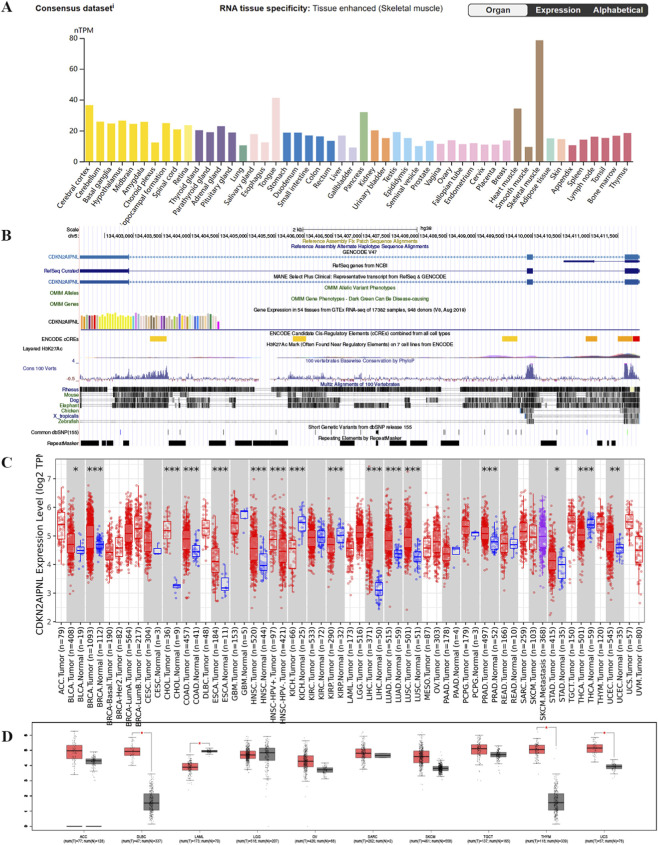 FIGURE 1
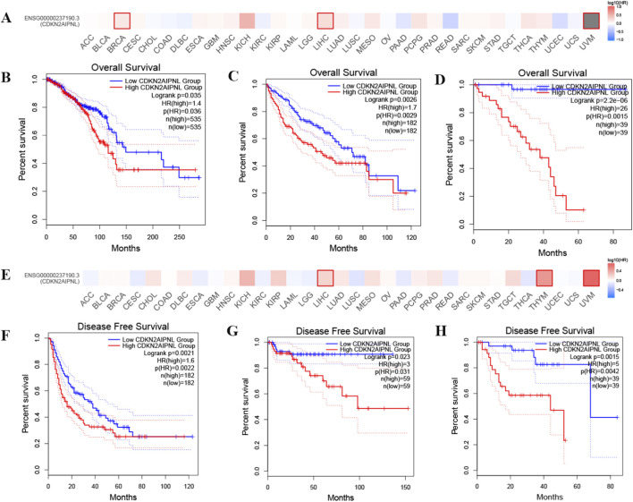 FIGURE 2
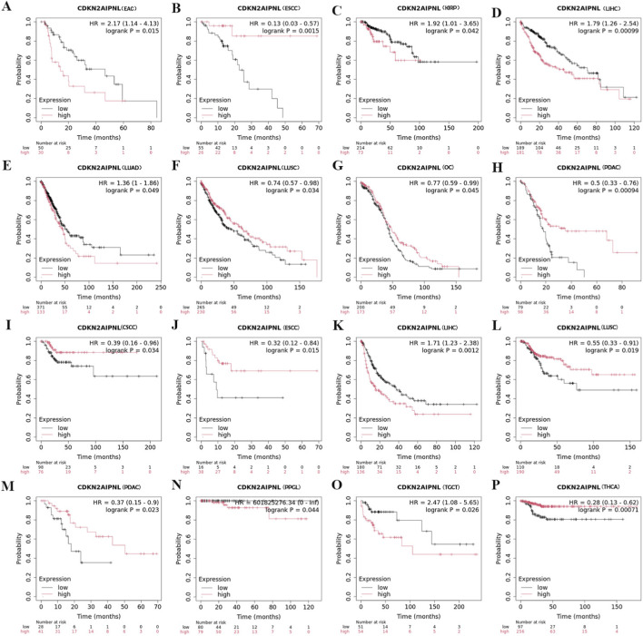 FIGURE 3
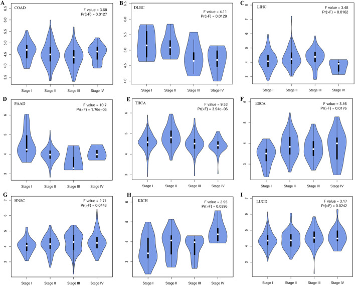 FIGURE 4
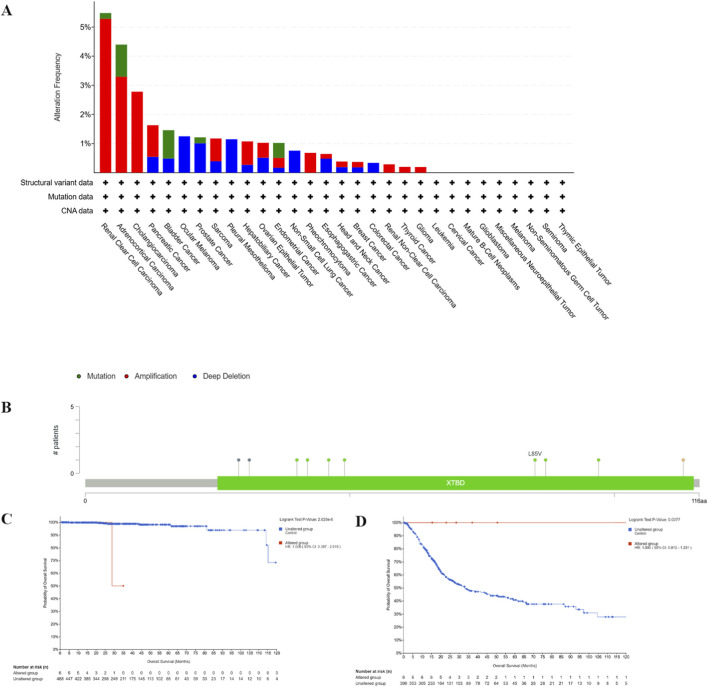 FIGURE 5
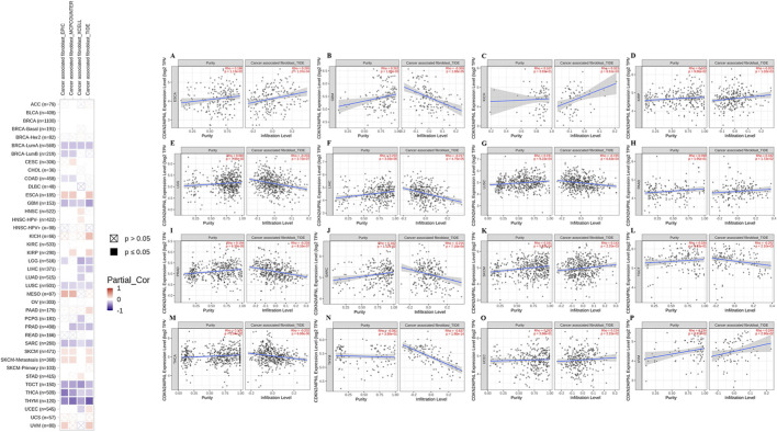 FIGURE 6
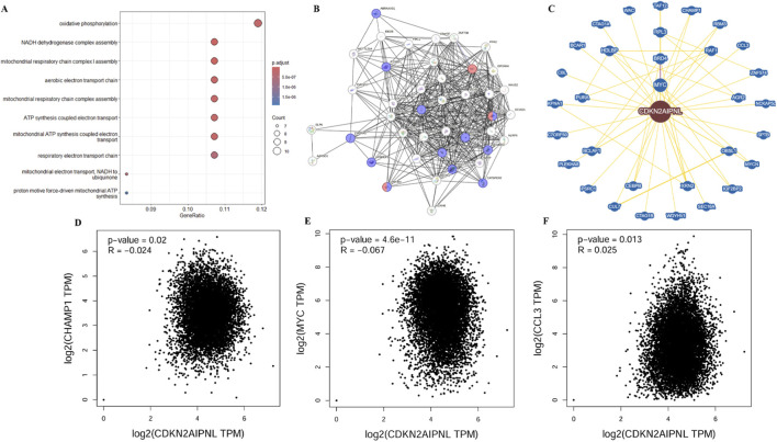 FIGURE 7
Peer Reviews
No public reviews on file for this paper yet. If you reviewed it on a platform where reviews are public (OpenReview, ICLR, NeurIPS, ICML), you can paste yours below so the community can read it here.
Videos
No videos yet. Explain this paper in a talk, walkthrough, or lecture? Add one.
Taxonomy
TopicsFerroptosis and cancer prognosis · Acute Myeloid Leukemia Research · Cancer-related Molecular Pathways
Introduction
According to data from the American Cancer Society, an estimated 611,720 people in the United States will die from cancer in 2024 (Siegel et al., 2024). Considering the population aging, this number is expected to increase further. Although targeted therapies and immunotherapies have demonstrated significant efficacy in treating various cancers, clinical outcomes exhibit substantial variability across different cancer types (Waldman et al., 2020; Xiao et al., 2023; Sonkin et al., 2024). Therefore, to broaden potential treatment options for malignant tumors, it is imperative to enhance our understanding of oncogenesis and tumor progression through the identification of oncogenes.
CDKN2A interacting protein N-terminal like (CDKN2AIPNL) is a homolog of the N-terminal domain of a protein that interacts with the CDKN2A gene. The CDKN2AIPNL gene is situated at position 5q31.1 on human chromosome 5, comprises three exons, and encodes a protein that is active in the nucleolus and nucleoplasm (details at https://www.ncbi.nlm.nih.gov/gene/91368#gene-expression). CDKN2AIPNL is expressed in a range of normal tissues, with particularly high expression levels in the thyroid and testis, and it is frequently classified as a tumor suppressor gene (Razvi et al., 2018). CDKN2AIPNL exhibits a paradoxical role in cancer progression: while acting as a tumor suppressor in myeloid malignancies by maintaining genomic stability (Visconte et al., 2017), it promotes tumor progression in solid cancers via metabolic reprogramming and immune evasion (Liu et al., 2019; Azimi et al., 2023). This functional duality underscores the need for systematic pan-cancer analyses.
CDKN2AIPNL is associated with the activity and stability of the 5′–3′ exoribonuclease XRN2 (Alexandrova et al., 2020). Research has demonstrated that CDKN2AIPNL is involved in regulating nucleic acid metabolism in colon cancer (Chodary Khameneh et al., 2022). Nonetheless, because CDKN2AIPNL remains a relatively understudied gene, its specific functions and mechanisms of action are not yet fully elucidated.
In this study, we systematically analyzed the expression status, prognostic value, genetic alterations, and molecular functions of CDKN2AIPNL, as well as its correlation with cancer-associated fibroblast infiltration across various tumor types.
Materials and methods
Gene expression analysis
To construct an mRNA expression map for CDKN2AIPNL, we utilized the Human Protein Atlas (HPA) database (https://www.proteinatlas.org/). We employed the “Gene DE” module of the Tumor Immune Estimation Resource (TIMER2) (http://timer.cistrome.org/) to evaluate the expression differences of CDKN2AIPNL between tumor and non-tumor tissues across various cancer types. In this module, we also examined CDKN2AIPNL expression across different molecular subgroups of breast cancer, as well as between HPV-positive and HPV-negative head and neck squamous cell carcinoma (HNSC) and between primary and metastatic skin cutaneous melanoma (SKCM). Additionally, we retrieved expression data for CDKN2AIPNL from the GEPIA2 database (http://gepia2.cancer-pku.cn/) to provide comparative validation of its expression levels. (Statistical significance was defined as p < 0.05).
Survival analysis
Overall survival (OS) and disease-free survival (DFS) Kaplan-Meier (KM) plots, along with survival significance plots for CDKN2AIPNL across all TCGA tumor types, were generated using the “Survival Analysis” module of GEPIA2 (http://gepia2.cancer-pku.cn/). Furthermore, the UCSC Xena browser (https://xenabrowser.net/), with high/low expression groups defined by median cutoffs was used to perform progression-free survival (PFS) analysis of CDKN2AIPNL using the TCGA Pan-Cancer dataset. The Kaplan-Meier plotter database was used to augment the prognostic analysis results. The expression threshold for high and low CDKN2AIPNL expression was set at 50%. For survival analysis, p-values were adjusted using the Benjamini–Hochberg method to control the false discovery rate (FDR) across multiple comparisons.
Genetic alteration analysis
Genetic alterations of CDKN2AIPNL were analyzed using cBioPortal (https://www.cbioportal.org/). Utilizing the TCGA Pan-Cancer Atlas study dataset, we calculated the frequency of CDKN2AIPNL gene mutations and copy number alterations via the “Cancer Type Summary” module. Additionally, the “Mutations” module was employed to create a mutation map for CDKN2AIPNL.
To assess the correlation between CDKN2AIPNL amplification status and prognosis in Prostate Adenocarcinoma (PRAD), molecular profiles were selected based on copy number alterations, and cases were divided into altered and unaltered groups to generate survival plots.
Similarly, for bladder urothelial carcinoma (BLCA), we evaluated the correlation between CDKN2AIPNL mutation status and prognosis by selecting molecular profiles based on mutations and categorizing cases into altered and unaltered groups to generate survival plots.
Immune cell infiltration analysis
The “Immune” module of TIMER2 (http://timer.cistrome.org/) was employed to investigate the correlation between CDKN2AIPNL expression and cancer-associated fibroblast infiltration using the Extended Multi-Dimensional Immune Profiling (EPIC) and Tumor Immune Dysfunction and Exclusion (TIDE) algorithms.
CDKN2AIPNL-related gene enrichment analysis
The STRING tool (https://string-db.org/) was utilized to construct a co-expression network for CDKN2AIPNL in Homo sapiens, employing the following parameters: (1) Active interaction sources: co-expression; (2) Meaning of network edges: evidence; (3) Maximum number of interactors: 50; and (4) Minimum required interaction score: low confidence (0.150).
The “Similar Gene Detection” module of GEPIA2 was used to extract 100 genes related to CDKN2AIPNL from the TCGA dataset, which have the most similar expression patterns with CDKN2AIPNL. The gene symbols of these 100 related genes were subsequently input into the ‘clusterProfiler’ R package for gene ontology (GO) pathway enrichment analysis, allowing for the identification of significantly enriched biological processes and pathways. Additionally, the ‘Correlation Analysis’ module of GEPIA2 facilitated pairwise gene correlation analysis, providing insights into the co-expression patterns of CDKN2AIPNL and its related genes.
CDKN2AIPNL-protein interaction analysis
The “Network” module of BioGRID (https://thebiogrid.org/) was used to create a CDKN2AIPNL-protein interaction network, with the layout set to “Concentric circles.”
Conservation status analysis of CDKN2AIPNL
The UCSC Genome Browser (http://www.genome.ucsc.edu/cgi-bin/hgTracks) was utilized to visualize the gene conservation of CDKN2AIPNL among vertebrates.
Results
Gene expression analysis of CDKN2AIPNL
Comprehensive analysis of datasets from the Human Protein Atlas (HPA), Genotype-Tissue Expression (GTEx), and FANTOM5 (Functional Annotation of Mammalian Genomes) demonstrated that CDKN2AIPNL is highly expressed in metabolically active tissues, including skeletal muscle, cardiac muscle, pancreas, and brain tissue, with a particularly pronounced expression in skeletal muscle (Figure 1A; Supplementary Figure S1). Additionally, single-cell RNA sequencing data further corroborated these findings, revealing significant expression of CDKN2AIPNL in cardiomyocytes, skeletal muscle cells, and smooth muscle cells (Figure 1A; Supplementary Figure S1). These observations suggest that CDKN2AIPNL exhibits low tissue specificity, indicating a broad functional role across various tissues. Furthermore, our analysis revealed that CDKN2AIPNL is relatively conserved among vertebrate species (Figure 1B), which underscores its potential functional significance across evolutionary contexts.
*Expression status of CDKN2AIPNL in different tumors and normal tissues and gene conservation of CDKN2AIPNL in vertebrates. (A) Tissue expression consensus CDKN2AIPNL based on HPA, GTEx, and FANTOM5 datasets. (B) Gene conservation analysis of CDKN2AIPNL among vertebrates visualized using UCSC genome browser. (C) Expression status of CDKN2AIPNL in different tumor types visualized through TIMER2. *p < 0.05; *p < 0.01; p < 0.001. (D) Expression of CDKN2AIPNL in several cancers and paired normal tissues in the GEPIA database.
We then investigated the expression pattern of CDKN2AIPNL in tumor tissues and found that the mRNA expression of CDKN2AIPNL exhibited high tumor specificity. The high tumor specificity of CDKN2AIPNL is not only beneficial for early screening of neoplastic diseases, improving the accuracy of early diagnosis and enabling earlier treatment for patients, but also makes CDKN2AIPNL a potential target for drug development. Notably, compared with the corresponding normal tissues, CDKN2AIPNL mRNA levels were significantly elevated in several tumor types (Figure 1C). Specifically, tumor tissues from Breast invasive carcinoma (BRCA)(T:N = 1093:112), Cholangiocarcinoma (CHOL)(T:N = 36:9), Colon adenocarcinoma (COAD)(T:N = 457:41), Esophageal carcinoma (ESCA)(T:N = 184:11), Head and Neck squamous cell carcinoma (HNSC)(T:N = 520:44), Liver hepatocellular carcinoma (LIHC)(T:N = 371:50), Lung adenocarcinoma (LUAD)(T:N = 515:59), Lung squamous cell carcinoma (LUSC)(T:N = 501:51), PRAD (T:N = 497:52), Uterine Corpus Endometrial Carcinoma (UCEC)(T:N = 545:35), and BLCA (T:N = 408:19) exhibited significantly higher CDKN2AIPNL expression compared to their respective normal tissues (Figure 1C). Furthermore, HPV-positive HNSC(T:N = 520:44) tissues showed markedly elevated CDKN2AIPNL expression compared to HPV-negative tissues (Figure 1C). Conversely, significantly reduced CDKN2AIPNL expression was noted in Kidney Chromophobe (KICH)(T:N = 66:25), Kidney Renal Papillary Cell Carcinoma (KIRP)(T:N = 290:32), and Thyroid Carcinoma (THCA)(T:N = 501:59) tumor tissues (Figure 1C).
To further elucidate the expression differences of CDKN2AIPNL in cancers lacking paired normal tissues, we utilized the GEPIA database, as indicated by the TIMER database. Figure 1D shows elevated CDKN2AIPNL expression in Lymphoid Neoplasm Diffuse Large B-cell Lymphoma (DLBC)(T:N = 47:337) (p = 0.003), Uterine Carcinosarcoma (UCS)(T:N = 57:78) (p = 0.004), reduced expression in Acute Myeloid Leukemia (LAML)(T:N = 173:70) (p = 0.01). Advanced tumor stages in LIHC and PAAD showed significantly elevated CDKN2AIPNL expression (p < 0.05, Figure 4), suggesting its role in malignant progression.
In summary, these findings suggest that CDKN2AIPNL may promote oncogenesis across various tumor types, highlighting the need for further investigation into its clinical significance.
Correlation between CDKN2AIPNL expression and cancer patient prognosis
To explore the potential prognostic value of CDKN2AIPNL based on the TCGA dataset, we employed the GEPIA2 and Kaplan-Meier plotter database to analyze the correlation between CDKN2AIPNL expression and prognosis in patients with different tumors (Figures 2A,E). In the GEPIA2, we found that lower CDKN2AIPNL expression was significantly associated with longer OS and DFS in LIHC (n = 364, OS: HR = 1.7, P = 0.0026; n = 364, DFS: HR = 1.6, P = 0.0021, Figures 2C,F) and UVM (n = 78, OS: HR = 26, P = 2.2e-6; n = 78, DFS: HR = 5, P = 0.0015, Figures 2D,H). In BRCA, lower CDKN2AIPNL expression was correlated with improved OS (n = 78, HR = 26, P = 2.2e-6, Figure 2B). Similarly, in THYM, lower CDKN2AIPNL expression was associated with better DFS (n = 118, HR = 3, P = 0.023, Figure 2G).
Correlation between CDKN2AIPNL expression and disease-free survival and overall survival in patients with different TCGA tumor types in the GEPIA2 database. GEPIA2 was used to construct survival plots (A,E) and perform OS (B–D) and DFS (F–H) analysis. Survival plots and Kaplan-Meier plots with significant results are displayed. The 95% confidence intervals for disease-free survival and overall survival are represented by red and blue dashed lines for the high CDKN2AIPNL and low groups, respectively.
In the Kaplan-Meier plotter database, we observed that in LIHC, high CDKN2AIPNL expression genes were associated with poorer OS and RFS (n = 370, OS: HR = 1.79, P = 0.0009; n = 164, DFS: HR = 0.39, P = 0.034, Figures 3D,K). In Esophageal squamous cell carcinoma (n = 81, OS: HR = 0.13, P = 0.015; n = 54, RFS: HR = 0.32, P = 0.015, Figures 3B,J), LUSC (n = 495, OS: HR = 0.74, P = 0.034; n = 300, RFS: HR = 0.55, P = 0.019, Figures 3F,L), and PAAD (n = 177, OS: HR = 0.5, P = 0.000094; n = 69, RFS: HR = 0.37, P = 0.023, Figures 3H,M), high expression was associated with better OS and RFS. Conversely, in Esophageal adenocarcinoma (n = 80, HR = 2.17, P = 0.015, Figure 3A), KIRP (n = 287, HR = 1.92, P = 0.042, Figure 3C), and LUAD (n = 504, HR = 1.36, P = 0.049, Figure 3E), high CDKN2AIPNL expression was associated with poorer OS. In contrast, high expression of CDKN2AIPNL was associated with better OS in OV (n = 373, HR = 0.77, P = 0.045, Figure 3G). Additionally, in PCPG (n = 216, HR = 1.71, P = 0.0012, Figure 3N) and TGCT (n = 159, HR = 0.37, P = 0.023, Figure 3O), high CDKN2AIPNL expression was associated with poorer OS. Interestingly, in CESC (n = 174, HR = 0.39, P = 0.034, Figure 3I) and THCA (n = 353, HR = 0.28, P = 0.00071, Figure 3P), low expression of CDKN2AIPNL meant poorer RFS. Collectively, these results indicate that CDKN2AIPNL expression is closely associated with the prognosis of various cancer types.
Correlation between the expression of CDKN2AIPNL and OS and RFS in patients with different TCGA tumor types in Kaplan-Meier plotter database. (A–H) Represents the relationship between CDKN2AIPNL expression and OS in different tumors, and (I–P) represents the relationship between CDKN2AIPNL expression and RFS in different tumors. Survival charts and Kaplan-Meier charts with significant results are displayed. 95% confidence intervals for disease-free survival and overall survival are indicated by red and blue dashed lines for high CDKN2AIPNL and low groups, respectively.
These results indicate that increased CDKN2AIPNL expression is associated with poor prognosis in various tumor types. To further investigate this relationship, we analyzed the correlation between CDKN2AIPNL expression and tumor pathological staging using the GEPIA2 database. We observed that low CDKN2AIPNL expression was significantly associated with advanced stages of COAD, DLBC, LIHC, PAAD, and THCA, which are cancers with higher metabolism (Figures 4A–E). In contrast, in tumors with lower metabolic rates, CDKN2AIPNL expression remained relatively stable across later stages (Figures 4F–I).
Correlation between CDKN2AIPNL expression and tumor pathological staging. The correlation between CDKN2AIPNL expression and pathological staging of COAD (A), DLBC (B), LIHC (C), PAAD (D), THCA (E), ESCA (F), HNSC (G), KICH (H), and LUCD (I) from the TCGA dataset. Log2 (TPM + 1) was applied on a logarithmic scale.
Genetic alterations
Next, we utilized cBioPortal to examine the genetic alterations of CDKN2AIPNL across various tumor types within the TCGA dataset. In KIRC, >5% of tumors harbored CDKN2AIPNL amplifications/mutations. (Figure 5A; Supplementary Table S1). In addition, more than 4% of ACC samples showed genetic alterations in CDKN2AIPNL (Figure 5A; Supplementary Table S1). As illustrated in Figure 5B, a total of 10 mutations in CDKN2AIPNL were detected across TCGA tumor samples, comprising 7 missense mutations, 2 truncating mutations, and 1 splice mutation (Supplementary Table S2). Notably, all mutations occurred within the XTBD region (amino acids 25-115) encoded by CDKN2AIPNL, making it the most frequently mutated region of the protein (Figure 5B).
Genetic alterations of CDKN2AIPNL in various tumor types of the TCGA. The types of CDKN2AIPNL gene alterations (A) and the mutation sites of CDKN2AIPNL (B) were generated by cBioPortal. The correlation between CDKN2AIPNL amplification status and PRAD (C) OS was analyzed through cBioPortal. The correlation between mutation status and BLCA (D) OS was analyzed.
Subsequently, we investigated the association between alterations in the CDKN2AIPNL gene and clinical outcomes in cancer patients. Amplifications in PRAD predicted poor OS (p = 0.008, Figure 5C; Supplementary Table S3). Conversely, patients with CDKN2AIPNL mutated BLCA demonstrated better OS outcomes (Figure 5D; Supplementary Table S4).
Cancer-associated fibroblast infiltration
Previous research has demonstrated that cancer-associated fibroblasts within the tumor stroma play a crucial role in regulating various tumor-infiltrating immune cells (Chen and Song, 2019). Consequently, we employed the EPIC and TIDE algorithms to investigate the correlation between cancer-associated fibroblast infiltration and CDKN2AIPNL expression across different malignant tumors. Our analysis revealed a positive correlation between CDKN2AIPNL expression and cancer-associated fibroblast infiltration in ESCA (n = 185), KICH (n = 66), KIRP (n = 290), UVM (n = 80), PAAD (n = 179), SKCM-Metastasis (n = 368), SKCM (n = 471), and UCEC (n = 545) (Figure 6).
Correlation between CDKN2AIPNL expression and cancer-associated fibroblast immune infiltration. (A–P) The correlation between CDKN2AIPNL expression and cancer-associated fibroblast immune infiltration in all tumor types of the TCGA was calculated using the EPIC and TIDE algorithms.
CDKN2AIPNL-related gene enrichment
To study the functional mechanism of CDKN2AIPNL in oncogenesis, we used GEPIA2 to extract the top 100 genes with similar expression patterns to CDKN2AIPNL from all tumor types in the TCGA dataset (Supplementary Table S5). GO enrichment analysis indicated that these genes are related to mitochondrial function and energy metabolism (Figure 7A). Subsequently, 50 genes co-expressed with CDKN2AIPNL were identified through the STRING tool to validate the results of the Gene Ontology enrichment analysis. As depicted in Figure 7B, these 50 genes are closely related; moreover, these genes are also enriched in unknown function and Retinal homeobox protein RAX/RAX2 (Supplementary Table S6). These findings led us to investigate the key proteins that CDKN2AIPNL may interact with in these biological processes. According to the BioGRID database, CDKN2AIPNL physically interacts with CHAMP1, XRN2, and MYC (Figure 7C), all of which are well-characterized genes encoding proteins involved in the regulation of energy metabolism, cell proliferation, differentiation, and tumorigenesis (Supplementary Figure S2). Additionally, we observed that the expression of CDKN2AIPNL is closely related to the expression levels of MYC, CHAMP1, and XRN2 (Figures 7D–F). Based on these results, we speculate that CDKN2AIPNL likely drives tumorigenesis via three axes: (1) MYC/XRN2-mediated metabolic reprogramming, (2) CAF-dependent immune suppression, and (3) genomic instability via mutation hotspots.
CDKN2AIPNL-related gene enrichment analysis. (A) GO enrichment analysis of the top 100 genes co-expressed with CDKN2AIPNL obtained from GEPIA2. (B) Co-expression network of 50 genes obtained with the STRING tool for CDKN2AIPNL co-expression. (C) CDKN2AIPNL-protein interactions obtained through BioGRID. (D-F) Correlation analysis between CDKN2AIPNL and CHAMP1, XRN2, and MYC from all tumor samples of the TCGA by GEPIA2.
Discussion
The advancement of gene bioinformatics algorithms, alongside the continuous improvement and updating of databases, has facilitated numerous pan-cancer analyses in recent years aimed at identifying tumor molecular biomarkers and elucidating their functional roles. In this study, we analyzed the prognostic value and oncogenic role of CDKN2AIPNL across various tumor types.
CDKN2AIPNL is located on chromosome 5q31.1 and encodes two transcripts, both of which produce proteins. Notably, only one transcript, NM_001008278.2, with a length of 1782 nucleotides, generates the 116-amino acid protein NP_001008279.1 (Gerhard et al., 2004). CDKN2AIPNL exhibits broad expression in various normal tissues, with the highest expression levels found in the thyroid (RPKM 3.8), followed closely by testicular tissue (RPKM 3.7) (Fagerberg et al., 2014). According to the Ensembl database, orthologs of CDKN2AIPNL have been identified in 489 species. The protein-coding sequence of CDKN2AIPNL is relatively conserved among vertebrates. Li-Min Liu et al. found that CDKN2AIPNL was significantly elevated in the mechanism of liver cancer occurrence and was associated with poor OS (Liu et al., 2019). Conversely, Visconte et al. identified a tumor suppressor role for CDKN2AIPNL in a cohort of 223 myeloid neoplasm cases based on whole-exome sequencing (WES) analyses (Visconte et al., 2017). Additionally, Ali Azimi et al. reported that CDKN2AIPNL expression levels were significantly downregulated in melanoma with distant metastasis, suggesting that CDKN2AIPNL may function as an inhibitor of distant metastatic processes (Azimi et al., 2023). However, the role of CDKN2AIPNL across various tumor types remains inadequately explored. Therefore, we systematically characterized CDKN2AIPNL in 33 TCGA tumor types by analyzing gene expression, genetic alterations, and immune infiltration features.
In this study, we observed that CDKN2AIPNL is widely expressed in various tissues, and CDKN2AIPNL is upregulated in most tumors. We further explored the relationship between CDKN2AIPNL overexpression and clinical parameters, including prognosis. Survival analysis shows that the effect of CDKN2AIPNL expression level on tumor prognosis is related to tumor type. Currently, research on the prognostic heterogeneity of the CDKN2AIPNL gene across different malignancies remains limited, and its underlying molecular mechanisms have not yet been fully elucidated. Future fundamental studies should focus on two key aspects: first, the role of CDKN2AIPNL in tumor microenvironment-specific immunoregulatory networks across various cancer types; second, the potential synergistic oncogenic effects between this gene and other somatic mutations. These investigations will provide critical theoretical insights into the tumor biological functions of CDKN2AIPNL. Notably, elevated CDKN2AIPNL expression correlated with poor prognosis in various tumor types, including Esophageal adenocarcinoma, KIRP, LIHC, LUAD, PCPG, TGCT, THYM, and UVM. Moreover, upregulation of CDKN2AIPNL was significantly associated with advanced cancer staging, indicating malignant progression. Increasing evidence suggests that genomic mutations contribute to tumor progression and influence chemotherapy responses (Gu et al., 2023; Chatrath et al., 2021). For instance, Yang et al. reported that mutations in BRCA1 and BRCA2 are significantly associated with patient survival rates, potentially due to unique responses to platinum-based therapy (De Talhouet et al., 2020). In our study, we found that CDKN2AIPNL mutations were most prevalent in KIRC (>5%), followed by ACC. In KIRC, CDKN2AIPNL has a high frequency of genetic alterations, mainly copy number amplification and missense mutations. These genetic alterations in CDKN2AIPNL may potentially influence the response of patients to PD-1/PD-L1 inhibitors, and mutations may lead to the escape of tumor cells from the immune system, which may affect the efficacy of the treatment. In the ACC, mutations in CDKN2AIPNL may affect patient response to targeted therapies such as mTOR inhibitors and TKI drugs. Our analysis of the impact of CDKN2AIPNL gene alterations on clinical outcomes revealed that CDKN2AIPNL amplification may serve as a risk factor for patients with PRAD, while mutations in CDKN2AIPNL may confer a protective effect in patients with BLCA. Collectively, these findings suggest that CDKN2AIPNL functions as an oncogene in the progression of various cancers and represents a promising predictor for applications in cancer prognosis, despite its previously recognized role as a tumor suppressor gene.
The interaction between immune cells and cancer cells plays a crucial role in shaping the tumor microenvironment and significantly influences the processes of tumor spread and metastasis (Adib et al., 2021; Zhang et al., 2024). Recent studies have reported that the tumor immune microenvironment is associated with the expression levels of various genes (Cai et al., 2024). In our analysis, we found that CDKN2AIPNL expression is positively correlated with the infiltration of CAFs in several types. Current evidence indicates that transforming growth factor-beta (TGF-β) and platelet-derived growth factor (PDGF) modulate diverse cellular components within the tumor microenvironment (including T lymphocytes, cancer-associated fibroblasts, and tumor-associated macrophages), thereby fostering an immunosuppressive phenotype and metastatic progression (Manzat Saplacan et al., 2017; Fasano et al., 2024). CDKN2AIPNL may exert either pro-tumorigenic or anti-tumorigenic effects by regulating this microenvironmental remodeling process. Furthermore, another study demonstrated that upregulation of CDKN2A significantly enhances tumor cell proliferation via activation of the JAK2/STAT3 signaling pathway (Yong et al., 2025). CAFs are integral components of the tumor stroma and have been associated with poor prognosis, chemotherapy resistance, and disease recurrence in various cancers (Tajaldini et al., 2022; Chen et al., 2021). CDKN2AIPNL interacts with CDKN2A to regulate the cell cycle and cell proliferation process (Cao et al., 2022; Shi et al., 2022). CDKN2A is an important regulator of the G1 phase of the cell cycle, which inhibits cell proliferation by preventing cells from moving from the G1 phase into the S phase through inhibiting the activity of CDK4/6 (Wang et al., 2024).CDKN2AIPNL may indirectly affect immune cells by regulating the function of CDKN2A proliferation (Luo et al., 2021). In addition, CDKN2AIPNL may also regulate cell proliferation by regulating signaling pathways such as PI3K/Akt and JAK/STAT (Hu et al., 2021). In fact, there are no direct research finding on the effect of CDKN2AIPNL on the proliferation of immune cells, but its mechanism of action in cell cycle regulation and signaling pathway modulation suggests that it may affect the proliferation of immune cells through multiple pathways. Further experimental studies are needed to explore the specific function and mechanism of action of CDKN2AIPNL in immune cells in order to provide deeper understanding of its role in immune regulation.
In summary, our work elucidates the potential impact of CDKN2AIPNL on tumor immunity and its prognostic value across different cancer types.
Using STRING and GEPIA2, we identified numerous genes co-expressed with CDKN2AIPNL in various tumors and other tissues. Gene enrichment analysis revealed that these co-expressed genes are closely related to cell cycle regulation, mitochondrial function, and cellular energy metabolism, consistent with findings from previous studies (Cargill et al., 2021; Liang et al., 2024; Li et al., 2022). Furthermore, our results indicate that CDKN2AIPNL physically interacts with MYC, CHAMP1, and XRN2 (García-Alonso et al., 2022). Importantly, the expression levels of MYC, CHAMP1, and XRN2 are closely correlated with CDKN2AIPNL expression. These genes are well-characterized and encode proteins that are pivotal in DNA repair, cell cycle regulation, and the regulation of mitochondrial function and cellular energy metabolism (Hino et al., 2021; Chu et al., 2024). Notably, MYC is a key transcription factor that plays a significant role in metabolic regulation. Recent studies have shown that PRMT5 activates lipid metabolic reprogramming via MYC, highlighting the importance of MYC in metabolic pathways (Liang et al., 2024). This suggests that the co-expression of CDKN2AIPNL with MYC may contribute to similar metabolic processes. Additionally, CHAMP1, which is involved in chromosome segregation and cell cycle regulation, may also influence metabolic regulation through homologous recombination (HR) repair, regulating the expression of the anti-apoptotic protein Mcl-1, and regulating cell division and growth (Li et al., 2022; Hino et al., 2021; Fujita et al., 2022). These findings validate our gene enrichment analysis results and pave the way for further exploration of the molecular functions of CDKN2AIPNL.
Limitations and prospects
This study also has several limitations. First, the sample sizes for some less common tumor types are relatively small, potentially leading to batch effects or inaccuracies in the results. Second, while this study provides preliminary insights linking CDKN2AIPNL to the progression of various tumors. Nevertheless, systematic functional experiments (Such as CRISPR-Cas9-mediated knockout, Western blot analysis, and cell cycle profiling) are still required to delineate the precise molecular mechanisms of CDKN2AIPNL in tumorigenesis and progression. Finally, Liu’s laboratory was one of the first laboratories involved in TCGA biomarker research and has developed various strategies in this field (Liu et al., 2024a; Li and Liu, 2022; Liu et al., 2024b). The broad and diverse nature of TCGA data makes it an important resource for cancer research, but there are limitations such as sample selection bias and tissue heterogeneity, which may affect the generalizability of the results (Liu et al., 2024c). Therefore, further basic studies need to be designed for the Therefore, further basic studies need to be designed to validate the relevant findings.
In conclusion, CDKN2AIPNL is widely overexpressed in various cancers, and both its expression and genetic alterations are statistically correlated with the clinical outcomes of affected patients. Moreover, immune infiltration analysis and CDKN2AIPNL-related gene enrichment analyses offer potential mechanisms through which CDKN2AIPNL may regulate tumor immunity, cell cycle dynamics, mitochondrial function, and cellular energy metabolism in cancer. Therefore, additional experimental and clinical studies are warranted to explore the practical applications of CDKN2AIPNL in cancer treatment and prognosis prediction.
The reference list from the paper itself. Each links out to its DOI / PubMed record.
- 1Adib E. Nassar A. H. Akl E. W. Abou Alaiwi S. Nuzzo P. V. Mouhieddine T. H. (2021). CDKN 2A alterations and response to immunotherapy in solid tumors. Clin. Cancer Res. 27 (14), 4025–4035. 10.1158/1078-0432.CCR-21-0575 34074656 PMC 8900067 · doi ↗ · pubmed ↗
- 2Alexandrova J. Piñeiro D. Jukes-Jones R. Mordue R. Stoneley M. Willis A. E. (2020). Full-length NF-κB repressing factor contains an XRN 2 binding domain. Biochem. J. 477 (4), 773–786. 10.1042/BCJ 20190733 32011671 PMC 7054742 · doi ↗ · pubmed ↗
- 3Azimi A. Patrick E. Teh R. Kim J. Fernandez-Penas P. (2023). Proteomic profiling of cutaneous melanoma explains the aggressiveness of distant organ metastasis. Exp. Dermatol 32 (7), 1072–1084. 10.1111/exd.14814 37082900 · doi ↗ · pubmed ↗
- 4Cai Y. Lu Z. Chen C. Zhu Y. Chen Z. Wu Z. (2024). An atlas of genetic effects on cellular composition of the tumor microenvironment. Nat. Immunol. 25 (10), 1959–1975. 10.1038/s 41590-024-01945-3 39223350 · doi ↗ · pubmed ↗
- 5Cao Y. Sun Q. Chen Z. Lu J. Geng T. Ma L. (2022). CDKN 2AIP is critical for spermiogenesis and germ cell development. Cell Biosci. 12 (1), 136. 10.1186/s 13578-022-00861-z 35989335 PMC 9394077 · doi ↗ · pubmed ↗
- 6Cargill K. R. Stewart C. A. Park E. M. Ramkumar K. Gay C. M. Cardnell R. J. (2021). Targeting MYC-enhanced glycolysis for the treatment of small cell lung cancer. Cancer Metab. 9 (1), 33. 10.1186/s 40170-021-00270-9 34556188 PMC 8461854 · doi ↗ · pubmed ↗
- 7Chatrath A. Ratan A. Dutta A. (2021). Germline variants that affect tumor progression. Trends Genet. 37 (5), 433–443. 10.1016/j.tig.2020.10.005 33203571 PMC 8043969 · doi ↗ · pubmed ↗
- 8Chen X. Song E. (2019). Turning foes to friends: targeting cancer-associated fibroblasts. Nat. Rev. Drug Discov. 18 (2), 99–115. 10.1038/s 41573-018-0004-1 30470818 · doi ↗ · pubmed ↗