On Hallucinations in Artificial Intelligence–Generated Content for Nuclear Medicine Imaging (the DREAM Report)
Menghua Xia, Reimund Bayerlein, Yanis Chemli, Xiaofeng Liu, Jinsong Ouyang, MingDe Lin, Georges El Fakhri, Ramsey D. Badawi, Quanzheng Li, Chi Liu

TL;DR
This paper discusses the risks of hallucinations in AI-generated nuclear medicine imaging and introduces the DREAM report to address these issues and improve clinical trust.
Contribution
The paper introduces the DREAM report, a framework for understanding and mitigating hallucinations in AI-generated nuclear medicine imaging.
Findings
Hallucinations in AI-generated nuclear medicine imaging can misrepresent anatomic and functional information.
The DREAM report provides recommendations for defining, detecting, and mitigating hallucinations in AIGC for NMI.
Abstract
Artificial intelligence–generated content (AIGC) has shown remarkable performance in nuclear medicine imaging (NMI), offering cost-effective software solutions for tasks such as image enhancement, motion correction, and attenuation correction. However, these advancements come with the risk of hallucinations, generating realistic yet factually incorrect content. Hallucinations can misrepresent anatomic and functional information, compromising diagnostic accuracy and clinical trust. This paper presents a comprehensive perspective on hallucination-related challenges in AIGC for NMI, introducing the DREAM report, which covers recommendations for definition, representative examples, detection and evaluation metrics, and attributions and mitigation strategies. This position statement paper aims to initiate a common understanding for discussions and future research toward enhancing AIGC…
Genes, proteins, chemicals, diseases, species, mutations and cell lines named across the full text — each resolved to its canonical identifier and authoritative record.
Click any figure to enlarge with its caption.
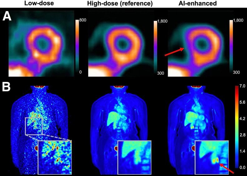 FIGURE 2
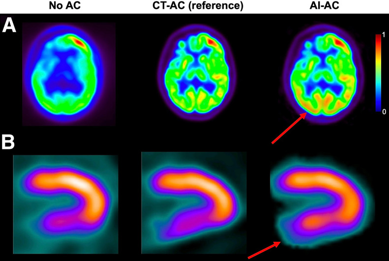 FIGURE 3
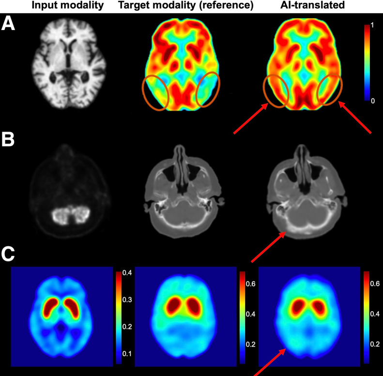 FIGURE 4
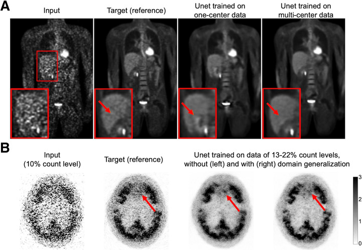 FIGURE 5
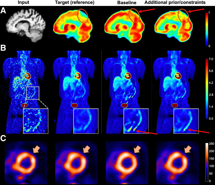 FIGURE 6
Peer Reviews
No public reviews on file for this paper yet. If you reviewed it on a platform where reviews are public (OpenReview, ICLR, NeurIPS, ICML), you can paste yours below so the community can read it here.
Videos
No videos yet. Explain this paper in a talk, walkthrough, or lecture? Add one.
Taxonomy
TopicsMedical Imaging Techniques and Applications · Adversarial Robustness in Machine Learning · Radiation Detection and Scintillator Technologies
Artificial intelligence–generated content (AIGC) has demonstrated significant potential in nuclear medicine imaging (NMI) over the past decade, achieving state-of-the-art performance across various tasks, based on a range of quantification metrics. Key applications include PET or SPECT image enhancement such as denoising, deblurring, and partial-volume correction (1); quantitative accuracy improvements such as motion correction, scatter correction, and attenuation correction (AC) (2); and cross-modality image translation such as generating PET images from CT or MRI and vice versa (3). These artificial intelligence (AI)–driven solutions offer the potential to replace traditional hardware-dependent approaches with more cost-effective software alternatives while also potentially reducing radiation exposure, easing clinical workloads, and optimizing imaging workflows.
Despite these advancements, hallucinations pose significant challenges to AIGC in NMI applications. Hallucinations can lead to cascading errors, including misdiagnosis, mistreatment, unnecessary interventions, medication errors, and ethical or legal concerns (4). These risks highlight the urgent need for robust hallucination detection frameworks and mitigation strategies before AIGC can be safely deployed in clinical practice.
Although recent surveys (5,6) have explored hallucinations in natural language processing, the medical imaging community still lacks a domain-specific and systematic analysis of hallucinations. To bridge this gap, this paper presents a comprehensive perspective on hallucination-related challenges in AIGC for NMI. We introduce the DREAM report, which outlines key aspects including the definition of hallucinations, representative examples, detection and evaluation methods, and attributions and mitigation recommendations, as illustrated in Figure 1, with the main components summarized in Table 1.
DEFINITION OF HALLUCINATIONS
The definition of hallucinations varies across publications and, in some cases, remains inconsistent or even contradictory. A precise and universally accepted definition has yet to be established (7). Although hallucinations were indistinguishable from general inaccuracies or errors in earlier studies (8,9), the term has gained renewed attention with the advent of large-scale and diffusion-based generative models (10,11). The enhanced generative capabilities introduce greater risks of fabricated content, prompting growing interest in establishing more rigorous and specific definitions of hallucinations.
In natural language processing, hallucinations are typically defined by their inconsistency with given targets (12). Factual hallucinations refer to AIGC that contradicts verifiable knowledge, whereas faithfulness hallucinations violate the instructions or source input. Other classifications include fact-conflicting, input-conflicting, and context-conflicting hallucinations (13). Additionally, Farquhar et al. (14) introduce a subset of hallucinations termed confabulations, referring to AIGC that are both incorrect and arbitrary. Here, arbitrary means that model outputs fluctuate unpredictably under identical inputs because of irrelevant factors such as random seed variations. This stochastic confabulation could be distinguished from systematic hallucinations in which AI claims are consistently incorrect, which may arise from flawed training data.
In medical imaging, the definition of hallucinations remains similarly ambiguous. Some studies interpret hallucinations narrowly as the addition of nonexistent tissue components (15), whereas others encompass both the addition and removal of image structures (16,17), such as the omission of lesions (18,19). Certain researchers emphasize the deceptive and realistic-looking appearance of hallucinations (20), whereas others expand the scope to include implausible or dreamlike content (21,22). There is also disagreement about whether hallucinations are unique to AI. A study on tomographic image reconstruction (23) defines hallucinations as false structures in reconstructed images, regardless of origin. In contrast, others argue that hallucinations are unique to AI (24).
In this paper, we focus specifically on AIGC in NMI. Errors or artifacts introduced by traditional imaging workflows (supplemental material, section I; supplemental materials are available at http://jnm.snmjournals.org) (25) are considered out of the scope. An extensive literature review indicates that most AIGC applications in NMI operate as image-to-image translation tasks. In such settings, implausible large-scale errors, such as the addition of organs or major structures, are rarely observed. We argue that these dreamlike errors are better defined as delusions, borrowing from the psychologic lexicon. Instead, hallucinations in AIGC for NMI are typically subtle but deceptive, manifesting as added small abnormalities or realistic-looking lesions that do not exist in reality. Other plausible AI-induced errors, such as the omission of real lesions (falsely replacing abnormal regions with normal structures) or pure quantification bias (uniform intensity shifts without creating new structures), are better interpreted as illusions: misinterpreting something rather than fabricating. These forms of errors, although clinically significant, fall outside the scope of hallucinations in this paper.
NOTEWORTHY
- Within this paper, hallucinations are defined as AI-fabricated abnormalities or artifacts that appear visually realistic and highly plausible yet are factually false and deviate from anatomic or functional truth.
- Hallucinations may occur across all AIGC applications in NMI.
- Recommended detection and evaluation methods include image-level comparisons, datasetwise statistical analysis, clinical task–based assessment (by human or model observers), and automated hallucination detectors trained on annotated benchmark datasets.
- Effective hallucination mitigation requires a comprehensive and multiperspective approach encompassing data quality, learning paradigms, and model design.
- Substantial adaptation and continued research are needed for robust detection, evaluation, and mitigation approaches tailored to AI hallucinations in NMI.
Given this context, within the scope of this paper we recommend a narrow AI hallucination definition in NMI: AI-fabricated abnormalities or artifacts that appear visually realistic and highly plausible yet are factually false and deviate from anatomic or functional truth (or, in the case of NMI when ground truth images are unavailable, represent structures not supported by the measurement).
REPRESENTATIVE EXAMPLES
In this section, we present visual examples of AIGC in representative NMI applications, illustrating scenarios in which hallucinations may arise during AI-driven processing.
Image Enhancement
Numerous studies have explored AI-driven translation from low-count/high-noise to high-count/low-noise images (26–28), demonstrating impressive performance with outputs that are visually compelling and often nearly indistinguishable from high-quality reference scans. However, hallucinations can occasionally emerge, distorting underlying anatomic or functional information. Figure 2 presents examples of AI-driven SPECT and PET denoising, highlighting cases in which AIGC unintentionally alters critical imaging details.
Examples of hallucinations in AI-driven image enhancement. (A) SPECT image denoising generated false-positive perfusion images (CC BY (27)). (B) PET image denoising generated false lesions, using method proposed by Dorjsembe et al. (28). Arrows indicate AI-introduced hallucinations.
AC
AI-based AC techniques have been proposed to estimate AC images directly from non-AC images (29,30), eliminating the need for CT-based attenuation maps and their associated radiation dose and enabling use in dedicated imaging systems without integrated CT. Figure 3 presents examples of AI-driven AC in both PET and SPECT imaging. Although the AIGC appears visually accurate, closer comparison with reference CT AC images reveals hallucinations.
Examples of hallucinations in AI-driven CT-free AC for PET imaging, showing generated false abnormality in brain region (reprinted with permission of (29)) (A) and for SPECT imaging, showing generated false-negative perfusion (reprinted with permission of (30)) (B). Arrows indicate AI-induced hallucinations that caused overestimation of tracer concentration.
Cross-Modality Translation
AI-driven cross-modality image translation synthesizes one imaging modality from another, enabling access to desired imaging information when only an alternative modality is available. For example, generating PET images from CT or MRI data (31,32) has been proposed to reduce the high costs and ionizing radiation exposure associated with PET. However, these potential benefits remain largely unproven in robust clinical trials, and the fundamental concept of inferring functional information from anatomic data (or vice versa) is inherently challenging and prone to significant pitfalls. In many conditions, functional abnormalities may appear before or independently of detectable structural changes, making such predictions unreliable. Cross-modality translation may be better suited as an auxiliary tool, such as for generating pseudo-CT scans from MRI for AC in PET/MRI workflows (33) or for augmenting training datasets with synthetic images to support AI model development.
Figure 4 presents examples of AI-driven cross-modality image translation, including PET–MRI (31), PET–CT (3), and PET–SPECT (34) conversions. Although these advancements highlight the promising capabilities of AI in cross-modality imaging, they also underscore the risk of hallucinations.
Examples of hallucinations in AI-driven cross-modality image translation. (A) PET/MRI translation (reprinted with permission of (31)). Although visually realistic, AI-generated PET image exhibits falsely increased glucose uptake in temporoparietal lobe, which could potentially lead to misdiagnosis of Alzheimer disease. (B) PET/CT translation (reprinted with permission of (3)). (C) PET/SPECT translation (CC BY (34)). Arrows highlight AI-induced hallucinations.
DETECTION AND EVALUATION METHODS
Although several commercial AI products, such as SubtlePET (Subtle Medical) and Precision DL (GE HealthCare), have already been cleared by the Food and Drug Administration for clinical use, the current Food and Drug Administration draft guidance on AI-enabled medical devices (January 2025) (35) does not explicitly reference the term hallucination. The guidance acknowledges that erroneous AI outputs can compromise device reliability and user trust and emphasizes a total-product-lifecycle approach spanning development, validation, and postmarket management. The guidance also recommends rigorous performance evaluation using metrics such as the area under the receiver operating characteristic curve and positive or negative likelihood ratios, among others. Although the Food and Drug Administration has jurisdiction over postdeployment monitoring, its oversight remains limited (e.g., adverse event reporting, required postmarket studies, and general device safety). Continuous performance monitoring in radiology practice is not mandated. To address this gap, the American College of Radiology has launched the Assess-AI initiative, with the long-term goal of influencing legislation to enable more robust postdeployment monitoring (36).
Within AI assessment and monitoring frameworks, hallucinations warrant dedicated and systematic attention. Instances identified during clinical use should be documented and reported through postmarket surveillance. In addition, establishing quantitative measures of hallucination could help define minimum acceptable thresholds for AI processing, such as the lowest-dose standards in AI-based denoising. Such thresholds would balance the extent of dose reduction with the risk of AI-induced hallucinations, ensuring that improved visual quality does not come at the cost of inaccurate representations.
To systematically assess hallucinations, we recommend adopting multifaceted metrics, which could draw on methodologies from related domains and be adapted to the specific context of NMI. Below, we outline several examples and preliminary ideas; however, further research is essential to develop accurate and widely accepted evaluation frameworks tailored to this field.
Image-Based Metric
When paired reference images are available, the hallucination index has been proposed to detect AI-generated spurious features (24). This index is computed as the Hellinger distance between the distribution of AIGC and a so-called zero-hallucination reference. The latter is generated by adding adaptive white gaussian noise to the reference image, with the noise power calibrated to match the signal-to-noise ratio of the AIGC. However, the original formulation was tailored specifically for Fourier diffusion-based models, in which noise power is inferred via a diffusion bridge between the output and reference images. To extend the applicability of the hallucination index across diverse AI models, potential adaptations informed by insights from previous work (37,38) can be considered. Further details are provided in section II of the supplemental material.
Radiomics analysis has also been explored as an evaluation tool (39). Most AI models currently used in NMI prioritize visual image quality, often relying on loss functions such as mean squared error. Although such models produce outputs that appear visually of high quality, they do not necessarily improve data quality for downstream tasks and may introduce subtle errors and hallucinations. Radiomics-based evaluation detects this issue by selecting clinically relevant regions of interest and extracting quantitative features from both the AIGC and the corresponding reference images. Statistical comparisons between the 2 feature sets can reveal inconsistencies, with significant discrepancies potentially indicating the presence of hallucinations. However, not all discrepancies in radiomic features necessarily reflect hallucinations. Other types of errors that fall outside the hallucination definition adopted in this paper, such as lesion omission or pure quantification bias (as discussed in the “Definition” section), may also produce radiomic differences. Therefore, further research is needed to identify radiomic features that are specifically sensitive to hallucinations while minimizing confounding from other nonhallucinatory errors.
It is also worth noting that both the hallucination index and radiomics analysis primarily capture underlying statistical discrepancies between AIGC and the reference. If AI-generated artifacts alter only the visual appearance without affecting the statistical or diagnostic characteristics of the data, they may not be detected by these methods. This observation supports the distinction that although an artifact may change the appearance of an image, a hallucination alters the underlying data statistics. This interpretation aligns with our proposed hallucination definition, in which hallucinations are considered a subset of artifacts—specifically, those that are visually plausible but deviate from anatomic or functional truth. It underscores the conceptual understanding that not all artifacts are hallucinations but that hallucinations generally manifest as visually misleading artifacts.
Dataset-Based Metric
In scenarios in which paired reference images are unavailable, the neural hallucination precursor was introduced to quantify hallucinations from a feature-space perspective (21), under the assumption that false content arises from misrepresented features. The metric measures the k-nearest neighbor distance between the intermediate feature embeddings of the AIGC and a hallucination-free feature bank preconstructed from a calibration dataset sampled from the training set. However, the approach is inherently model-dependent, as the feature bank is defined and obtained by a specific model architecture. This dependence limits its applicability for comparing hallucination levels across different AI models, as each model may use distinct feature extraction mechanisms that shape the learned feature distributions differently.
To compare different models in the absence of reference images, the concept of no-gold-standard evaluation (40) may offer insights. Originally developed for assessing conventional quantitative imaging techniques, this method could be adapted for AIGC evaluation. It models a linear stochastic relationship between measured values and unknown true values, which are assumed to follow a 4-parameter beta distribution. Model parameters are estimated by maximizing the likelihood of the observed data, and the noise-to-slope ratio derived from these estimates is used to quantify the precision of each method (40). For AIGC evaluation, quantitative values could be defined as metrics such as the mean or maximum activity concentration within specific regions of interest in the AIGC. However, 2 key challenges arise. First, the assumed linearity between true and measured values may not hold for nonlinear generative models. Second, and as previously noted, this metric may capture general errors rather than hallucinations specifically. Adapting it to isolate hallucinations from other nonhallucinatory deviations will require substantial methodologic refinement.
Clinical Task–Specific Metric
Clinically relevant tools for hallucination detection and evaluation are essential for real-world deployment. One strategy assesses hallucinations indirectly through downstream segmentation or classification performance (41). Another relies on direct expert evaluation, in which medical professionals assess AIGC using disease-specific image features (42) or rate them on a 5-point Likert scale (section III of the supplemental material) (43). However, such scalar ratings alone are insufficient to capture the complexity of hallucinations. A more informative strategy could pair the Likert score with bounding box annotations that localize suspected hallucinations, accompanied by concise descriptive text (e.g., “a false, small lesionlike hot spot at the apex of the liver”). This offers greater granularity than scalar ratings while remaining more feasible for clinicians than full voxelwise segmentation. Nonetheless, these evaluations often require access to reference images; without them, even experienced readers may be misled by hallucinations. Furthermore, because it is impractical for physicians to review all generated cases, determining an adequate and representative sample size for hallucination evaluation remains a key challenge.
Automatic Hallucination Detector
Automatic hallucination detectors (44), trained on hallucination benchmark datasets (4), have recently been explored in large (vision) language models to reduce the burden of human evaluation (section IV of the supplemental material). However, to the best of our knowledge, no hallucination-annotated benchmark dataset currently exists for NMI applications. This underscores an urgent need for the research community to collaboratively develop such a resource, ideally through multiinstitutional efforts. A promising approach involves leveraging crowdsourcing platforms to collect a diverse set of AI-generated NMI images that exhibit hallucinations, along with expert annotations. These annotations could be guided by standardized criteria, such as bounding boxes to indicate hallucination locations, brief descriptive text, and Likert-scale ratings for severity, as discussed in the “Clinical Task–Specific Metric” section. Of course, the final annotation protocol will require further discussion and consensus to ensure consistency and clinical relevance. The dataset could be designed for continuous expansion, accommodating new contributions over time.
ATTRIBUTIONS AND MITIGATION RECOMMENDATIONS
Most AIGC applications in NMI can be formulated as image-to-image estimation tasks, in which the objective is to learn a mapping function from a source domain S to a target domain T, denoted as . Given a training dataset with marginal distributions and , the goal is to identify an optimal approximation: , where is the loss function and H the hypothesis space (21). Hallucinations arise when the learned mapping function deviates from the true underlying mapping G. The mechanisms and attributions of hallucinations are complex and multifaceted, and mitigation strategies must be tailored to their specific causes, encompassing data quality, training paradigms, and model architecture (15,41).
Data Perspective
Domain Shift
Domain shift, a mismatch between data distribution used for training and testing (i.e., test sample ), is widely recognized as a key contributor to hallucinations (6,17). Since generative AI models rely heavily on learned statistical priors, any deviation between training and testing distributions can result in unpredictable outputs, increasing the risk of hallucinations. For instance, overrepresentation of certain patterns in training data (e.g., lesions frequently occurring in the liver) may lead the model to erroneously hallucinate such features in test samples where they do not exist (10,45). Conversely, underrepresentation of certain pathologic scenarios may impair the model’s performance on out-of-distribution samples, resulting in synthesized artifacts that do not correspond to actual medical conditions. A model trained primarily on healthy subjects, for instance, may hallucinate features when applied to rare diseases by extrapolating from incomplete or biased representations (19,22).
To mitigate hallucinations caused by domain shift, several strategies can be considered. First, guidelines on use should clearly define the intended scope and limitations of AI models, to prevent hallucinations caused by inappropriate or unintended applications. Second, improving the quality, quantity, and diversity of training data by including a wider range of scanners, imaging protocols, and patient populations can reduce hallucination risk. Figure 5A illustrates how richer and more comprehensive training datasets effectively decrease hallucinated artifacts. In a study by Zhou et al. (46), a federated learning network trained on datasets from 3 institutions outperformed a network trained on a single institution dataset. Third, when large-scale datasets are unavailable for training, domain adaptation techniques become useful. For instance, in a study by Liu et al. (47), an adversarial domain generalization method was used to handle PET denoising across arbitrary noise levels with training data limited to a narrow noise range. This method used a continuous discriminator to classify noise levels, thereby minimizing distribution shifts in latent feature representations across different noise domains. As shown in Figure 5B, the model incorporating this technique demonstrated reduced hallucinations compared with that trained without it. Fourth, transfer learning offers another effective solution by leveraging publicly pretrained models followed by fine-tuning on local data, striking a balance between generalization and specialization. For example, in another study by Liu et al. (48), a pretrained ^18^F-FDG PET denoising model was fine-tuned on only 3 ^89^Zr immuno-PET scans, enabling effective adaptation to a tracer-scarce target task. Under similar ideas, the concept of continuous dataset updating has been proposed, with models being regularly fine-tuned with newly acquired data to keep up with evolving clinical scenarios (13). However, such methods come with additional training costs and the potential risk of catastrophic forgetting. Fifth, retrieval-augmented generation provides an inference-time solution that improves output quality without retraining. For example, Shi et al. (49) reformulated complex medical questions into search-optimized synthetic queries, retrieving external knowledge from online databases to improve output quality. However, unlike language tasks, retrieval-augmented generation for NMI is currently limited because of the lack of well-structured, publicly available visual knowledge sources.
Examples of hallucination mitigation through use of richer and higher-quality training data for 18F-FDG whole-body PET denoising (reprinted with permission of (46)) (A) and domain generalization technique for 18F-MK-6240 brain PET denoising (adapted with permission of (47)) (B). Arrows indicate regions where hallucinations occur and their mitigation after applying corresponding proposed strategies.
As discussed, each potential mitigation strategy presents advantages and limitations, particularly when applied to NMI. Substantial adaptation and continued research are needed to tailor these approaches to the unique challenges of this field.
Data Nondeterminism
The mapping function is inherently nondeterministic because of aleatoric uncertainty in the dataset (21). This nondeterminism arises from random variability in data acquisition processes, including measurement noise and stochastic fluctuations during image formation. The intrinsic ill-posedness of the estimation problem , given the dataset results in one-to-many mappings for , where multiple plausible solutions may exist and many of them do not reflect the true observations. Consequently, this ambiguity can give rise to hallucinations.
To mitigate hallucinations caused by nondeterministic mappings, several strategies may be considered. First, optimizing data acquisition can help produce high-quality and consistent datasets, thereby establishing a more reliable foundation for model training. However, implementing this in practice is challenging, as it requires access to high-performance scanners and the ability to execute ultra-high-quality imaging protocols, particularly difficult for modalities such as SPECT and planar imaging. Second, applying rigorous data preprocessing, such as systematic data cleaning, can reduce inconsistencies and improve overall data fidelity. Nonetheless, substantial effort is required, and defining clear, objective criteria for determining whether the data meet quality standards remains a complex challenge.
In summary, although addressing aleatoric uncertainty at the data level holds promise for reducing hallucination risks, its practical implementation is often constrained by the high costs of hardware and the operational complexity of data acquisition.
Input Perturbations or Imperfect Prompts
Even in well-trained and high-performing AI models, hallucinations may still arise because of input perturbations or suboptimal prompts (50). Prompt engineering seeks to improve output accuracy by optimizing the structure and content of input instructions (20). Carefully formulated prompts that clearly define response boundaries and expectations help reduce ambiguity and guide the model toward more precise and reliable outputs (13). For instance, Yu et al. (51) introduced structured text prompts that explicitly specify organs and anatomic structures in the image, thereby enhancing the anatomic fidelity of denoised PET results. The accuracy of prompts plays a critical role in the model success. Similarly, Liu et al. (52) used dual prompts, one indicating noise count level and another providing a general denoising directive, to improve PET denoising across varying count levels.
Learning Perspective
The inherent probabilistic nature of AI models makes hallucinations inevitable to some extent, analogous to the concept of epistemic uncertainty. AI models rely on pattern recognition and statistical inference from training data, without a true understanding of meaning or facts. Consequently, hallucinations emerge as a fundamental limitation of data-driven learning systems (21). This inevitability arises from underspecification, in which many candidate solutions within the Rashomon set can equally satisfy the training objective, that is, , where is the validation criteria and τ a predefined threshold. The Rashomon set comprises all models that achieve near-optimal performance within the possible space H (21). Despite fitting the data well, these solutions may not align with the true underlying function, expressed as . Without a theoretic basis to prefer one solution over another, the randomly selected function may deviate from ground truth, particularly in cases of small datasets or underconstrained generative frameworks such as unsupervised learning.
To mitigate this kind of hallucination, several techniques can be considered. First, ensemble model averaging or feature averaging, which aggregates outputs or latent features from multiple runs of models with similar architectures (i.e., multiple qualified candidates), can reduce uncertainty and produce more stable results with fewer hallucinated artifacts. For example, in translating non-AC low-dose PET images into AC standard-dose PET images, Chen et al. (53) averaged outputs from three 2.5-dimension diffusion models across axial, sagittal, and coronal views, achieving better results than a single model run. Likewise, injecting random noise into inputs and averaging the resulting vision features across multiple runs have been shown to suppress spurious signals and improve reliability in medical image translation tasks (54). However, these averaging strategies incur high computational cost due to the need for multiple model runs. Second, user-guided interactive alignment may be especially valuable in the safety-critical context of NMI. This human-in-the-loop strategy uses iterative expert feedback to guide model learning toward better understanding of real-world facts (55). Practically, it involves incorporating human knowledge to interactively select the most plausible solution from a pool of candidates. Although effective, this method is labor-intensive and subject to interobserver variability. Third, to alleviate human workload, automated fast-checking systems have been developed to simulate expert feedback and interactions (13,56). These systems leverage predefined rules, statistical heuristics, or learned hallucination detectors (as discussed in the “Automatic Hallucination Detector” section), to flag potentially erroneous content in model outputs. Serving as an auxiliary verification layer or adversarial critic, these systems enhance the reliability and interpretability of AI-generated outputs. Nevertheless, the effectiveness of this approach depends on the accuracy and robustness of the checking system itself.
Model Perspective
Another key contributor to hallucinations may be the AI model’s limited capacity for visual understanding and feature learning, directly impacting the reliability of its outputs.
Improving the perceptual capability of vision encoders can be achieved through more context-appropriate architectural designs and the integration of additional perceptual information, such as semantic maps or multimodality representations. For example, a pathology-aware translation model was proposed for PET/MRI image translation (31). It used adaptive group normalization layers to integrate multimodal conditions, including demographic information, cognitive scores, and Alzheimer disease biomarkers. These fused multimodal priors enhanced the preservation of pathologic features in the generated PET images, compared with the baseline model without such conditioning, as illustrated in Figure 6A. In addition, incorporating strong anatomic and functional constraints, through either auxiliary encoders or specialized loss functions, has shown promise in reducing hallucinations by guiding more robust feature extraction. For example, in a study by Xia et al. (57), an anatomically and metabolically informed diffusion model was introduced for PET denoising. This model incorporated lesion and organ segmentation maps as auxiliary constraints to regularize the denoising process, improving structural fidelity in generated PET images (Fig. 6B). Similarly, in a study by Rahman et al. (58), a task-specific loss term was added to a baseline SPECT denoising model, incorporating performance on perfusion defect detection as an auxiliary supervision signal. This addition helped suppress hallucinations in the denoised outputs, as shown in Figure 6C. Although effective, these approaches often incur additional computational cost due to the integration of complex priors and regularization mechanisms.
Examples of hallucination mitigation using additional constraints or priors. (A) MRI-to-PET translation incorporating multimodal and clinical prior information (reprinted with permission of (31)). (B) PET denoising using anatomic and metabolic priors to regularize generation (57). (C) SPECT denoising guided by task-specific loss function informed by perfusion defect detection (CC BY (58)). Circles and arrows indicate regions where hallucinations occur and their mitigation after applying corresponding proposed strategies.
FUTURE WORK
Despite the recommendations and insights outlined in this paper, continued research and substantial work are needed. The strategies presented here may encounter limitations when applied to specific NMI scenarios. Moreover, if AI is intended to enhance clinical workflow efficiency, any associated detection or mitigation strategies must be designed to minimize time burden during deployment. Additional discussion on future directions and potential implementations is provided in section V of the supplemental material.
CONCLUSION
Hallucinations in AIGC for NMI remain a critical challenge. In this paper, we introduce the DREAM report, which offers a comprehensive perspective on hallucinations in AIGC for NMI. These hallucinations may arise from biased or nondeterministic data, the intrinsic probabilistic nature of deep learning, or limited visual feature understanding by models. Effective detection and evaluation require multifaceted frameworks, incorporating image-based, dataset-based, and clinical task–based metrics, as well as the development of automated detectors trained on hallucination-annotated datasets. Mitigation strategies must be tailored to the specific causes of hallucinations and should involve enhancements in data quality, learning methodologies, and model architectures. This DREAM report serves as a starting point for discussions in the field, highlighting the need for continued and extensive research.
DISCLOSURE
This work was supported by the National Institutes of Health (NIH) under grants R01CA275188 and P41EB022544. No other potential conflict of interest relevant to this article was reported.
The reference list from the paper itself. Each links out to its DOI / PubMed record.
- 1Balaji V Song TA Malekzadeh M Heidari P Dutta J. Artificial intelligence for PET and SPECT image enhancement. J Nucl Med. 2024;65:4–12.37945384 10.2967/jnumed.122.265000 PMC 10755520 · doi ↗ · pubmed ↗
- 2Lee JS. A review of deep-learning-based approaches for attenuation correction in positron emission tomography. IEEE Trans Radiat Plasma Med Sci. 2020;5:160–184.
- 3Armanious K Jiang C Fischer M. Med GAN: medical image translation using GA Ns. Comput Med Imaging Graph. 2020;79:101684.31812132 10.1016/j.compmedimag.2019.101684 · doi ↗ · pubmed ↗
- 4Chen J Yang D Wu T. Detecting and evaluating medical hallucinations in large vision language models [preprint]. https://arxiv.org/abs/2406.10185. Accessed October 16, 2025.
- 5Rawte V Sheth A Das A. A survey of hallucination in large foundation models [preprint]. https://arxiv.org/abs/2309.05922. Accessed October 16, 2025.
- 6Liu H Xue W Chen Y. A survey on hallucination in large vision-language models [preprint]. https://arxiv.org/abs/2402.00253. Accessed October 16, 2025.
- 7Sun Y Sheng D Zhou Z Wu Y. AI hallucination: towards a comprehensive classification of distorted information in artificial intelligence-generated content. Humanit Soc Sci Commun. 2024;11:1278.
- 8Zhang W Wang YX. Hallucination improves few-shot object detection. In: 2021 IEEE/CVF Conference on Computer Vision and Pattern Recognition (CVPR). IEEE; 2021:13003–13012.