Anaplastic Thyroid Carcinoma With Horner Syndrome and Unusual Metastasis Reveals Masked Papillary Thyroid Carcinoma Following Neoadjuvant Therapy
Zain Akhtar, Humberto López Castillo, Deepa Taneja

TL;DR
A rare case of anaplastic thyroid cancer revealed a hidden papillary thyroid cancer after treatment, highlighting unusual tumor behavior and symptoms.
Contribution
This case report presents a rare instance of two independent thyroid tumors with distinct origins in a single patient.
Findings
Anaplastic thyroid carcinoma presented with Horner syndrome and unusual metastasis.
Neoadjuvant therapy revealed a masked non-BRAF-mutated papillary thyroid carcinoma.
Comorbid thyroid malignancies with independent origins are exceptionally rare.
Abstract
Anaplastic thyroid carcinoma (ATC) is a rare thyroid cancer that has the propensity to be hyperactive and is characterized by the dedifferentiation of follicular cells leading to pleomorphism. This report highlights a 68-year-old man’s progression of ATC with an unusual presentation and secondary symptoms revealed to be masking an independent tumor cell origin residual papillary thyroid carcinoma following targeted treatment. A 68-year-old man presenting with a history of aortic aneurysm and hyperlipidemia was referred for type 2 diabetes mellitus complications. He had a successful aneurysm repair; however, follow-up imaging revealed a thyroid mass, cystic lesions in the pancreas and liver, and extensive lymphadenopathy, and he endorsed symptoms of Horner syndrome. Fine-needle aspiration suggested ATC, and sequencing confirmed a BRAF V600E allelic variant. He began neoadjuvant BRAF/MEK…
Genes, proteins, chemicals, diseases, species, mutations and cell lines named across the full text — each resolved to its canonical identifier and authoritative record.
Click any figure to enlarge with its caption.
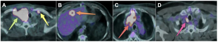 Figure 1
Peer Reviews
No public reviews on file for this paper yet. If you reviewed it on a platform where reviews are public (OpenReview, ICLR, NeurIPS, ICML), you can paste yours below so the community can read it here.
Videos
No videos yet. Explain this paper in a talk, walkthrough, or lecture? Add one.
Taxonomy
TopicsThyroid Cancer Diagnosis and Treatment · Thyroid and Parathyroid Surgery · Cancer Diagnosis and Treatment
Highlights
- •Papillary thyroid carcinoma is generally masked by anaplastic thyroid carcinoma (ATC)
- •Papillary thyroid carcinoma can persist despite treatment if the allelic variant is different than ATC
- •Horner syndrome is a rare complication of ATC prior to thyroidectomy
- •Liver and pancreas metastasis is rarely seen in ATC
Clinical RelevanceThis case reports a rare presentation of anaplastic thyroid carcinoma originating from a BRAF V600E sequence variant that was revealed to be masking a papillary thyroid carcinoma of independent tumor origin. This case emphasizes the need to identify each cancerous site’s origin for targeted treatment and identify causes of rare secondary symptoms.
Introduction
Anaplastic thyroid carcinoma (ATC) is the rarest yet most fatal thyroid cancer, being responsible for nearly 50% thyroid cancer–related deaths.1 This hyperactive malignancy is characterized by pleomorphic and undifferentiated cells, metastasis, and lymph node positivity commonly in the cervical region.1^,^2 The most prevalent symptoms of ATC include dyspnea, dysphagia, and voice hoarseness arising from local invasion of neighboring structures, although clinical presentation remains variable.2 Horner syndrome (HS) is a neurologic disruption that normally results in unilateral ptosis, miosis, and anhidrosis. Although it develops similarly, HS is highly unlikely to develop from ATC.3
It has been proposed that ATC develops from a preexisting differentiated thyroid cancer or nodule; however, its progression following treatment has not been observed to revert to a lower stage of thyroid cancer, ultimately presenting with a coexisting thyroid malignancy.1^,^4 We report a case of a patient who underwent targeted therapy for ATC, ultimately unmasking a comorbid wild-type papillary thyroid carcinoma (PTC), along with a rare manifestation of HS.
Case Report
A 68-year-old Hispanic man with past medical history of thoracic aortic aneurysm, chronic kidney disease, hyperlipidemia, and hypertension was referred for weight and type 2 diabetes management. Initial blood test results and measurements are found in Table 1. Outside of fatigue, he did not endorse symptoms indicative of a thyroid mass (eg, voice hoarseness, difficulty swallowing, or dyspnea), and his thyroid-stimulating hormone levels were unremarkable. After several months of using semaglutide 0.50 mg weekly, the patient’s parameters pertaining to management of diabetes and weight improved (Table 1), making him a suitable candidate for thoracic aortic aneurysm repair. Other prior medications included statins, antihypertensives, and loop diuretics.Table 1. Laboratory Test ResultsLaboratory test7 mo preoperatively2 mo preoperatively4 mo s/pReference valuesHbA1C (%)6.66.06.8<7% for well controlFasting glucose (mg/dL)159968365-99BMI (kg/m^2^)52.9247.5942.27See noteTSH (mIU/L)…0.891.860.40-4.50Free T_3_ (pg/mL)…3.72.92.3-4.2Abbreviations: BMI = body mass index (a value of >40 signifies Class 3 obesity); HbA1C = glycated hemoglobin; s/p = status post thoracic aorta aneurysm repair; TSH = thyroid-stimulating hormone; T_3_ = triiodothyronine.
Three months after undergoing successful aortic aneurysm repair and being initiated on apixaban 5 mg twice daily, cardiology ordered a routine postoperative computed tomography angiography (CTA), which incidentally revealed an rapidly enlarging mass from the inferior aspect of the right thyroid lobe measuring 5.6 × 9.4 cm (previously 3.5 × 2.2 cm), a new 2.1-cm hypodense mass in the left hepatic lobe suggestive of hepatic metastasis, and a 2.9 × 1.4–cm cystic lesion in the pancreatic head (previously 2.2 cm). Subsequent imaging confirmed increased progression of metastases (Fig. A through D).FigA, Positron emission tomography (PET)/computed tomography (CT) scan 2 weeks following fine-needle aspiration (FNA) diagnosis, confirming hypermetabolic activity in right thyroid (green arrow) as well as left supraclavicular lymph node (yellow arrow). The trachea was displaced due to enlarged thyroid mass. B, PET/CT scan 2 weeks following FNA diagnosis showing intense fluorodeoxyglucose (FDG)-avid lesion along with central necrosis located on the left side of the liver (orange arrow). C, PET/CT scan 2 weeks following FNA diagnosis, emphasizing increased FDG uptake causing mediastinal para-aortic lymphadenopathy along with thyroid mass extending inferiorly into the mediastinum (red arrow). D, PET/CT scan 1 month status post right thyroid lobectomy, emphasizing increased FDG uptake in right paratracheal lymph node (pink arrow) stemming from residual papillary thyroid carcinoma.
A fine-needle aspiration biopsy of the thyroid mass was performed after the CTA, initially suggesting poorly differentiated thyroid carcinoma (PDTC). Updated immunohistochemistry (Table 2) along with pleomorphic epithelioid cells and significant nuclear atypia confirmed ATC with a pathologic T4aN1bM1 stage IVC. Next-generation sequencing (NGS) was immediately started, identifying the BRAF V600E sequence variant (variant allele fraction of 0.532).Table 2. Immunohistochemical Results From Fine-Needle Aspiration of Right Thyroid MassImmunohistochemical markersExpressionMOC-31+CK5/6++p40NegativePAX8+ThyroglobulinNegativep53+++SOX10NegativeSmooth muscle actinNegativeS-100NegativeMIB-1 - Ki-67 proliferationApproximately 25%TTF-1++, immunopositivity; ++, focal weak to moderate immunopositivity; and +++, diffuse overexpression.
In the interim, the patient endorsed symptoms of hemoptysis, ptosis, miosis, and right-sided facial anhidrosis, the latter 3 consistent with HS. A magnetic resonance imaging scan of the brain was ordered and unremarkable, thus ruling out possible brain metastasis or oculomotor or trigeminal nerve impairment. Physical examination showed a noticeable cervical mass, and positron emission tomography scans revealed aggressive cancer progression causing bilateral cervical, supraclavicular, and mediastinal lymphadenopathy (Fig. B), alongside hypermetabolic bilateral pulmonary nodules consistent with ATC metastasis.
Following confirmation of the BRAF V600E variant, the patient discontinued both apixaban and semaglutide and was initiated on dabrafenib 150 mg twice daily and trametinib 2 mg daily. The patient’s hemoptysis and HS symptoms stopped within weeks. In addition to affected lymph nodes, areas with ATC metastasis (ie, liver, pancreas, and lung) had noticeably reduced metabolic activity, and the thyroid mass shrank to 2.3 × 5.3 cm, allowing for surgical resection. The patient’s appearance and physical examination noticeably improved during the targeted treatment.
The patient consented to a right thyroid lobectomy nearly 7 months after beginning neoadjuvant therapy. The final pathology report of the resected thyroid lobe revealed a residual tall cell variant PTC. Further positron emission tomography scans (Fig. C) revealed lymphadenopathy in the right paratracheal lymph node and level II lymph node. The residual PTC did not test positive for BRAF mutation burden in the NGS panel.
The patient’s thyroid markers have remained stable without supplementation but show diminished follicular cell characteristics (Table 3). The patient is currently involved in multidisciplinary discussion among his care team, considering a total thyroidectomy due to residual cancer.Table 3. Thyroid Markers Following LobectomyLaboratory test1 wk s/p3 mo s/pReference valuesTSH (mIU/L)2.082.140.40-4.50Free T_3_^(pg/mL)^3.23.12.3-4.2Thyroglobulin (ng/mL)0.51.03-40TgAb (IU/mL)210250≤1Abbreviations: s/p ^=^ status post right thyroid lobectomy; TgAb = thyroglobulin antibodies; TSH = thyroid-stimulating hormone; T_3_ = triiodothyronine.
Discussion
Although the process of ATC forming from a preexisting thyroid nodule or other follicular cell thyroid cancer has been recognized in the literature, we present a case where the successful targeted treatment of ATC led to unmasking a hidden comorbid PTC of independent tumor cell origin. Historically, the preexisting ATC following treatment is either resolved, unresolved leading to fatal outcomes, or comorbid with other thyroid cancers, such as PTC or PDTC.5^,^6 Despite the appearance of the residual PTC, the patient did not have any prior history of thyroid cancer, had only 1 suspicious nodule at time of ATC diagnosis, and was not observed to have multiple unique (differentiated and undifferentiated) thyroid malignancies at once prior to surgical resection. No thyroid imaging (eg, ultrasound or scans) was performed prior to postoperative CTA.
The patient using semaglutide (a glucagon-like peptide-1 receptor agonist) 10 months prior to thyroid cancer diagnosis poses a theoretical risk in developing a thyroid malignancy, as seen in animal studies. However, human data have been inconclusive in correlating to a higher incidence of any type of thyroid cancer.7^,^8
The immunohistochemistry of the patient’s fine-needle aspiration, as seen in Table 2, confirms immunopositivity of PAX8, diffuse p53 expression, and a relatively high Ki-67 proliferation index. This, along with thyroglobulin absence, supported the diagnosis of ATC.6 Although the immunopositivity of TTF-1 is atypical in ATC, it may persist if its positivity is present focally, the tissue displays prominent pleomorphism, and squamoid components of epithelioid cells are present, all of which occurred in the following case, thus ruling out the presence of PDTC in the patient.6^,^9
BRAF V600E sequence variations are very commonly observed in ATC.10 With it being the only positive variant identified, combination therapy of dabrafenib (BRAF inhibitor) and trametinib (inhibitor of mitogen-activated protein kinase or MEK) was used. This treatment has been shown to increase the median overall survival from a historically reported <6 months to approximately 14.5 months.11 The median overall survival is noted to be approximately 3 months for those diagnosed with stage IVC ATC.12 The patient’s first dose of BRAF/MEK inhibitors was recorded to be 10 months after his first visit, expected to be approaching over a year. Analysis of the residual PTC revealed that it did not test positive for the BRAF V600E allelic variant, explaining why the targeted therapeutic approach was ineffective toward it. Residual PTCs are generally excised because of total thyroidectomies being performed; however, there is very little literature discussing the significance of dealing with the cancer after a lobectomy.13 There is discussion of the patient undergoing radiation therapy due to proximal metastasis and lymphadenopathy due to the PTC.
The presence of HS in a preoperative setting is a rare complication of thyroid etiology with scant literature addressing the matter.3 There is much more discussion on HS development postoperatively, related to thyroid cancers in cases where either a neck dissection, thyroidectomy, or cervical lymphadenectomy is required. It is estimated the 1.3% of cases of HS stem from thyroid pathology, which does not exclude postoperative complications.14^,^15 ATC is expected to be the cause of <0.1% of all HS cases in a preoperative scenario. The patient’s enlarged thyroid mass (maximum dimensions at 5.6 × 9.4 cm) led to preganglionic neuron damage to his right oculosympathetic nerve pathway. It is recommended to treat HS by addressing the underlying cause first, and the patient’s HS was resolved after the reduction in size of his thyroid mass following BRAF/MEK inhibitor therapy. HS can be misdiagnosed with cranial nerve impairment (either oculomotor or trigeminal) and was ruled out following patient’s brain magnetic resonance imaging.16
Approximately 50% of ATC cases present with metastasis at diagnosis.17 The patient presented with common sites of metastasis in his lungs (78%) and cervical lymph nodes (51%) but also observed 2 other infrequent sites of metastatic progression in his liver (Fig. D) featuring (20%) and pancreas (4%).18 These 2 sites were concerning due to the patient’s history of comorbid diabetes and hyperlipidemia. All metastatic sites were resolved shortly after BRAF/MEK inhibitor treatment, and the patient has resumed his statin usage while adding empagliflozin/metformin to his regimen for further diabetic management.
ATC manifests and presents in a multitude of ways that should be clinically recognized, with increasing number of possibilities in malignancy progression. This case report highlights the complexity of diagnosing comorbid thyroid malignancies in a patient whose ATC was incidentally diagnosed with a routine CTA, ultimately masking the presence of any underlying cancer. NGS panel should be initiated early on in diagnosis of thyroid cancer, potentially salvaging the development of the cancer to rare metastatic sites depending on mutation burden. Although rare, clinicians should be aware of conditions caused by ATC tumor compression to cervical neuronal pathways such as HS. See Supplementary Figure “Visual Timeline.”
Conclusion
ATC presents with various unexpected modalities and can form due to aggressive dedifferentiation of follicular cells; however, coexisting thyroid malignancies from independent origins are rare and often indistinguishable. This case highlights the significance of early NGS testing and coordinated multidisciplinary care to detect masked malignancies and guide targeted therapy. Such approaches can help clinicians tailor treatment for aggressive thyroid cancers while providing researchers with data to refine diagnostics and optimize therapeutic strategies for better patient outcomes.
Disclosure
The authors have no conflicts of interest to disclose.
The reference list from the paper itself. Each links out to its DOI / PubMed record.
- 1Anaplastic thyroid cancer Limaiem F.Kashyap S.Naing P.T.Mathias P.M.Giwa A.O.https://www.ncbi.nlm.nih.gov/books/NBK 53817930844206 · pubmed ↗
- 2Rao S.N.Smallridge R.C.Anaplastic thyroid cancer: an update Best Pract Res Clin Endocrinol Metab 371202310167810.1016/j.beem.2022.10167835668021 · doi ↗ · pubmed ↗
- 3Donaldson J.F.Rodriguez-Gomez I.A.Parameswaran R.Rapidly enlarging neck masses of the thyroid with Horner’s syndrome: a concise clinical review Surgeon 13220151101152507393210.1016/j.surge.2014.06.010 · doi ↗ · pubmed ↗
- 4Tuttle R.M.Haugen B.Perrier N.D.Updated American Joint Committee on Cancer/tumor-node-metastasis staging system for differentiated and anaplastic thyroid cancer (eighth edition): what changed and why?Thyroid 27620177517562846358510.1089/thy.2017.0102 PMC 5467103 · doi ↗ · pubmed ↗
- 5Maniakas A.Dadu R.Busaidy N.L.Evaluation of overall survival in patients with anaplastic thyroid carcinoma, 2000-2019 JAMA Oncol 692020139714043276115310.1001/jamaoncol.2020.3362 PMC 7411939 · doi ↗ · pubmed ↗
- 6Xu B.Fuchs T.Dogan S.Dissecting anaplastic thyroid carcinoma: a comprehensive clinical, histologic, immunophenotypic, and molecular study of 360 cases Thyroid 30102020150515173228402010.1089/thy.2020.0086 PMC 7583343 · doi ↗ · pubmed ↗
- 7Pasternak B.Wintzell V.Hviid A.Glucagon-like peptide 1 receptor agonist use and risk of thyroid cancer: Scandinavian cohort study BMJ 3852024 e 07822510.1136/bmj-2023-078225 PMC 1100466938683947 · doi ↗ · pubmed ↗
- 8Bjerre Knudsen L.Madsen L.W.Andersen S.Glucagon-like peptide-1 receptor agonists activate rodent thyroid C-cells causing calcitonin release and C-cell proliferation Endocrinology 15142010147314862020315410.1210/en.2009-1272 · doi ↗ · pubmed ↗