Two new species of Periconia and a new record of Paramonodictys from Miscanthus sinensis in Guizhou Province, China

Abstract
Genes, proteins, chemicals, diseases, species, mutations and cell lines named across the full text — each resolved to its canonical identifier and authoritative record.
Click any figure to enlarge with its caption.
 Figure 1
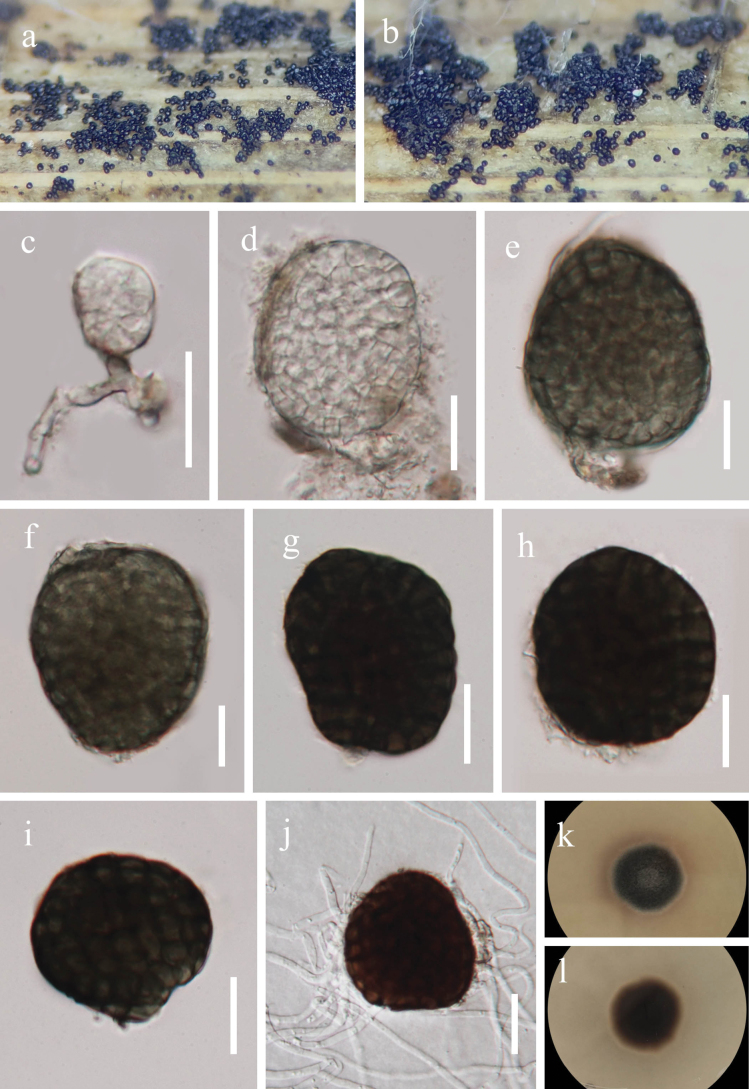 Figure 2
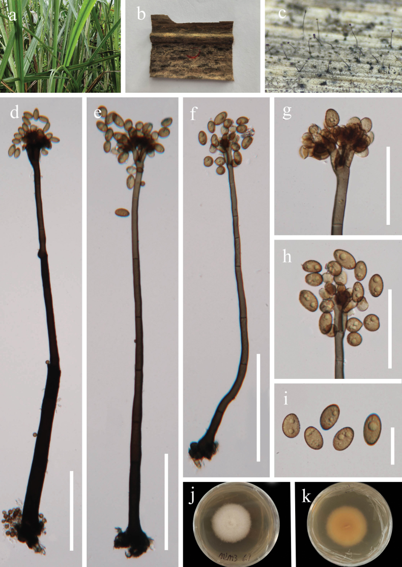 Figure 3
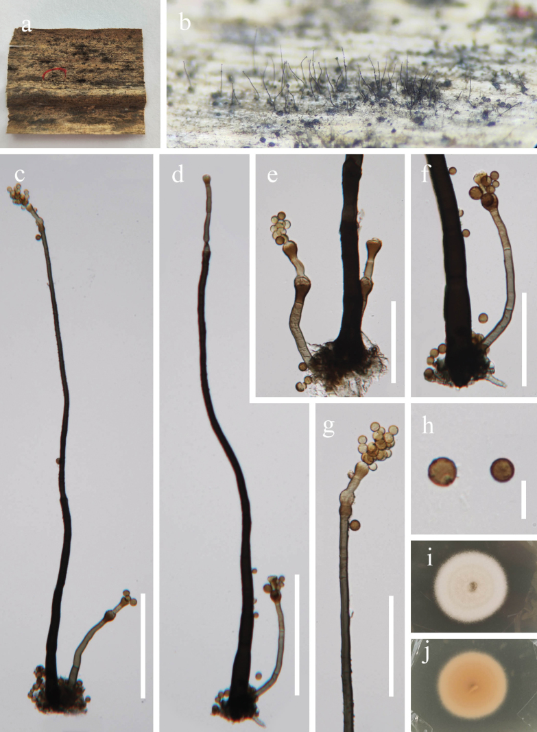 Figure 4
| Taxon | Strain | GenBank Accessions | |||
|---|---|---|---|---|---|
|
|
|
| |||
|
| CBS 266.62T |
|
|
|
|
|
| CBS 473.64 |
|
|
|
|
|
| KUNCC 10782 |
|
|
|
|
|
| KUNCC 10783 |
|
|
|
|
|
| KUNCC 10788T |
|
|
|
|
|
| BWL59 | – |
|
| – |
|
|
|
|
|
|
|
|
| – |
|
|
| |
|
| ZHKUCC 24-1234 | – |
|
| – |
|
| KUMCC 21-0343T |
|
|
|
|
|
| CGMCC 3.24437T |
|
|
|
|
|
| UESTCC 23.0085 |
|
|
|
|
|
|
|
|
|
| |
|
| KUMCC 21-0337T |
|
|
|
|
|
| KUMCC 21-0347 |
|
|
|
|
|
| CBS 321.79T | – |
|
| – |
|
| KUMCC 19-0174 | – |
|
|
|
|
| MFLUCC 19-0145T | – |
|
|
|
|
| NCYUCC 19-0186 | – |
|
|
|
|
| KUMCC 21-0470 |
|
|
|
|
|
| MFLUCC 21-0155T |
|
|
|
|
|
| MFLUCC 16-0912T | – |
|
|
|
|
| SNT19A |
|
|
|
|
|
| KUMCC 20-0265T |
|
|
|
|
|
| CBS 381.55 | – |
|
| – |
|
| ZHKUCC 24-1141 | – |
|
| – |
|
| CBS 129526T | – |
|
| – |
|
| MFLUCC 17-2292 |
|
|
|
|
|
| MFLUCC 19-0134 | – |
|
|
|
|
| MFLUCC 20-0172T | – |
|
| – |
|
| NCYUCC 19-0314 | – |
|
| – |
|
| UESTCC 22.0132 |
|
|
|
|
|
| UESTCC 22.0137 |
|
|
|
|
|
| UESTCC 22.0138 |
|
|
|
|
|
| LAMIC 110/16T | – |
|
| – |
|
| CPC 46238T | – |
|
| – |
|
| MFLU 19-2784T | – |
|
| – |
|
| CGMCC 3.23930T |
|
|
|
|
|
| UESTCC 22.0140 |
|
|
|
|
|
| UESTCC 22.0142 |
|
|
|
|
|
| KUMCC 21-0471 |
|
|
|
|
|
| MFLUCC 21-0164 |
|
|
|
|
|
| KUMCC 20-0266T |
|
|
|
|
|
| UESTCC 22.0133 |
|
|
|
|
|
| UESTCC 22.0144 |
|
|
|
|
|
| CBS 263.37 | – |
|
|
|
|
| ENCB 140251T | – |
|
| – |
|
| IOM 325319.2 | – |
|
| – |
|
| MFLUCC 17-1399 | – |
|
|
|
|
| MFLUCC 17-1679 | – | – |
|
|
|
| MFLUCC 15-0451 |
|
|
|
|
|
| MFLUCC 15-0453 | – |
|
|
|
|
| MFLUCC 15-0457T |
|
|
|
|
|
| CGMCC 3.23927T |
|
|
|
|
|
| CPC 32138T | – |
|
| – |
|
| MFLUCC 17-2584T |
| – |
|
|
|
|
|
|
|
| |
|
| MFLU 15-0058T |
|
|
|
|
|
| CBS 510.77 |
|
|
|
|
|
| KUNCC 23-13482T | – |
|
|
|
|
| MFLUCC 17-0087T |
|
|
| – |
|
| ZHKUCC 23-0995T |
|
|
|
|
|
| ZHKUCC 23-0996 |
|
|
|
|
|
| CBS 144017T | – |
| – | – |
|
| MFLUCC 21-0153 |
|
|
|
|
|
| CGMCC 3.23929T |
|
|
|
|
|
| CPC 45904T | – |
|
| – |
|
| CBS 322.79T | – |
|
| – |
|
| GZAAS 25-0735T |
|
|
|
|
|
| GZAAS 25-0733 |
|
|
|
|
|
|
|
|
|
|
|
|
|
|
|
|
|
|
|
| CBS 139698T |
|
|
|
|
|
| KUNCC 23-13549 | – |
|
| – |
|
| KUNCC 23-13550T | – |
|
|
|
|
| ZHKUCC 24-2101 |
|
|
|
|
|
| ZHKUCC 24-2102 |
|
|
|
|
|
| UESTCC 23.0456 |
|
|
|
|
|
| UESTCC 24.0232 |
|
|
|
|
|
| CBS 298.66 | – |
|
| – |
|
| CBS 583.66 | – |
|
| – |
|
| CGMCC 3.23931T |
|
|
|
|
|
| UESTCC 22.0145 |
|
|
|
|
|
| UESTCC 22.0146 |
|
|
|
|
|
| MFLUCC 18-0679 |
|
|
|
|
|
| CBS 292.36 | – |
|
| – |
|
|
|
|
|
| |
|
|
|
|
|
| |
|
| CBS 135663 |
|
|
| – |
|
| MFLUCC 15-0245 | – |
|
| – |
|
|
|
|
|
|
|
|
|
|
|
|
|
|
|
| KUNCC 24-17924 |
|
|
|
|
|
| KUNCC 24-17925 |
|
|
|
|
|
| KUNCC 24-17926 |
|
|
|
|
|
| KUNCC 24-17927 |
|
|
|
|
|
| CGMCC 3.25599T |
|
|
|
|
|
| CPC 37903T | – |
|
| – |
|
| ZHKUCC 24-1140 |
|
|
|
|
|
| CBS 149514T | – |
|
| – |
|
| MFLUCC14-0400T |
| – |
|
|
|
| CGMCC 3.23928T |
|
|
|
|
|
| CBS H-25197T | – |
|
| – |
|
| CBS 209.64T | – |
|
| – |
|
| DLUCC 0850 | – |
|
|
|
|
| H4151 |
|
|
|
|
|
| MAFF 243874 |
|
|
|
|
|
| UESTCC 22.0135 |
|
|
|
|
|
| UESTCC 22.0147 |
|
|
|
|
|
| CBS 139699T |
|
|
|
|
|
| JCM 13164 |
|
|
|
|
|
| JCM 13165 |
|
|
|
|
|
| CBS 320.79T | – |
|
| – |
|
| GJ374T |
|
|
| – |
|
|
|
|
|
| |
|
| CGMCC 3.25598T |
|
|
|
|
|
| CGMCC 3.23932T |
|
|
|
|
|
| MFLUCC 16-1098T | – |
|
|
|
|
| MFLUCC 17-0065T |
|
|
| – |
|
| KUMCC 20-0262T |
|
|
|
|
|
| CBS 120374T | – |
| – | – |
|
| MFLUCC 17-2158T |
|
|
|
|
|
| UESTCC 22.0136 |
|
|
|
|
|
| UESTCC 22.0148 |
|
|
|
|
|
| ZHKUCC 23-0999T |
|
|
|
|
|
| ZHKUCC 23-1000 |
|
|
|
|
|
| GZAAS25-0707T |
|
|
|
|
|
| GZAAS25-0734 | – |
|
| – |
|
| ZHKUCC 23-0997T |
|
|
|
|
|
| ZHKUCC 23-0998 |
|
|
|
|
|
| SICAUCC 23-0047T |
|
|
|
|
|
| SICAUCC 23-0147 |
|
|
|
|
|
| KUNCC 23-14259T | – |
|
|
|
|
| KUNCC 23-16910 | – |
|
|
|
- —Science and Technology Foundation of Guizhou Province 501100004001 https://ror.org/00kwnh405 http://doi.org/10.13039/501100004001
- —National Natural Science Foundation of China 501100001809 https://ror.org/01h0zpd94 http://doi.org/10.13039/501100001809
Peer Reviews
No public reviews on file for this paper yet. If you reviewed it on a platform where reviews are public (OpenReview, ICLR, NeurIPS, ICML), you can paste yours below so the community can read it here.
Videos
No videos yet. Explain this paper in a talk, walkthrough, or lecture? Add one.
Taxonomy
TopicsPlant Pathogens and Fungal Diseases · Plant and fungal interactions · Yeasts and Rust Fungi Studies
Introduction
Miscanthus belongs to the subtribe Saccharinae, tribe Andropogoneae, subfamily Panicoideae, and family Poaceae (Ge et al. 2017). It is a self-incompatible C4 perennial grass with high photosynthetic efficiency, high biomass yield, and strong environmental adaptability, and it is widely utilized as a biomass energy source and industrial feedstock (Robertson et al. 2017; Wang et al. 2021; Fu et al. 2023). The genus is mainly distributed in eastern Asia, Southeast Asia, and the Pacific islands, with a few species extending into tropical Africa (Sacks et al. 2012). In addition, Miscanthus hosts a diverse fungal community. During a fungal survey conducted in the Maolan National Nature Reserve, Guizhou Province, China, several saprophytic fungi resembling Periconia and Paramonodictys were isolated from dead leaves of Miscanthus sinensis.
Periconia (Periconiaceae, Pleosporales, Dothideomycetes, Ascomycota; Hyde et al. 2024) was introduced by Tode (1971) with Pe. lichenoides as the type species. The sexual morph is characterized by globose ascomata with a central ostiole, fissitunicate asci with short stalks, and broadly fusiform, 1-septate, hyaline ascospores with an entire sheath (Tanaka et al. 2015; Yang et al. 2022a). The asexual morph features macronematous, mononematous conidiophores; monoblastic to polyblastic, terminal or intercalary conidiogenous cells; and ellipsoidal to oblong, catenate or solitary, smooth or verruculose conidia (Ellis 1971; Markovskaja and Kačergius 2014; Hyde et al. 2018; Calvillo-Medina et al. 2020; Su et al. 2023; Pem et al. 2024). Members of Periconia are globally distributed and occur as endophytes, saprobes, and pathogens on various hosts, with most associated with graminaceous plants, but they also occur on submerged wood in freshwater habitats (Leukel 1948; Ellis 1971; Romero et al. 2001; Cai et al. 2003; Carmarán and Novas 2003; Markovskaja and Kačergius 2014; Yang et al. 2022a; Su et al. 2023; Shen et al. 2025; Tian et al. 2025; Zhang et al. 2025a).
Paramonodictys (Parabambusicolaceae, Pleosporales, Dothideomycetes, Ascomycota) was introduced by Hyde et al. (2020) and typified with P. solitarius. The asexual morph is hyphomycetous, characterized by superficial stromata that are subcylindrical or truncated-cone-shaped, lacking conidiophores, bearing monoblastic conidiogenous cells, and producing muriform, globose to subglobose, darkly pigmented conidia (Hyde et al. 2020; Xu et al. 2023; Zhang et al. 2023). The sexual morph remains unknown. All currently known species of Paramonodictys are saprophytic, occurring in freshwater and terrestrial habitats on hosts such as Mangifera, Paeonia, and various types of dead wood (Hyde et al. 2020; Yang et al. 2022b; Li et al. 2023; Xu et al. 2023; Zhang et al. 2023).
During our survey of fungal diversity associated with Miscanthus in Maolan, Guizhou Province, we collected three hyphomycetous taxa from the surface of dead leaves of Miscanthus sinensis. Based on morphological characteristics and phylogenetic analyses of combined ITS, LSU, SSU, and tef1-α sequence datasets, we identified two new species, Periconia guizhouensis and Pe. miscanthusensis, and recorded a new host association for Paramonodictys globosa. Full descriptions, photoplates illustrating macro- and micromorphological characteristics, and phylogenetic trees showing the placement of the new and known taxa are provided in this study.
Materials and methods
Sampling and examination of specimens
Specimens were collected from the Maolan National Nature Reserve, Libo County, Guizhou Province, China, at elevations ranging from 490–580 m (25°16'20"–25°16'25" N, 108°2'20"–108°3'60" E). Samples were placed in sterile, moistened plastic bags and transported to the laboratory for detailed examination. Fresh materials were dissected and observed for morphological characteristics, including the structures of conidiophores, conidiogenous cells, and conidia, using stereo microscopes (SMZ 745 and SMZ 800N, Nikon, Tokyo, Japan) and an ECLIPSE Ni compound microscope (Nikon, Tokyo, Japan). Measurements were obtained using Tarosoft Image Framework software, and photoplates were assembled using Adobe Photoshop 2025 (version 26.5; Adobe Systems, CA, USA).
Single-spore isolation was conducted following the protocol of Senanayake et al. (2020). The resulting cultures were incubated at 28 °C under continuous light, and colony morphology was observed and recorded systematically. Voucher specimens were deposited in the Herbarium of the Kunming Institute of Botany (Herb. HKAS), Chinese Academy of Sciences, and living cultures were preserved in the Guizhou Culture Collection (GZCC). Registration numbers from Index Fungorum and the Facesoffungi database (Jayasiri et al. 2015) were obtained for each taxon. The new fungal species were introduced following the guidelines of Chethana et al. (2021).
DNA extraction, PCR amplification, and sequencing
Fresh mycelia were scraped from PDA plates using sterile toothpicks and transferred into 1.5 mL microcentrifuge tubes. Genomic DNA was extracted using the Ezup Column Fungi Genomic DNA Purification Kit according to the manufacturer’s instructions. PCR amplification targeted four loci: the internal transcribed spacer (ITS), large subunit rDNA (LSU), small subunit rDNA (SSU), and translation elongation factor 1-alpha (tef1-α), using the primer pairs ITS5/ITS4 (White et al. 1990), LR0R/LR5 (Vilgalys and Hester 1990), NS1/NS4 (White et al. 1990), and TEF1-983F/TEF1-2218R (Carbone and Kohn 1999), respectively.
PCR amplifications were performed in 25 μL reaction mixtures containing 21 µL of 1.1× T3 Super PCR Mix (Tsingke Biotechnology Co., Ltd., Chengdu), 1 µL of each primer, and 2 µL of genomic DNA. Reactions were run on a JS-G9612 PCR thermocycler (Shanghai Peiqing Technology Co., Ltd., Shanghai, China). Cycling conditions for ITS, LSU, SSU, and tef1-α followed the protocols described by Ma et al. (2023). PCR products were visualized on 1% agarose gels and subsequently purified and sequenced by Tsingke Biotechnology Co., Ltd. (Chengdu, China). Newly generated sequences were submitted to GenBank (https://ncbi.nlm.nih.gov/WebSub/).
Phylogenetic analyses
BioEdit v.7.0.5.3 (Hall 1999) was used to examine raw sequence chromatograms for overall quality, including the detection of base-calling errors, ambiguities, and possible contamination. Forward and reverse reads were assembled using SeqMan v.7.0.0 (DNASTAR, Madison, WI, USA; Swindell and Plasterer 1997). Reference sequences for phylogenetic analyses were obtained from GenBank (Table 1) and downloaded using the One-click Fungal Phylogenetic Tool (OFPT) (Zeng et al. 2023). Each gene region was aligned using the online MAFFT v.7 server, and the alignments were subsequently trimmed and refined with trimAl (Capella-Gutiérrez et al. 2009; Katoh and Standley 2013). Multi-gene alignments were combined using SequenceMatrix 1.7.8 (Vaidya et al. 2011). Maximum likelihood (ML) analyses were performed using the IQ-TREE web server (http://iqtree.cibiv.univie.ac.at/), with the best-fit substitution models automatically tested. Ultrafast bootstrap (BS) analysis was implemented with 1,000 replicates. Maximum likelihood bootstrap values (ML-BS) equal to or greater than 75% are marked near each node. Bayesian inference analyses were conducted using MrBayes v.3.2.7a on the CIPRES web portal (http://www.phylo.org/portal2/, accessed on 9 November 2025). The optimal nucleotide substitution models inferred for each locus were TNe+R3 for LSU, GTR+F+R3 for ITS, TNe+R2 for SSU, and GTR+F+I+G4 for tef1-α.
The phylogenetic tree was visualized and edited using FigTree v.1.4.0, and the final graphic design and figure layouts were completed using Adobe Photoshop 2024 and Adobe Illustrator 2021 (Adobe Systems, San Jose, CA, USA).
Results
Phylogenetic analysis
Partial ITS, LSU, SSU, and tef1-α nucleotide sequences were used to determine the phylogenetic positions of the new collections. The dataset comprised sequences from 134 isolates representing 71 Periconia and six Paramonodictys species. Massarina cisti (CBS 266.62) and M. eburnea (CBS 473.64) were selected as the outgroup taxa. The concatenated sequence matrix included ITS (1–511 bp), LSU (512–1,349 bp), SSU (1,350–2,347 bp), and tef1-α (2,348–3,240 bp), with gaps included. The concatenated ITS, LSU, SSU, and tef1-α datasets were analyzed using both maximum likelihood (ML) and Bayesian inference (BI) methods, which yielded similar tree topologies.
Phylogenetic relationships among Pleosporales species were well resolved in the multi-gene phylogenetic analysis. The resulting phylogenetic tree confirmed that the two newly obtained isolates of Periconia guizhouensis (GZCC 25-0766 and GZCC 25-27613) formed a distinct clade. They clustered with Pe. aquatica (MFLUCC 16-0912), Pe. imperatae (CGMCC 3.23931, UESTCC 22.0145, UESTCC 22.0146), Pe. motuoensis (KUNCC24-17925, KUNCC24-17924, KUNCC24-17927, KUNCC24-17926), Pe. prolifica (CBS 209.64), Pe. spodiopogonis (CGMCC 3.23932), and Pe. submersa (MFLUCC 16-1098), supported by 100% MLB/1.00 BYPP. Likewise, two isolates of Pe. miscanthusensis (GZCC 25-0767 and GZCC 25-27614) clustered together and were sister to Pe. neominutissima (CBS 149514), supported by 85% ML-BS (Fig. 1). Additionally, Paramonodictys globosa (GZCC 25-0768) grouped with P. globosa (GZCC 23-0594, BWL59, and ZHKUCC 24-1234), receiving 88% ML-BS/1.00 BYPP support.
Taxonomy
Paramonodictys
globosa
Taxon classificationFungiPleosporalesParabambusicolaceae
Jian Ma, Y.Zhe Zhang & Y.Z. Lu
CB390802-B774-554D-AC26-273AEF29B362
Fungal Names: FN 571668
Facesoffungi Number: FoF19046
Paramonodictys globosa Jian Ma, Y.Zhe Zhang & Y.Z. Lu, in Zhang, Chen, Ma, Lu, Chen and Liu, Frontiers Microbiol. 14 (no. 1253239): 9 (2023).
Description.
Saprobic on dead leaves of Miscanthus sinensis. Sexual morph: Undetermined. Asexual morph: Hyphomycetous. Colonies on natural substrate effuses, superficial, black. Mycelium partly immersed, branched, septate, smooth, hyaline to pale brown. Conidiophores are absent. Conidiogenous cells holoblastic, integrated, terminal, cylindrical, short, hyaline to brown, often arising directly on the superficial mycelium. Conidia 35–65 × 35–50 µm (x̄ = 50 × 44 μm, n = 30), solitary, irregular subglobose or globose, hyaline to olivaceous brown to dark brown, muriform, thickened and darkened at the septa.
Maximum likelihood (ML) majority-rule consensus tree based on ITS, LSU, SSU, and tef1-α sequence data. ML bootstrap support values (ML-BS ≥ 75%) and Bayesian posterior probabilities (BYPP ≥ 0.95) are indicated below or above the nodes. Ex-type strains are shown in bold and marked with T, and newly generated sequences are shown in red.
Paramonodictys globosa (GZAAS 25-07813) a, b. Colonies on host surface; c. Conidiophores, conidiogenous cells, and conidia; d–i. Conidia; j. Germinated conidium; k, l. Colony on PDA (above and below). Scale bars: 20 μm (c–j).
Cultural characteristics.
Conidia germinating on PDA medium within 24 h and germ tube produced from around of conidium. Colonies on PDA medium reaching 15 mm diam in 10 days at 28 °C in natural light, dense, circular, with entire edge, olivaceous brown; reverse brown.
Material examined.
China, • Guizhou Province, Qiannan Buyi and Miao Autonomous Prefecture, Libo County, Guizhou Maolan National Nature Reserve, 25°16'23"N, 108°3'56"E, on dead leaves of Miscanthus sinensis, 31 May 2025, Xingjuan Xiao, MLM5 (GZAAS 25-07813), living culture GZCC 25-0768.
Notes.
Paramonodictys globosa was introduced by Zhang et al. (2023) from submerged decaying wood in a freshwater stream in Guangxi Zhuang Autonomous Region, China. Our collection was obtained from dead leaves of Miscanthus sinensis in a terrestrial habitat in Guizhou, China. Morphologically, our specimen is similar to P. globosa (HKAS 129169) in having solitary, irregularly subglobose or globose, muriform conidia with thickened and darkened septa (35–65 × 35–50 µm vs. 34–65 × 24–60 µm) (Zhang et al. 2023). Moreover, comparisons of ITS, LSU, and tef1-α sequences revealed differences of only 4 bp (1 gap, total 606 bp), 4 bp (0 gaps, total 822 bp), and 16 bp (no gaps, total 887 bp) between our strain GZCC 25-0768 and the ex-type strain (GZCC 23-0594) of P. globosa, respectively. Based on the combined morphological characteristics and molecular evidence, we identify our collection as Paramonodictys globosa, representing the first report of this species from M. sinensis.
Periconia
guizhouensis
Taxon classificationFungiPleosporalesPericoniaceae
L.J. Zhang, Y.Z. Lu & X.J. Xiao sp. nov.
0908E488-FA7B-5766-9D6B-F6B2CBDB4A47
Index Fungorum: IF904704
Facesoffungi Number: FoF19047
Etymology.
Referring to the collecting location at Guizhou Province, where the holotype was discovered.
Holotype.
GZAAS 25-07814
Description.
Saprobic on dead leaves of Miscanthus sinensis. Sexual morph: Undetermined. Asexual morph: Colonies on natural substrate effuse, superficial, hairy, dark brown to black. Mycelium immersed, branched, septate, smooth, brown to dark brown. Conidiophores 400–750 × 12–22 μm (x̄ = 630 × 18 µm, n = 20), macronematous, mononematous, erect, straight to slightly curved, solitary, cylindrical, apically branched, brown to dark brown, multi-septate, smooth-walled. Conidiogenous cells 6–16 × 5–10 μm (x̄ = 10 × 7 µm, n = 20), polyblastic, terminal at the apex, ovoid to subglobose, light to dark brown. Conidia 12–18 × 7–10 μm (x̄ = 15 × 8.5 µm, n = 30), solitary or in short chains, globose to fusiform, guttulate, smooth to verrucose, aseptate, pale brown to dark brown.
Periconia guizhouensis (GZAAS 25-07814, holotype). a. Host of Miscanthus sinensis; b. Colonies on a decaying leaf of Miscanthus sinensis; c. Conidiophores on host surface; d–f. Conidiophore, conidiogenous cells, and conidia; g, h. Conidiogenous cells and conidia; i. Conidia; j, k. Colony on PDA (above and below). Scale bars: 100 μm (d–f); 50 μm (g, h); 20 μm (i).
Cultural characteristics.
Conidia germinated on PDA medium within 12 h, and germ tubes produced from around of conidium. Colonies on PDA medium reaching 33 mm diam in 10 days at 28 °C in natural light, dense, circular, with entire edge, white reverse pale brown.
Material examined.
China, • Guizhou Province, Qiannan Buyi and Miao Autonomous Prefecture, Libo County, Guizhou Maolan National Nature Reserve, 25°16'23"N, 108°3'56"E, on dead leaves of Miscanthus sinensis, 31 May 2025, Xingjuan Xiao, MLM3 (GZAAS 25-07814, holotype), ex-type living culture GZCC 25-0766, other living culture GZCC 25-27613.
Notes.
In the phylogenetic tree, Periconia guizhouensis (GZCC 25-0766 and GZCC 25-27613) formed a distinct lineage within Periconia and clustered with Pe. prolifica (CBS 209.64) and Pe. aquatica (MFLUCC 16-0912) (Fig. 1). The ITS and LSU sequences of Pe. guizhouensis (GZCC 25-0766, type) differ from those of Pe. prolifica (CBS 209.64, type) by 23 bp (498/535 bp, 14 gaps) and 3 bp (854/857 bp, 0 gaps), respectively; Pe. prolifica lacks SSU and tef1-α sequences. Periconia guizhouensis (GZCC 25-0766, type) and Pe. aquatica (MFLUCC 16-0912, type) differ by 12 bp (438/452 bp, 2 gaps) in ITS, 3 bp (786/789 bp, 0 gaps) in LSU, and 25 bp (809/834 bp, 0 gaps) in tef1-α; Pe. aquatica lacks an SSU sequence. Morphologically, Pe. prolifica shares similar characteristics with our collection in having subglobose, aseptate conidia. However, Pe. guizhouensis can be readily distinguished by its verrucose conidia, whereas the conidia of Pe. prolifica are consistently smooth-walled (Bunning and Griffiths 1982; Markovskaja and Kačergius 2014). Periconia guizhouensis differs from Pe. aquatica in that the latter has unbranched, smaller conidiophores (400–750 × 12–22 μm vs. Conidiophores 370–454 × 8.5–11 μm) and smaller conidia (12–18 × 7–10 μm vs. 10–12 × 6–7 µm) with more obviously minutely verruculose to shortly echinulate ornamentation (Hyde et al. 2017). Moreover, Periconia guizhouensis can be easily distinguished from Pe. imperatae, Pe. motuoensis, Pe. spodiopogonis, and Pe. submersa by its globose to fusiform, guttulate, aseptate conidia with inconspicuous verrucosity (Hyde et al. 2017; Su et al. 2023; Sun et al. 2025). Therefore, based on morphological characteristics and molecular evidence, Periconia guizhouensis is introduced as a new species.
Periconia
miscanthusensis
Taxon classificationFungiPleosporalesPericoniaceae
L.J. Zhang, Y.Z. Lu & X.J. Xiao sp. nov.
43E00B18-B573-52AA-80C7-DADEAB68B746
Index Fungorum: IF904705
Facesoffungi Number: FoF19048
Etymology.
The specific epithet miscanthusensis refers to Miscanthus, the grass genus from which the type strain was isolated.
Holotype.
GZAAS 25-07815
Description.
Saprobic on dead leaves of Miscanthus sinensis. Sexual morph: Undetermined. Asexual morph: Colonies on natural substrate effuse, superficial, hairy, dark brown to black. Mycelium immersed, branched, septate, smooth, brown. Conidiophores 350–900 × 10–20 μm (x̄ = 540 × 14 µm, n = 20), macronematous, mononematous, erect, straight to slightly curved, cylindrical, unbranched, brown to dark brown, multi-septate, smooth-walled. Typically, a single longer conidiophore is present on the stroma, accompanied by 1–2 shorter conidiophores 70–125 × 4–17 μm (x̄ = 98 × 7 µm, n = 20). Conidiogenous cells 7–14 × 6–12 μm (x̄ = 11 × 9 µm, n = 20), polyblastic, proliferous, terminal at the apex, globose to subglobose, light to dark brown. Conidia 4–9 × 4–7 μm (x̄ = 6 × 5.5 µm, n = 30), compact chains, globose, smooth to verrucose, aseptate, pale brown to dark brown.
Periconia miscanthusensis (GZAAS 25-07815, holotype). a. Colonies on a decaying leaf of Miscanthus sinensis; b. Conidiophores on host surface; c–f. Conidiophore, conidiogenous cells, and conidia; g. Conidiogenous cells and conidia; h. Conidia; i, j. Colony on PDA (above and below). Scale bars: 100 μm (c, d); 50 μm (e–g); 10 μm (h).
Cultural characteristics.
Conidia germinated on PDA medium within 12 h, and germ tubes produced from around of conidium. Colonies on PDA medium reaching 18 mm diam in 10 days at 28 °C in natural light, dense, circular, with entire edge, white reverse pale brown.
Material examined.
China, • Guizhou Province, Qiannan Buyi and Miao Autonomous Prefecture, Libo County, Guizhou Maolan National Nature Reserve, 25°16'23"N, 108°3'56"E, on dead leaves of Miscanthus sinensis, 31 May 2025, Xingjuan Xiao, MLM4 (GZAAS 25-07815, holotype), ex-type living strain GZCC 25-0767, other living culture GZCC 25-27614.
Notes.
Phylogenetic analyses revealed that our strains (GZCC 25-0767 and GZCC 25-27614) formed a distinct clade and were sister to Periconia neominutissima (CBS 149514) (Fig. 1). Pairwise nucleotide comparisons between Pe. miscanthusensis (GZCC 25-0767, type) and Pe. neominutissima (CBS 149514, type) showed sequence similarities of 95% (480/503 bp, 9 gaps) in ITS and 99% (835/843 bp, 0 gaps) in LSU, indicating significant genetic differences between the two species. Morphologically, Pe. miscanthusensis resembles Pe. miscanthusensis in having apically branched conidiophores that produce acropetal chains and globose, verrucose, aseptate conidia. However, our species differs by possessing longer conidiophores (350–900 × 10–20 μm vs. 700 × 12–15 µm), larger conidiogenous cells (7–14 × 6–12 μm vs. 6–8 × 4–5 µm), and a single prominent conidiophore accompanied by 1–2 shorter conidiophores (Crous et al. 2023). Therefore, based on morphological characteristics and molecular evidence, Periconia miscanthusensis is introduced as a novel species.
Discussion
Nannizzi (1934) established Periconiaceae with Periconia as the type genus, but the family was overlooked in modern taxonomic treatments, and its members were traditionally placed in Massarinaceae. Tanaka et al. (2015) revived this family based on phylogenetic analyses and accepted four genera, Bambusistroma, Flavomyces, Noosia, and Periconia. Later, Yang et al. (2022b) re-evaluated Bambusistroma and Noosia based on morphological comparisons and multi-gene phylogenetic analyses and subsequently synonymized both genera under Periconia. Consequently, Periconiaceae currently comprises only two genera, Flavomyces and Periconia (Hyde et al. 2024). However, because Flavomyces lacks clear morphological diagnostic traits and clusters with Periconia species in phylogenetic analyses, its generic status remains uncertain and requires further clarification. According to Species Fungorum (https://www.speciesfungorum.org/Names/Names.asp, accessed on 20 November 2025), Periconia contains 206 epithets. Among these, 29 species have been transferred to other genera within Dothideomycetes, Pezizomycetes, Sordariomycetes, and Ascomycota genera incertae sedis, and seven species have been synonymized under other Periconia species (Persoon 1801; Mason and Ellis 1953; Benjamin and Hesseltine 1959; Ellis 1971; Illman and White 1984; Okada et al. 2000; Partridge and Morgan-Jones 2002; Schubert et al. 2007; Linnakoski et al. 2012; Yang et al. 2022b; Zhang et al. 2025b).
During the past three years, 30 species of Periconia have been reported (Su et al. 2023; Cai et al. 2024; Liao et al. 2024; Phookamsak et al. 2024; He et al. 2025; Sun et al. 2025; Wijesinghe et al. 2025; Zhang et al. 2025a; Zhang et al. 2025b). Most species in the genus have been accepted solely based on morphological characters. Molecular data are available for only 68 species, and for the majority of these species, only ITS and LSU sequences have been generated, whereas more informative markers (e.g., mtSSU, rpb1, rpb2, tef1-α, and tub2, remain unavailable (Yang et al. 2022b; Liao et al. 2024; Sun et al. 2025; Wijesinghe et al. 2025; Zhang et al. 2025b). Moreover, several Periconia species exhibit noticeable intraspecific variation, including P. byssoides, P. cortaderiae, P. prolifica, and P. pseudobyssoides (Yang et al. 2022b). Therefore, future studies require additional phylogenetic markers to more accurately resolve species boundaries within this highly diverse genus (Yang et al. 2022b; Liao et al. 2024; He et al. 2025; Zhang et al. 2025a).
Most Periconia species have been recorded from temperate and tropical regions (Numata et al. 1997; Kolomiets et al. 2008; Sarkar et al. 2019; Gunasekaran et al. 2021; Liu et al. 2024; Madagammana et al. 2024; He et al. 2025). The genus is widely distributed and has been reported as endophytes, plant pathogens, and saprobes across terrestrial, mangrove, marine, and freshwater habitats (Odvody et al. 1977; Romero et al. 2001; Alias and Jones 2000; Goga 2000; Kohlmeyer 1977; Luo et al. 2004; Liu et al. 2017a, b; Hyde et al. 2017; Jayasiri et al. 2019; Sarkar et al. 2019; Shen et al. 2025; Bao et al. 2025; Hongsanan et al. 2025; Tian et al. 2025). In addition to their ecological diversity, species of this genus produce secondary metabolites with diverse biological activities, including antimicrobial, anti-human immunodeficiency virus (HIV), cytotoxic, and anti-inflammatory effects (Numata et al. 1997; Teles et al. 2006; Bhilabutra et al. 2007; Wu et al. 2015a, b; Liu et al. 2017b; Azhari and Supratman 2021). These findings underscore the ecological and biochemical significance of Periconia and highlight the need for continued taxonomic and ecological investigations of newly discovered species.
Periconia prolifica has been reported from a wide variety of substrates in Saudi Arabia, including driftwood in Jeddah, mangrove wood, and material from the Red Sea coast, as well as seawater and sea foam along the Arabian Gulf coast (Aleem 1979; Bunning and Griffiths 1982; Bokhary et al. 1992; Vishwakiran et al. 2001; Hodhod 2013; Hodhod et al. 2023). It has also been recorded from diverse geographical regions worldwide, including Brazil, China, Egypt, Ghana, Indonesia, Japan, Kuwait, Malaysia, Mexico, Saudi Arabia, South Africa, and the USA, on substrates such as decayed intertidal mangrove wood and seedlings (Aleem 1979; Vrijmoed et al. 1982; Vishwakiran et al. 2001; Abdel-Wahab 2005; Pang et al. 2011; Hodhod et al. 2023). However, its taxonomic position has long been controversial. The sexual morph of Pe. prolifica was originally classified in Halosphaeria as H. cucullata (Kohlm.) Kohlm. (Kohlmeyer 1972) but was later transferred to Okeanomyces (Halosphaeriaceae, Microascales) by Pang et al. (2004) based on its conidiogenetic features. In this study, the ex-type strain of Pe. prolifica formed a sister clade with our newly collected species, Pe. guizhouensis (GZCC 25-0766 and GZCC 25-27613) (Fig. 1). Morphologically, our species produces acropetal conidial chains from conidiogenous cells on conidiophores, whereas Pe. prolifica forms basipetal conidial chains directly on the wood substrate (Bunning and Griffiths 1982; Carmarán and Novas 2003; Hodhod et al. 2023). These morphological differences may reflect adaptations to distinct habitats. Thus, both multi-gene phylogenetic analyses and detailed morphological observations are essential for robust fungal taxonomy.
Supplementary Material
XML Treatment for Paramonodictys globosa
XML Treatment for Periconia guizhouensis
XML Treatment for Periconia miscanthusensis
The reference list from the paper itself. Each links out to its DOI / PubMed record.
- 1Abdel-Wahab MA (2005) Diversity of marine fungi from Egyptian Red Sea mangroves. Botanica Marina 48. 10.1515/bot.2005.047 · doi ↗
- 2Aleem AA (1979) A contribution to the study of seagrasses along the Red Sea Coast of Saudi Arabia. Aquatic Botany 7: 71–78. 10.1016/0304-3770(79)90009-3 · doi ↗
- 3Azhari A, Supratman U (2021) The chemistry and pharmacology of fungal genus Periconia: A review. Scientia Pharmaceutica 89: 1–34. 10.3390/scipharm 89030034 · doi ↗
- 4Bao DF, Zhang JY, Lu YZ, Tian XG, Hyde KD, Liu NG, Xiao YP, Luo ZL, Karunarathna SC, Samarakoon MC, Xiao XJ, Ma J, Yang Y, Han JJ, Zhao GL, He ZJ, Wen TC, Liu ZH, Kang JC (2025) Endophytic fungi associated with medicinal ferns in Guizhou Province, China I: Morpho-molecular characterization of culturable endophytic fungi associated with Dicranopteris spp. Mycosphere: Journal of Fungal Biology 16(1): 2259–2455. 10.5943/mycosphere/16/1/13 · doi ↗
- 5Benjamin CR, Hesseltine C (1959) Studies on the genus Phycomyces. Mycologia 51: 751–771. 10.1080/00275514.1959.12024858 · doi ↗
- 6Bhilabutra W, Techowisan T, Peberdy JF, Lumyong S (2007) Antimicrobial activity of bioactive compounds from Periconia siamensis CMUGE 015. The Journal of Microbiology 2: 749–755. 10.3923/jm.2007.749.755 · doi ↗
- 7Bunning S, Griffiths D (1982) Spore development in Periconia species: IP >P. Prolifica and P. macrospinosa. Transactions of the British Mycological Society 78: 147–159. 10.1016/S 0007-1536(82)80087-9 · doi ↗
- 8Cai T, He SC, Yu FM, Li CJY, Wang ZY, Ma C, Zhang Y, Zhao Q (2024) Periconia shannanensis sp. nov. (Periconiaceae, Pleosporales) from Xizang, China. Phytotaxa 664: 249–262. 10.11646/phytotaxa.664.4.2 · doi ↗